Observing the growth of individual actin laments in cell extracts by time-lapse atomic force microscopy
←
→
Page content transcription
If your browser does not render page correctly, please read the page content below
FEBS 27566                                                                                                        FEBS Letters 551 (2003) 25^28

   Observing the growth of individual actin ¢laments in cell extracts by
                   time-lapse atomic force microscopy
 Tiina Lehtoa;b , Marta Miaczynskab , Marino Zerialb , Daniel J. Mu«llera;b; , Fedor Severinb;
                                         a
                                           BIOTEC, Technical University Dresden, 01609 Dresden, Germany
               b
                   Max Planck Institute of Molecular Cell Biology and Genetics, Pfotenhauerstrasse 108, D-01307 Dresden, Germany

                                                  Received 30 June 2003; accepted 13 July 2003

                                                       First published online 14 August 2003

                                                            Edited by Amy McGough

                                                                            locomotion, cytokinesis and certain types of organelle motility
Abstract High-resolution atomic force microscopy (AFM) was
applied to directly observe the dynamic assembly of single actin            inside the cell (see [3] for review). So far imaging of individual
¢laments in HeLa cell extracts in vitro. The F-actin ¢laments               actin ¢lament dynamics in vitro has been performed using
established a dynamic network and formed di¡erent types of                  light microscopy [4]. In contrast to optical microscopy, the
junctions and branches. The connections of this network were                exceptionally high signal-to-noise ratio of the atomic force
X-, Y- or T-shaped. It was found that the actin ¢laments were               microscope (AFM [5]) allows the observation of single unla-
densely covered by endosomes and vesicles from the cell extract,            beled macromolecules in a heterogeneous environment [6].
which are thought to stabilize their structures. Using time-lapse           AFM imaging of biological samples is performed in bu¡er
AFM, the growth, shrinkage, branching and the interaction of                solution and at ambient temperatures [7], which allows the
actin ¢laments with endosomes could be characterized. Our re-               imaging of living cells [8] and single proteins at work [9,10].
sults indicate that the majority of F-actin ¢laments are static in
                                                                            Such experiments can be performed over a time range of
HeLa extract and that only a minor fraction of ¢laments under-
go dynamic changes. Furthermore, the AFM imaging approach                   several hours without destruction of individual proteins or
not only provides unique insights into the assembly and dynam-              disturbing their inherent assembly. We report here an ap-
ics of actin networks; it also builds an avenue to study in vitro           proach to study the dynamics of individual actin ¢laments
assays of complex biological systems.                                       in the cytosol by AFM. The presented method allows visual-
. 2003 Federation of European Biochemical Societies. Pub-                   izing the growth/shrinkage of individual ¢laments in physio-
lished by Elsevier B.V. All rights reserved.                                logically relevant bu¡er solution, their formation of networks
                                                                            and their association with membrane organelles.
Key words: Actin; Atomic force microscopy; Branching ;
Cell extract; Endosome                                                      2. Materials and methods

                                                                            2.1. Sample preparation
1. Introduction                                                                HeLa cytosol and endosomes were prepared as described in [11]
                                                                               First, 20 Wl of endosomes were adsorbed to the support for 5 min.
                                                                            Then the liquid was aspirated and 50 Wl of HeLa cytosol was added.
   Fluorescence microscopy, frequently used in modern bio-                  To verify that the observed ¢laments were F-actin we tried to inhibit
logical research, requires labeling the molecules of particular             the ¢lament growth by 5 Wg/ml cytochalasin D. Alternatively, we
interest with £uorophores. These chemically ¢xed labels have                omitted ATP from the reaction. In both cases, no ¢laments were
                                                                            detected, indicating that the observed ¢laments were polymerized
the potential to in£uence the native behavior of the labeled                from actin.
molecule [1,2]. Chemical reactions limit the lifetime of the
£uorophore and thereby the visualization process. Most im-                  2.2. AFM
portantly however, molecules other than the labeled ones will                 The AFM (Nanoscope III, Digital Instruments, Santa Barbara,
                                                                            CA, USA) was operated in bu¡er solution using the standard £uid
not be visualized by optical methods. To overcome this limi-
                                                                            cell, without an O-ring. Imaging was performed in tapping mode. The
tation multicolor imaging by the use of various £uorophores                 oxide-sharpened Si3 N4 cantilevers (OMCL TR400PS, Olympus, To-
allows simultaneous imaging of di¡erent biological structures.              kyo, Japan) employed had a nominal force constant of 0.09 N/m and
However, the number of colors that are simultaneously detect-               a resonance frequency close to 9 kHz in water. The microscope was
able is presently limited to a few and the selection of struc-              operated at conditions (tapping frequency close to resonance and
                                                                            amplitude) which allowed high precision control of the imaging pro-
tures that may be observed has to be done carefully.                        cess and the applied force to be 9 100 pN [12]. All samples were
   Actin in cells polymerizes into ¢laments (F-actin). In non-              imaged and manipulated in bu¡er solution at 21‡C.
muscle cells F-actin is organized mostly in bundles (stress
¢bers) and the di¡use network close to the cell cortex. The
dynamic growth behavior of F-actin provides the basis for cell              3. Results and discussion

                                                                            3.1. Observing the polymerization of individual actin ¢laments
*Corresponding author. Fax: (49)-351-210 2020.                                 Within the past decade AFM has become an established
**Corresponding author. Fax: (49)-351-210 2020.                             method to image biological objects under native conditions
E-mail addresses: mueller@mpi-cbg.de (D.J. Mu«ller),
severin@mpi-cbg.de (F. Severin).
                                                                            at (sub-)nanometer resolution. In many AFM preparations
                                                                            free particles and proteins resulting from the bu¡er solution
Abbreviations: AFM, atomic force microscopy                                 easily contaminate the AFM tip [13], thereby signi¢cantly

0014-5793 / 03 / $22.00 I 2003 Federation of European Biochemical Societies. Published by Elsevier B.V. All rights reserved.
doi:10.1016/S0014-5793(03)00867-6

                                              FEBS 27566 27-8-03 Cyaan Magenta Geel Zwart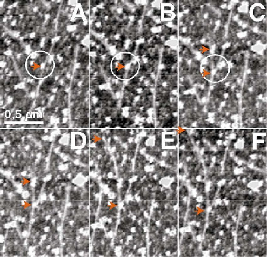
26 T. Lehto et al./FEBS Letters 551 (2003) 25^28
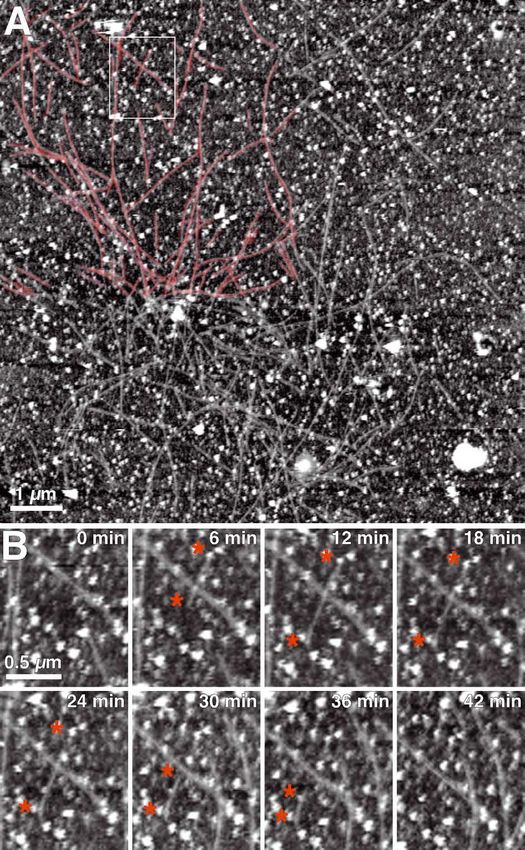
Fig. 1. Observing actin growth by time-lapse AFM. A: Shortly after the injection of HeLa cytosol individual actin ¢laments were observed on
the supporting mica surface. The support was heavily decorated with endosomes and the macromolecules resulting from the cytosol. In the top
left part of the image the ¢laments are highlighted in red. B: Dynamics of an individual actin ¢lament. 0 min, enlarged region of A. After a
time of 6 min the same surface area was imaged and showed an actin ¢lament being polymerized. Asterisks mark the ¢lament ends. One end
of the ¢lament is continuously growing (12 and 24 min). Suddenly, the polymerization of the lower end stops while the other ¢lamentous end
depolymerizes (30 min). This e¡ect continues until the entire ¢lament disappears (42 min). Apparently, other actin ¢laments in the immediate
neighborhood were not a¡ected by this process and independently continued their individual dynamics. Topographs exhibit a vertical height
scale of 50 nm.
limiting the resolution of the AFM topography or causing prevent contamination of the scanning AFM tip by uncon-
imaging artifacts [14]. To visualize the actin polymerization trolled interaction with macromolecules resulting from the so-
in cytosol, we titrated the dilution of the cytosol to ensure a lution. At a total protein concentration of 0.8 mg/ml individ-
su⁄cient protein concentration for actin to polymerize and to ual actin ¢laments were observed in the AFM topography
FEBS 27566 27-8-03 Cyaan Magenta Geel ZwartT. Lehto et al./FEBS Letters 551 (2003) 25^28 27
ments. The z-data are color-coded: the lighter color corre-
sponds to the thicker objects. Most of the ¢laments shown
in Fig. 1 are of uniform color intensity and estimated to be 8^
10 nm which ¢ts well with the thickness of an individual actin
¢lament.
Time-lapse AFM topographs (Fig. 1B) show individual ac-
tin ¢laments growing in a heterogeneous manner. While most
of the ¢laments (85%, 279 ¢lament ends scored) remained
mainly unchanged in their length, others grew at variable
rates. Occasionally, individual ¢laments disappeared indicat-
ing that they depolymerized. Fig. 2A shows the polymeriza-
Fig. 2. Analyzing actin ¢laments in cell extract. A: Polymerization tion rates of various ¢laments observed over the time range of
rate of all growing actin ¢laments. The average growth was
more than 5 h. Interestingly, most actin ¢laments interrupted
0.091 R 0.043 Wm/min and that of shrinking 30.051 R 0.031 Wm/min.
B: Polymerization of a single actin ¢lament recorded over a time their polymerization for a while and then continued to grow
range of 115 min. The ¢lament reversibly changes its state from or to shrink again. According to the rates found it is possible
growing (polymerizing) to being static. to distinguish three classes to describe the polymerization pro-
cess: an inactive phase with no or nearly no polymerization, a
polymerization phase exhibiting an average rate of 0.091 R
(Fig. 1). Knowing that the contents of actin in HeLa cytosol is 0.043 Wm/min (n = 36; mean value R S.D.) and a depolymeri-
approximately 3% of the total protein [15], we estimated the zation phase showing a rate of 30.051 R 0.031 Wm/min. Those
concentration of actin in our assay to be around 0.6 WM. That values are close to the ones found by Fujiwara et al. at com-
the observed ¢laments were F-actin was con¢rmed by the parable concentrations with puri¢ed actin [4]. Fig. 2B shows
¢nding that addition of cytochalasin D to the assay prevented the growth behavior of a single actin ¢lament over the time
the formation of the ¢laments (data not shown). It is impor- range of 5 h. From this analysis, it becomes clear that the
tant to mention that AFM provides three-dimensional data actin ¢laments exhibit the possibility to switch between the
on the scanned object. Images presented in Fig. 1 also carry three growth phases of depolymerization, polymerization
the information on the thickness (z-dimension) of the ¢la- and inactivity (Fig. 2B and data not shown).
Fig. 3. Branching of an actin ¢lament. A: Selected area of actin ¢laments. The white circle indicates a region of an actin ¢lament being associ-
ated with an endosome. B: Same area imaged after a time course of 6 min. C,D: After 12 and 18 min, however, an actin ¢lament grows from
the endosomes labeling the actin ¢lament. Apparently, the growing actin ¢lament interacts with another endosome. After this, the actin ¢lament
continued its growth (E, t = 24 min and F, t = 30 min). Topographs exhibit a vertical height scale of 50 nm. Arrowheads indicate the ends of
the ¢lament.
FEBS 27566 27-8-03 Cyaan Magenta Geel Zwart28 T. Lehto et al./FEBS Letters 551 (2003) 25^28
3.2. Branching and junction of actin ¢laments mechanisms of actin and endosomal function. Improved time
Some of the branching and cross-linking proteins such as resolution of the AFM will be available by establishing fast-
the Arp2/3 complex are contained in endosomes [3,16,17]. speed AFM currently enabling to record V20 images per
Moreover, it was recently shown that membrane organelles second.
and endosomes in particular can induce actin polymerization
[16]. To study the behavior of the actin network in the pres- Acknowledgements: We are grateful to Isabel Richter for technical
ence of a de¢ned membrane organelle population we included assistance and to Tony Hyman and Laurence Pelletier for comments
puri¢ed endosomes [11] in our assay. on the manuscript.
From the actin networks, it is possible to extract the
branched and crosslinked ¢laments (Fig. 2). Among these References
examples, branching angles varied between 40‡ and 90‡, show-
ing a broad distribution. Similar data were obtained by cry- [1] Schmidt, T., Schu«tz, G.J., Baumgartner, W., Gruber, H.J. and
oelectron tomography ([18]). These observations are consis- Schindler, H. (1996) Proc. Natl. Acad. Sci. USA 93, 2926^2929.
[2] Weiss, S. (1999) Science 283, 1676^1683.
tent with the presence of a variety of actin-cross-linking and
[3] Pollard, T.D., Blanchoin, L. and Mullins, R.D. (2000) Annu.
actin-branching proteins that connect actin ¢laments to each Rev. Biophys. Biomol. Struct. 29, 545^576.
other at variable angles. Although the ¢laments grew onto the [4] Fujiwara, I., Takahashi, S., Tadakuma, H., Funatsu, T. and
mica surface without being exposed to any mechanical stress, Ishiwata, S. (2002) Nat. Cell Biol. 4, 666^673.
some of the ¢laments were bent close to their site of attach- [5] Binnig, G., Quate, C.F. and Gerber, C. (1986) Phys. Rev. Lett.
56, 930^933.
ment. This presumably was caused by some unidenti¢ed actin- [6] Mu«ller, D.J. and Anderson, K. (2002) Trends Biotechnol. 20,
binding proteins that link one end of a ¢lament to the side of S45^49.
another one. In most cases, the branches and junctions of [7] Drake, B. et al. (1989) Science 243, 1586^1588.
actin ¢laments were covered by vesicle-like structures (Fig. [8] Domke, J., Parak, W.J., George, M., Gaub, H.E. and Rad-
macher, M. (1999) Eur. Biophys. J. 28, 179^186.
3) which are assumed to present endosomes. These observa-
[9] Engel, A. and Mu«ller, D.J. (2000) Nat. Struct. Biol. 7, 715^
tions are consistent with the recent studies addressing the role 718.
of actin in the endocytic pathway [17]. Endosomes interacting [10] Mu«ller, D.J., Janovjak, H., Lehto, T., Kuerschner, L. and An-
with actin ¢laments can induce their branching, bending or derson, K. (2002) Prog. Biophys. Mol. Biol. 79, 1^43.
mediate their interaction with the cell membrane [17]. [11] Gorvel, J.P., Chavrier, P., Zerial, M. and Gruenberg, J. (1991)
Cell 64, 915^925.
We have shown that time-lapse AFM can be applied to [12] Mo«ller, C., Allen, M., Elings, V., Engel, A. and Mu«ller, D.J.
study in vitro complex biological systems at high resolution. (1999) Biophys. J. 77, 1050^1058.
The AFM topographs revealed molecular insights into the [13] Amrein, M. and Mu«ller, D.J. (1999) Nanobiology 4, 229^256.
formation of actin networks, growth, shrinkage, branching, [14] Schwarz, U.D., Haefke, H., Reimann, P. and Guntherodt, H.J.
junction, and into the interaction of actin with endosomes. (1994) J. Microsc. 173, 183^197.
[15] Heacock, C.S., Eidsvoog, K.E. and Bamburg, J.R. (1984) Exp.
Although the wealth of information potentially revealed by Cell Res. 153, 402^412.
AFM may show important and novel insights into in vitro [16] Merri¢eld, C.J., Moss, S.E., Ballestrem, C., Imhof, B.A., Giese,
systems, technical improvements may soon allow more de- G., Wunderlich, I. and Almers, W. (1999) Nat. Cell Biol. 1, 72^
tailed insights. Technological developments will concentrate 74.
[17] Qualmann, B., Kessels, M.M. and Kelly, R.B. (2000) J. Cell Biol.
on the combination of high-resolution £uorescence microsco- 150, F111^6.
py and AFM. Individual constituents of the cell extract will be [18] Medalia, O., Weber, I., Frangakis, A.S., Nicastro, D., Gerisch,
labeled with £uorescence markers to reveal insights into the G. and Baumeister, W. (2002) Science 298, 1209^1213.
FEBS 27566 27-8-03 Cyaan Magenta Geel ZwartYou can also read