Organizzazione cromosomi - EUCARIOTI - Elearning
←
→
Page content transcription
If your browser does not render page correctly, please read the page content below
Organizzazione cromosomi

          EUCARIOTI

          Gli eucarioti contengono un numero variabile di cromosomi.
          Ciascun cromosoma eucariotico contiene una sola molecola
          di DNA lineare a doppio filamento

               L’impacchettamento del DNA nel nucleo
               eucariotico è dinamico, cioè cambia durante il ciclo
               cellulare restando sotto forma di cromatina durante
               tutta l’interfase per condensarsi ulteriormente
               durante la divisione cellulare (mitotica o meiotica)
               a formare i cromosomi
               H. sapiens possiede un genoma di 3,4 x 10⁹ bp
               il nucleo eucariotico è di circa 2 µm

                  elevato livello di compattamento

                                                                 1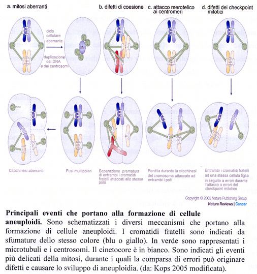
Dimensioni dei cromosomi
Triturus                                Homo sapiens
cristatus

                                   Drosophila
                                   melanogaster

                                 30µM

                                                       2
Dimensioni dei cromosomi: il Muntjac
Muntiacus reevesi

Muntiacus muntjak

                                            3
5 avvolgimenti            2nm
                della doppia elica

                sezione della              11nm
                cromatina
Lunghezza del
cromosoma
                fibra di 30nm con i
                nucleosomi
                strettamente               30nm
                impacchettati

                parte di una sezione di   300nm
                cromosoma

                sezione condensata
                di un cromosoma           700nm
                metafasico

                cromosoma                 1400nm
                metafasico                  4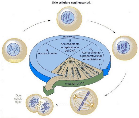
Livello 1
fattore di impacchettamento = 6

                                  5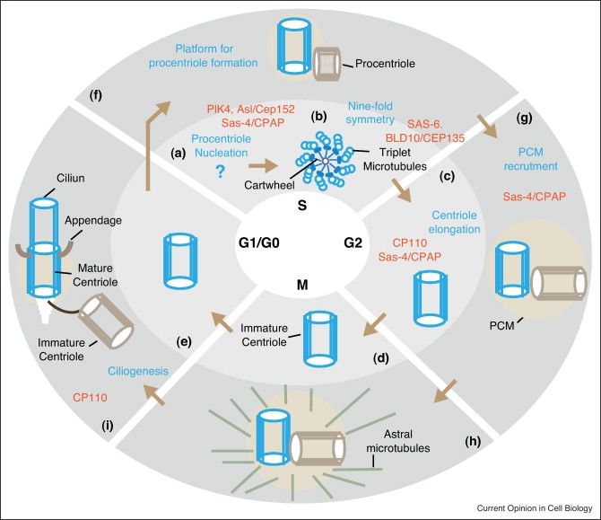
The core histones

A. Structure of nucleosomal histones.
B. Amino-terminal tails of core histones. The numbers indicate amino acid position.
The post-translational modifications are indicated (red ac = acetylation sites ;
blue p = phosphorylation sites ; green m = methylation sites ; purple rib = ADP ribosylation).
S: Serina; K: Lisina; E: Acido Glutammico                                                   6
Il DNA cromosomico che si avvolge intorno agli istoni si può dividere in due regioni:
ü    DNA core di lunghezza invariabile di 146 bp, relativamente resistente alla digestione da parte di nucleasi

ü    DNA linker la cui lunghezza può variare da 8 bp a 114 bp in maniera specie-specifica, tessuto-specifica o anche
     genoma-specifica

                                                               Più del 90% del DNA è stato trovato in associazione
                                                                                con i nucleosomi

                                                          Istoni
    Gli istoni subiscono modificazioni durante il ciclo cellulare,che sono:

                 ➟ transienti,
                 ➟ associate a cambiamenti strutturali della cromatina durante la replicazione e la trascrizione,
                 ➟ associate anche al grado di condensazione della cromatina.

                    •    Acetilazione (Lys)
                    •    Metilazione (Lys, Arg, His)
                    •    Fosforilazione (Ser, Thr le più comuni, His e Asp le meno stabili)

                                                                                                                     7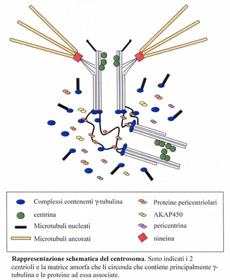
Livello 1      ➟fattore di impacchettamento = 6
                                                         Livello 2
                      I nucleosomi si associano a formare una struttura più compatta del diametro di
                       30 nm visibile al microscopio elettronico (fattore di impacchettamento = 40)

                                                    Livello 3

Il livello successivo di avvolgimento ➛ formazione di domini di DNA ad ansa simili a
                                        quelli osservati nei cromosomi dei procarioti
fattore di impacchettamento ➛ eucromatina = 1000-2000
                             ➛ eterocromatina e cromatina interfasica = 10000                      Livello 4

                                                                                                               8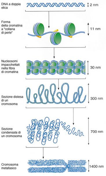
Livello 1
fattore di impacchettamento = 6

 Livello 2
fattore di impacchettamento = 40

 Livello 3
 fattore di impacchettamento
 eucromatina     = 1000-2000
 eterocromatina = 10000

 Livello 4

Unica e definitiva ipotesi ???
                                   9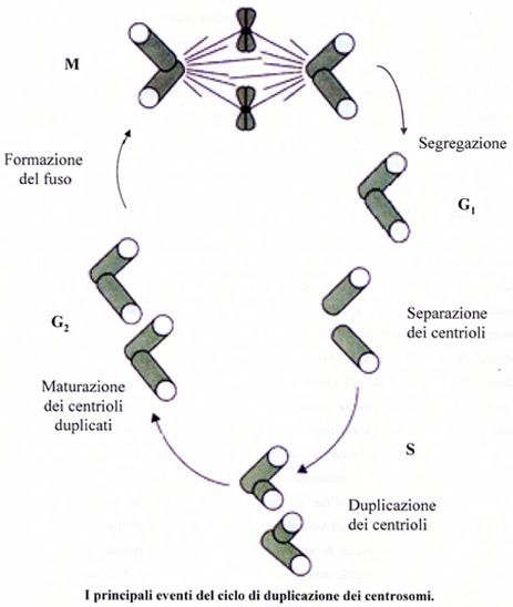
Maeshima et al., Chromatin as dynamic 10-nm fibers. Chromosoma, 123(3):225-237, 2015. 10.1007/s00412-014-0460-2

Abstract
DNA is wrapped around core histones, forming a nucleosome fiber (10-nm fiber).
What is the structure of chromatin?
This fiber has long been assumed to fold into a 30-nm chromatin fiber and
subsequently into helically folded larger fibers or radial loops.
➛ However, several recent studies, including our cryo-EM and X-ray scattering
 analyses, demonstrated that chromatin is composed of irregularly folded 10-nm
 fibers, without 30-nm chromatin fibers, in interphase chromatin and mitotic
 chromosomes.

➛ This irregular folding implies a chromatin state that is physically less constrained,
which could be more dynamic compared with classical regular helical folding structures.

Consistent with this, recently, we uncovered by single nucleosome imaging large
nucleosome fluctuations in living mammalian cells (∼50 nm/30 ms). Subsequent
computational modeling suggested that nucleosome fluctuation increases chromatin
accessibility, which is advantageous for many “target searching” biological processes
such as transcriptional regulation.

This review provides a novel view on chromatin structure in which chromatin consists
of dynamic and disordered 10-nm fibers.
                                                      cryo-EM: Microscopia elettronica a freddo
                                                      X-ray scattering: diffrazione a raggi X                 10
Maeshima et al., Chromatin as dynamic 10-nm fibers. Chromosoma, 123(3):225-237, 2015. 10.1007/s00412-014-0460-2
Fig. 1 Old and novel views of chromatin structure.
The right panel shows the novel
hypothesis of irregularly folded
nucleosome fibers
A long DNA molecule with a
diameter of ∼2 nm is wrapped
around a core histone octamer
and forms a nucleosome with a
diameter of 11 nm
(Alberts et al. 2007).
The nucleosome has long been
assumed to fold into 30-nm
chromatin fibers (left) and
subsequently into the higher
order organization of
interphase nuclei or mitotic
chromosomes.
novel hypothesis of irregularly 11
folded nucleosome fibersMaeshima et al., Chromatin as dynamic 10-nm fibers. Chromosoma, 123(3):225-237, 2015. 10.1007/s00412-014-0460-2
Fig. 2 Two classical models of 30-nm chromatin fibers and higher order chromatin structures
a One-start helix (solenoid),
b two-start helix (zigzag).
(Top) A scheme of the two different
topologies of chromatin fibers is
shown (Robinson and Rhodes 2006).
Positions from the first (N1) to the
eighth (N8) nucleosome are labeled.
c Two classical higher order chromatin
structure models:
➛ the hierarchical helical folding model
(Sedat and Manuelidis 1978) and
➛ the radial loop model (Laemmli et al. 1978).
In the radial loop model, many loop structures
of the 30-nm fiber (red) wrap around the
scaffold structure (gray) (Laemmli et al. 1978),
which consists of condensin and topoisomerase IIα
(Maeshima and Laemmli 2003)
12Maeshima et al., Chromatin as dynamic 10-nm fibers. Chromosoma, 123(3):225-237, 2015. 10.1007/s00412-014-0460-2
Fig. 3 Small angle X-ray scattering (SAXS)
analysis of chromatin structure.
a Experimental design.
The chromosome pellet in a quartz capillary
tube was exposed to synchrotron X-ray beams,
and the scattering patterns were recorded
using the imaging plate (Nishino et al. 2012).
b When non-crystal materials were irradiated
with X-rays, scattering at small angles
generally reflected periodic structures.
(Images a and b were reproduced from Joti et
al. 2012, with some modifications).
c (Upper left) Typical SAXS patterns of
purified mitotic HeLa chromosome fractions.
Three peaks at ∼6, ∼11 (weak), and ∼30 nm
were detected (arrows).
(Upper right) After the removal of ribosome
aggregates, the 30-nm peak disappeared,
whereas the other peaks remained.
(Bottom) A model whereby the 30-nm peak
in SAXS results from regularly spaced
ribosome aggregates and not from the
chromosomes.
(Image c was reproduced from Nishino et al.
13
2012, with some modification).Maeshima et al., Chromatin as dynamic 10-nm fibers. Chromosoma, 123(3):225-237, 2015. 10.1007/s00412-014-0460-2
Fig. 4 Polymer melt model.
low-salt
a Under low-salt conditions, nucleosome fibers
could form 30-nm chromatin fibers via intra-fiber
nucleosome associations.
An increase in salt [cation (+)] concentration results
in inter-fiber nucleosomal contacts that interfere
with intra-fiber nucleosomal associations, leading
to a polymer melt scenario.
Note that in these illustrations, we show a
highly simplified two-dimensional nucleosome
model.
Arrows and dotted lines show repulsion forces
and interactions, respectively.
b During the melting process, the 30-nm chromatin
fibers become irregularly folded nucleosome fibers
14Maeshima et al., Chromatin as dynamic 10-nm fibers. Chromosoma, 123(3):225-237, 2015. 10.1007/s00412-014-0460-2
Fig. 5 Higher order structure of interphase chromatin.
a Condensed chromatin domains.
Active chromatin regions are transcribed on
the surfaces of chromatin domains with
transcriptional complexes (purple spheres)
and RNA polymerase II (green spheres).
NPC: Nuclear Pore Complex,
NE: Nuclear Envelope.
b (Left) Condensed chromatin is more
resistant to radiation damage or chemical
attack.
(Right) Reactive radicals arising from the
radiolysis of water molecules by irradiation
can damage decondensed chromatin;
decondensed chromatin is also more
accessible to chemicals (labeled “Ch”)
15Maeshima et al., Chromatin as dynamic 10-nm fibers. Chromosoma, 123(3):225-237, 2015. 10.1007/s00412-014-0460-2
Conclusions
The traditional view of chromatin is changing from one of static
regular structures including 30-nm chromatin fibers to a dynamic
irregular folding structure of 10-nm nucleosome fibers.
Although the term “irregular” or “disordered” might give the
impression that the organization is functionally irrelevant, the
irregular folding results in less physical constraint and increased
dynamism, increasing the accessibility of the DNA.
This dynamic state may be essential for various genome functions,
including transcription, replication, and DNA repair/recombination.
Another paper (Eltsov et al., ELCS in ice: cryo-electron microscopy of nuclear envelope-limited
chromatin sheets. Chromosoma. 123(3): 303-312, 2014 June. doi: 10.1007/s00412-014-0454-0
(published after this article went to press).
The authors studied nuclear Envelope-Limited Chromatin Sheets (ELCS) by cryo-EM.
They found that the 30-nm chromatin fibers could only be observed following aldehyde fixation;
none were seen in cryo-sections, suggesting that the 30-nm chromatin fibers in ELCS
visualized by conventional EM could be an artifact structure.
16Gibcus et al., A pathway for mitotic chromosome formation. Science, 359(6376):eaao6135, February 9, 2018. DOI: 10.1126/science.aao6135
Tracking mitotic chromosome formation
How cells pack DNA into fully compact, rod-shaped chromosomes during mitosis has fascinated cell biologists for more
than a century.
Gibcus et al.(2018) delineated the conformational transition trajectory from interphase chromatin to mitotic
chromosomes minute by minute during the cell cycle.
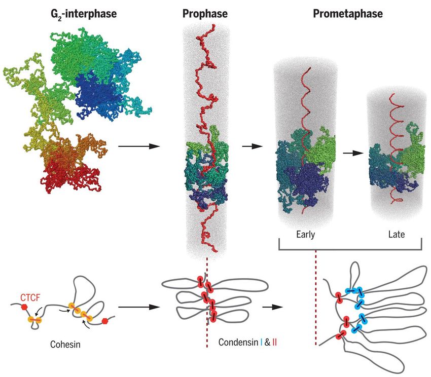
The mitotic chromosome is organized in a spiral staircase architecture in which chromatin loops emanate radially from
a centrally located helical scaffold.
We integrate genetic, genomic, and computational approaches to characterize the key steps in mitotic chromosome
formation from the G2 nucleus to metaphase, and we identify roles of specific molecular machines, condensin I and II,
in these major conformational transitions.
The molecular machines condensin I and II play distinct roles in these processes:
➾ condensin II is essential for helical winding, whereas
➾ condensin I modulates the organization within each helical turn.
CONCLUSION
We describe a pathway of mitotic chromosome folding that unifies many previous observations.
In prophase, condensins mediate the loss of interphase organization and the formation of arrays of consecutive loops.
In prometaphase, chromosomes adopt a spiral staircase–like structure with a helically arranged axial scaffold of
condensin II at the bases of chromatin loops.
The condensin II loops are further compacted by condensin I into clusters of smaller nested loops that are
additionally collapsed by chromatin-to-chromatin attractions.
The combination of nested loops distributed around a helically twisted axis plus dense chromatin packing achieves the
10,000-fold compaction of chromatin into linearly organized chromosomes that is required for accurate chromosome
segregation when cells divide.Gibcus et al., A pathway for mitotic chromosome formation. Science, 359(6376):eaao6135, February 9, 2018. DOI: 10.1126/science.aao6135
A pathway for mitotic chromosome formation.
A pathway for mitotic chromosome
formation.
In prophase, condensins mediate
the loss of interphase chromosome
conformation, and loop arrays are
formed.
In prometaphase, the combined
action of condensin I (blue spheres
in the bottom diagram) and II (red
spheres) results in helically
arranged nested loop arrays.
CTCF: fattore di trascrizione coinvolto
nella regolazione della trascrizione,
attività da ‘isolatore’ (insulator),
etc
Svolge ruolo fondamentale come
CTCF (11-zinc finger protein) o CCCTC-binding factor regolatore della architettura 3D
della cromatinaGeneral steps in chromatin assembly
➛ Assembly begins with the incorporation of the H3/H4
tetramer (1),
➛ followed by the addition of two H2A-H2B dimers (2) to form
a core particle. The newly synthesized histones utilized are
specifically modified; typically, histone H4 is acetylated at
Lys5 and Lys12 (H3-H4*).
➛ Maturation requires ATP to establish a regular spacing,
and histones are de-acetylated (3).
➛ The incorporation of linker histones is accompanied by folding
of the nucleofilament. Here the model presents a solenoid
structure in which there are six nucleosomes per gyre (4).
➛ Further folding events lead ultimately to a defined domain
organization within the nucleus (5).
19Eagen et al., Stable Chromosome Condensation Revealed by Chromosome Conformation Capture. Cell, 163(4): 934-946, 2015.
doi:10.1016/j.cell.2015.10.026
Highlights
• Hi-C of polytene chromosomes reveals an
equivalence of polytene bands with TADs
(Topologically Associating Domains)
• TADs are conserved between polytene and
diploid cells
• Fully extended and up to 10-fold compacted
fibers constitute euchromatin
• Up to 30-fold compacted fibers represent
heterochromatin of the nuclear periphery
In Brief
Analysis of polytene bands, which are
shown to correspond to topologically
associating domains in interphase nuclei,
reveals two stable forms of folded
chromatin within euchromatic regions of
diploid cells that are distinct from more
highly structured heterochromatin.
Hi-C, an extension of 3C (Chromosome Conformation Capture)
that probes the three-dimensional architecture of
whole genomes 20Eagen et al., Stable Chromosome Condensation Revealed by Chromosome Conformation Capture. Cell, 163(4): 934-946, 2015.
doi:10.1016/j.cell.2015.10.026
SUMMARY
Chemical cross-linking and DNA sequencing have revealed regions
of intra-chromosomal interaction, referred to as
Topologically Associating Domains (TADs),
interspersed with regions of little or no interaction, in interphase nuclei.
TADs and the regions between them correspond with the
bands and interbands of polytene chromosomes of Drosophila.
We further establish the conservation of TADs between polytene
and diploid cells of Drosophila.
Two states of folding,
➛ fully extended fibers containing regulatory regions and promoters, and
➛ fibers condensed up to 10-fold containing coding regions of active
genes,
constitute the euchromatin of the nuclear interior.
✔ Chromatin fibers condensed up to 30-fold, containing coding
regions of inactive genes, represent the heterochromatin of
the nuclear periphery.
A convergence of molecular analysis with direct observation
reveals the architecture of interphase chromosomes.
21Eagen et al., Stable Chromosome Condensation Revealed by Chromosome Conformation Capture. Cell, 163(4): 934-946, 2015.
doi:10.1016/j.cell.2015.10.026
SUMMARY
Chemical cross-linking and DNA sequencing have revealed regions
of intra-chromosomal interaction, referred to as
Topologically Associating Domains (TADs),
interspersed with regions of little or no interaction, in interphase nuclei.
TADs and the regions between them correspond with the
bands and interbands of polytene chromosomes of Drosophila.
We further establish the conservation of TADs between polytene
and diploid cells of Drosophila.
Two states of folding,
➛ fully extended fibers containing regulatory regions and promoters, and
➛ fibers condensed up to 10-fold containing coding regions of active
genes,
constitute the euchromatin of the nuclear interior.
✔ Chromatin fibers condensed up to 30-fold, containing coding
regions of inactive genes, represent the heterochromatin of
the nuclear periphery.
A convergence of molecular analysis with direct observation
reveals the architecture of interphase chromosomes.
22Eagen et al., Stable Chromosome Condensation Revealed by Chromosome Conformation Capture. Cell, 163(4): 934-946, 2015.
doi:10.1016/j.cell.2015.10.026
Figure 7. Chromosome Condensation in the Interphase Nucleus
Left: thin section electron micrograph of a
nucleus (Cross and Mercer, 1993), with lightly
staining euchromatin in the nuclear interior,
surrounded by darkly staining heterochromatin,
concentrated at the nuclear periphery.
Right: cartoon representation of white, gray, and
black chromatin, showing proposed relationships
to heterochromatin, euchromatin, and the
nuclear envelope (yellow).
Active TADs in the euchromatin are nearby
other active TADs and inactive TADs in the
heterochromatin are nearby other inactive TADs,
resulting in gray-gray and black-black TAD-TAD
interactions.
The actual pattern of chromatin folding
is unknown and indicated only schematically.
23Eagen et al., Stable Chromosome Condensation Revealed by Chromosome Conformation Capture. Cell, 163(4): 934-946, 2015.
doi:10.1016/j.cell.2015.10.026
The organization of the interphase nucleus in Drosophila is relevant to the mouse and to humans, where
TADs organize chromosomes into spatial modules connected by short chromatin segments (Dixon et al.,
2012).
Furthermore, biochemical fractionation of open chromatin fibers from human cells revealed that the
fibers are cytologically decondensed (Gilbert et al., 2004), and it is now apparent that these fibers are
likely in the fully extended state.
The packing ratios, DNA sequences, functional states, and chromosomal protein patterns of the
differentially staining areas of the interphase nucleus are thus determined.
Genome-wide amplification and alignment in the polytene state reveals interphase chromosome structure
at the level of light microscopy, likely applicable to the diploid state in all monocentric metazoans.
24Ciclo Cellulare di una cellula eucariote (durata variabile a seconda dell’organismo e del tessuto)
2n 2n
2c 4c
2n
2c
25Schema dei principali checkpoint
del ciclo cellulare
2627
Avidor-Reiss, Building a centriole. Current Opinion in Cell Biology, 2012. http://dx.doi.org/10.1016/j.ceb.2012.10.016

                                                                                                         PCM: PeriCentriolar Material

Figure 1. Building of a centriole. Depiction of the structural and molecular events taking place during the formation of
one of the centrioles in a cell (depicted in blue) through two consecutive cell cycles. During the first cell cycle (light
gray background, A–E), the basic structure of the centriole is formed. During second cell cycle (darker gray background,
F–I), the immature centriole acquires functions in a step-by-step manner until it become fully mature and functional (H).
A second centriole formed near the original centriole is depicted in light brown. Major events in the formation of the
centriole are noted in blue. Key proteins are indicated in orange. Centrioles are depicted as they would appear from a
cross section (B) and a side view (C–I).
29
You can also read