Open MRI Offers Imaging Alternative for Obese Patients, Patients With Claustrophobia - Open MRI Offers Imaging Alternative for ...
←
→
Page content transcription
If your browser does not render page correctly, please read the page content below
Open MRI Offers Imaging Alternative for Obese Patients,
Patients With Claustrophobia
For some patient populations, entering and remaining in the narrow bore of conventional MRI systems
can be difficult.
The open MRI design helps to address these challenges by providing additional space on either side
of the patient.
In a recently launched open MRI imaging suite at MGH, ambient audio and video features help to
create a patient-centered scanning environment.
M agnetic resonance imaging (MRI) has advanced medical imaging by providing a noninvasive and safe means of
seeing inside the body. For all its advantages, its use can be constrained by its physical limitations. To be scanned,
patients must first comfortably fit into a relatively narrow tube, or “bore,” which can be difficult for some patient
populations, most notably obese patients and patients who experience claustrophobia and anxiety. In more recent
years, open MRI has begun to address this issue with a design that leaves more room for the patient.
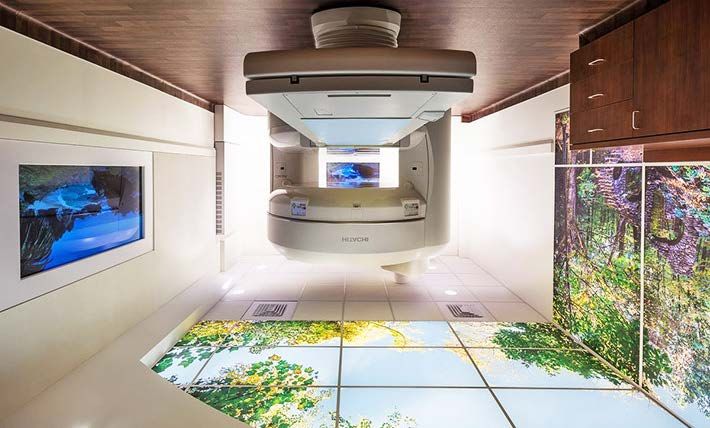
Figure 1. The recently installed open MRI imaging suite at Mass General Imaging - Chelsea serves patient populations who
have difficulty with the narrow bore of conventional MRI.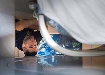
Overcoming the obstacles of claustrophobia and anxiety
Claustrophobia and anxiety are widely reported issues during MR imaging. Patients who experience claustrophobia
often have trouble with the narrow bore of an MR scanner. The idea of entering and remaining in the bore for the
duration of the scan can induce high levels of anxiety, which can negatively impact the session in a variety of ways.
It can bring about increased patient movement, leading to motion artifacts that impact the diagnostic quality of the
scan; sedatives may be required to calm the patient; and in many cases the session cannot be completed, due
either to patient refusal or to premature termination of the scan following a claustrophobic event.

The cumulative effect is substantial. A 2015 review and meta-analysis by Munn et al. looked at 18 studies reporting
the number of claustrophobic reactions during MRI scans. It found that approximately 1 in 100 scans (1.18%) was
terminated early as a result of the reactions. While this statistic is significant, it does not capture the number of
patients who experience claustrophobia and anxiety but are able to complete the scan with difficulty. The overall
impact on diagnostic yield, clinical workflow and healthcare costs can be considerable.

Some types of exams may be more likely than others to induce claustrophobia and anxiety. In the Munn et al.
study, stress cardiac MR scans appeared to have the highest rate of premature termination, which could be due to
the length and requirements of the scan as well as to patients’ claustrophobic reactions. The authors found similar
rates with head, neck and thorax scans, in contrast with abdomen/pelvis and lower extremity scans, which showed
a much lower incidence of termination. These results were not surprising, as the former scans require inserting the
patient into the scanner headfirst, a position more likely to provoke a claustrophobic event.

In more recent years, new, patient-centered scanner designs have helped to reduce the number of claustrophobic
events during scans. Short-bore and wide-bore configurations allow, respectively, a smaller portion of the patient’s
body inside the scanner and more space between the patient and the walls of the bore. Open MRI removes the
bore altogether. With this configuration, the magnets are above and below the patient, with enough open space on
either side that the patient may not feel as confined as in a conventional MRI scanner.

The growing challenge of scanning overweight and obese patients
A second group that may benefit from open MRI is patients who cannot fit comfortably inside a conventional MRI
scanner because of their body type, including many obese patients.

Obesity is a serious concern in the US. According to data from the National Health and Nutrition Examination
Survey (NHANES), 2013–2014, more than two in three adults in the US are considered overweight or obese. This
statistic has immediate implications for health care—the growing prevalence of obesity has led to increased
incidence of diabetes, heart disease and particular types of cancer—but it has also had an indirect impact on
hospitals. With greater numbers of overweight and obese patients, hospitals are facing a need for larger beds,
wheelchairs and operating tables. Similarly, they are finding that, because of the size of their bores, conventional
MRI scanners often cannot accommodate these patients.

The introduction of short-bore and wide-bore scanners has helped to address this growing problem, either by
requiring only a portion of the body to be inserted into the scanner or by increasing the amount of space in the
bore. Open MRI is also providing new opportunities to image overweight and obese patients, because of its more
accommodating open configuration and because the design allows for a wider MRI table that can support greater
weights. Such tables are included with many open MRI systems.

It is important to note that size and weight are not the only determining factors in whether a patient will fit
comfortably inside an MRI scanner. In some cases, his or her abdomen is configured in such a way as to prevent
imaging inside the bore. For this reason, clinicians will generally use the term body habitus when discussing
patients for whom conventional systems do not offer enough space.

                                                         2
The benefits and limitations of open MRI
In late 2017, Mass General Imaging in Chelsea, MA installed an open MRI scanner in a newly designed imaging
suite. The scanner now serves the various patient populations that can have difficulty with conventional MRI:
patients who experience claustrophobia and anxiety as well as overweight and obese patients. In addition to
providing more space, the bore-less design offers an unobstructed 270-degree view for both the patient and the
technologist, allowing for easy observation and monitoring so the technologist can readily respond to the patient’s
needs. The patient table is approximately six inches wider than a conventional MRI table and can support patients
up to 600 pounds.
The newly designed imaging suite includes ambient
audio and video features that are designed to help
soothe the patient during the imaging session. In 2011,
in the first randomized controlled trial on claustrophobia
in MRI, Enders et al. found claustrophobic event rates of
more than 25% for both short-bore and open MRI
scanners. In part, this statistic reflects the fact that
patients imaged with short-bore and open MRI are a
high-risk population: many were likely referred to the
scanners because they had claustrophobic reactions
with conventional MRI. But it also demonstrates the
need for a more patient-centered MR scanner
environment. The imaging suite at Mass General
Imaging – Chelsea represents an attempt to create such
an environment.
Figure 2. The open MRI design also helps to facilitate
monitoring of and communication with the patient.
Operating at 1.2 Tesla, the open MRI magnet has lower
field strength than those of most conventional MRI
systems. Because of the design—with the magnets above and below the patient instead of encircling him or her—
the field strength is not homogeneous across the scanned areas. For this reason, the clinicians involved with open
MRI at Mass General have developed protocols for the system that match the diagnostic quality of scans performed
on conventional systems elsewhere in the hospital. Some exams are not performed on the open MRI because
clinicians cannot ensure the same diagnostic quality as that with conventional scanners. These include breast and
prostate exams, as well as exams that require high-field MRI.
Open MRI is still not widely available, in part because of the limitations noted above. However, for institutions that
have the volume to keep an open MRI scanner occupied, the technology offers a means to serve patient
populations that otherwise might not receive the imaging care they need.
Scheduling
Open MRI is performed at Mass General Imaging - Chelsea. Appointments can be made through Epic (inside the
Partners network) or Physician Gateway (outside the Partners network) or by calling 617-887-3500.
Further Information
For further information about open MRI, please contact Mukesh Harisinghani, MD, Director, Abdominal MRI,
Department of Radiology, Massachusetts General Hospital. We would like to thank Dr. Harisinghani, Dr. Raul Uppot,
MD, Division of Interventional Radiology, Department of Radiology, Massachusetts General Hospital, and Wayne
Marshall, Operations Manager, Mass General Imaging – Chelsea, for their advice and assistance in preparing this
article.
3References
Enders J, Zimmermann E, Rief M, et al. (2011). Reduction of claustrophobia with short-bore versus open magnetic
resonance imaging: a randomized controlled trial. PLoS One 6(8):e23494.
Munn Z, Moola S, Lisy K, et al. (2015). Claustrophobia in magnetic resonance imaging: A systematic review and
meta-analysis. Radiography 21:e59-e63.
Uppot R, Sahani D, Hahn PF, et al. (2007). Impact of obesity on medical imaging and image-guided intervention.
AJR Am J Roentgenol 188(2):433-440.
©2017 MGH Department of Radiology
Gary Boas, Author
Raul N. Uppot, M.D., EditorYou can also read