Perichondritis Case Report - Journal of Urgent Care Medicine
←
→
Page content transcription
If your browser does not render page correctly, please read the page content below
Case Report
Perichondritis
Urgent message: With the popularity of piercing of the ear cartilage,
urgent care providers need to be on the alert for perichondritis and to
treat it promptly.
SHAILENDRA K. SAXENA, MD, PHD, and MIKAYLA SPANGLER, PHARM D, BCPS
Case Presentation
A
26-year-old female presented with complaints of a
swollen right pinna for 2 weeks. The swelling progres-
sively worsened over time. In addition, she also com-
plained of severe pain of the right pinna, with an inten-
sity of 7/10, with no radiation and no aggravating or
relieving factors. One week previously, she had been
examined in an urgent care facility and was given a 10-
day course of amoxicillin for presumed acute otitis
externa. However, the infection continued to worsen.
There was no history of trauma to the ear and no other
significant medical history.
Physical Exam
© corbis.com
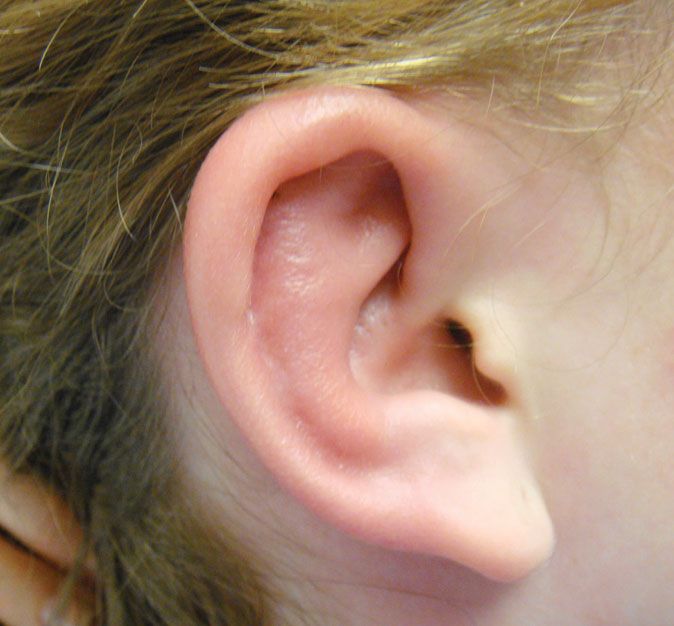
On examination, the patient had an inflamed, erythe-
matous and tender right pinna (Figure 1). The pre- and
post-auricular lymph nodes were enlarged and tender.
Examination of the rest of the ear was normal and tilage covered by a layer of connective tissue called
hearing was not impaired. She was afebrile and all other perichondrium.1 The blood supply to the ear arises
systemic examinations were normal. from the posterior auricular and superficial temporal
arteries.
Diagnosis
Perichondritis Differential Diagnoses
Table 1 lists differential diagnoses. The patient had no
Anatomy of the External Ear exposure to swimming and no involvement of deeper
The external ear consists of the pinna, a fan-like projec- structures or soft tissues. With involvement of the
tion that works to collect sound, and the external pinna, diagnosis of perichondritis was favored.
acoustic meatus. The pinna is composed of elastic car- Perichondritis is an infection of the skin and soft tis-
sues surrounding the cartilage of the external ear, includ-
ing the pinna. The tissues of the pinna receive less
Shailendra K. Saxena is an Associate Professor in the Department of humoral circulation, therefore, any injury or infection
Family Medicine, Creighton University School of Medicine, Omaha, NE.
Mikayla Spangler is an Assistant Professor in the Creighton University takes longer to heal, and any edema and exudates take
School of Pharmacy and Health Professions and School of Medicine, longer to be absorbed, increasing the likelihood of sec-
Department of Family Medicine. ondary infection and abscess formation.2
w w w. j u c m . c o m JUCM T h e J o u r n a l o f U r g e n t C a r e M e d i c i n e | O c t o b e r 2 0 1 3 19
CASE REPORT: PERICHONDRITIS

Figure 1.                                                                                                         Surgery and burns can also cause
                                                                                                               perichondritis, and there have been
                                                                                                               rare cases of the infection occurring
                                                                                                               secondary to furunculosis, chron-
                                                                                                               ic ear discharge, congenital intra-
                                                                                                               auricular sinuses, allergy, diffuse
                                                                                                               lesions following herpes zoster, or
                                                                                                               insect bites.2,3 Perichondritis can
                                                                                                               also develop secondary to malig-
                                                                                                               nant otitis externa.3

                                                                                                               Symptoms
                                                                                                              Perichondritis usually presents first
                                                                                                              as a dull pain that increases in
                                                                                                              severity, accompanied by redness
                                                                                                              and swelling.2 The redness usually
                                                                                                              surrounds an area of injury, such as
                                                                                                              a cut or scrape. The infection begins
                                                                                                              in the helix and anti-helix, and
                                                                                                              resembles cellulitis, a simple skin
                                                                                                              infection; however, it quickly wors-
                                                                                                              ens and involves the perichondrium.
                                                                                                              In severe cases, an abscess can devel-
                                                                                                              op, peeling the perichondrium off
                                                                                                              the cartilaginous layer and resulting
                                                                                                              in necrosis of the cartilage1,4 and
 The pinna is inflamed, erythematous, and tender.                                                             deformation of the ear, known as
                                                                                                              “cauliflower ear.”2 In such advanced
Table 1. Differential Diagnosis                                                                               cases, the patient may be febrile, and
                                                                                         there may be fluid draining from the wound.
 • Simple otitis externa (“swimmer’s ear”)
 • Malignant external otitis                                                             Exams and Tests
 • Cellulitis                                                                            Perichondritis is diagnosed based on the patient’s med-
 • Perichondritis                                                                        ical history and by examination of the ear. If there is a
 • Abscess                                                                               history of trauma to the ear and the ear is red and ten-
                                                                                         der, perichondritis is the most likely diagnosis. There
Causes                                                                                   may be a change in the normal shape of the ear.
The most common bacteria that cause perichondritis are
Pseudomonas aeruginosa. Staphylococcus aureus, Escherichia                               Treatment
coli and Proteus species, with P. aeruginosa being the                                   Treatment consists of broad antibiotic coverage, either
most common culprit.3 Perichondritis is usually a result                                 by mouth or directly into the bloodstream via an intra-
of secondary infection of the ear after traumatic injury.                                venous line. Because most of the cases are associated
   In recent years, penetrating injuries to the ear such as                              with P. aeruginosa bacteria, empiric treatment would
acupuncture and cartilage piercing have increasingly                                     include a fluoroquinolone, as these drugs are the only
becoming causes of perichondritis.4 In fact, ear piercing                                oral treatment effective against these bacteria.4 If there
through the cartilage is probably the most significant                                   is a trapped collection of pus or abscess formation, sur-
risk factor today.5 The cartilage itself is relatively avas-                             gical intervention, such as needle aspiration or incision
cular, and trauma via piercing devascularizes it even fur-                               and drainage, may be necessary to drain the fluid and
ther, providing a good medium for bacteria that could                                    remove any dead skin and cartilage. Recent studies have
be introduced by the piercing needle or gun.5                                            also shown success with the newly developed method

20   JUCM T h e J o u r n a l o f U r g e n t C a r e M e d i c i n e | O c t o b e r 2 0 1 3                                       w w w. j u c m . c o m
CASE REPORT: PERICHONDRITIS

of aspiration, injection of streptomycin and hyaluroni-           Conclusion
dase directly into the infected site, and finally triamci-        Although ear piercing was not the cause of the perichon-
nolone to restrict inflammation.2                                 dritis in our patient, the culture of ear piercing in young
                                                                  adults has increased recently. If it is not done properly
Complications                                                     and sterile techniques are not used carefully, young
If antibiotics are taken early, full recovery from peri-          adults may end up having the complication of peri-
chondritis is expected. In more advanced cases, the               chondritis. It is important for urgent care physicians to
infection can involve the ear cartilage. This is called           be familiar with this common condition and treatment
“chondritis,” and with such an infection, part of the ear         should be started as early as possible to prevent perma-
may die and need to be surgically removed. A peri-                nent damage to a soft cartilage. ■
chondrial abscess may also develop. If so, plastic surgery
                                                                  References
will be needed to restore the ear to its normal shape.2-4         1. Moore KL, Dalley AF. Clinically Oriented Anatomy, Fifth Edition. Philadelphia: Williams
                                                                  & Wilkins, 2006:1022-1024.
                                                                  2. Pattanaik S. Effective, simple treatment for perichondritis and pinna haematoma. J
Prevention                                                        Laryngol Otol. 2009;123:1246-1249.
The best way to prevent perichondritis is to avoid pierc-         3. Prasad HK, Sreedharan S, Prasad HS, Meyyappan MH, Harsha KS. Perichondritis of the
                                                                  auricle and its management. J Laryngol Otol. 2007;121:530-534.
ing one’s ear through the cartilage, as opposed to the ear        4. Davidi E, Paz A, Duchman H, Luntz M, Potasman I. Perichondritis of the auricle: analy-
lobe. The popularity of cartilage piercing has led to a sig-      sis of 114 cases. Isr Med Assoc J. 2011;13:21-24.
                                                                  5. van Wijk MP, Kummer JA, Kon M. Ear piercing techniques and their effect on cartilage,
nificant increase in the number of perichondritis and             a histologic study. J Plast Reconstr Aesthet Surg. 2008;61:S104-S109.
chondritis cases.5

w w w. j u c m . c o m                                   JUCM T h e J o u r n a l o f U r g e n t C a r e M e d i c i n e | O c t o b e r 2 0 1 3       21

                                                                              Urgent Care Clinic
                         the wood
                         insurance
                                                                                 Medical
                         group                                                 Professional
   The Wood Insurance Group, a leading
                                                                                 Liability
   national insurance underwriter, offers
   significantly discounted, competitively
                                                                                Insurance
   priced Medical Professional Liability                       Our Total Quality Approach includes:
   Insurance for Urgent Care Medicine.
   We have been serving the Urgent Care                        ! Preferred Coverage Features
   community for over 25 years, and our                          – Per visit rating (type & number)
   UCM products were designed specifically                       – Prior Acts Coverage
   for Urgent Care Clinics.                                      – Defense outside the limit
                                                                 – Unlimited Tail available
                   Contact Us at:                                – Exclusive “Best Practice” Discounts
         4835 East Cactus Road, Suite 440
                                                                 – Protects the Clinic and Providers
              Scottsdale, Arizona 85254                        ! Exceptional Service Standards
       (800) 695-0219 • Fax (602) 230-8207                       – Easy application process
               David Wood at Ext 270                             – Risk Mgmt/Educational support
     E-mail: davidw@woodinsurancegroup.com                       – Fast turnaround on policy changes
                                                                 – Rapid response claim service
You can also read