Rapid emotional processing in relation to trauma-related symptoms as revealed by magnetic source imaging
←
→
Page content transcription
If your browser does not render page correctly, please read the page content below
Erschienen in: BMC Psychiatry ; 14 (2014). - 193
Schalinski et al. BMC Psychiatry 2014, 14:193
http://www.biomedcentral.com/1471-244X/14/193

 RESEARCH ARTICLE                                                                                                                                  Open Access

Rapid emotional processing in relation to
trauma-related symptoms as revealed by
magnetic source imaging
Inga Schalinski*, James Moran, Maggie Schauer and Thomas Elbert

  Abstract
  Background: Traumatic stress leads to functional reorganization in the brain and may trigger an alarm response.
  However, when the traumatic event produces severe helplessness, the predominant peri-traumatic response may
  instead be marked by a dissociative shutdown reaction. The neural correlates of this dissociative shutdown were
  investigated by presenting rapidly presented affective pictures to female participants with posttraumatic stress
  disorder (PTSD), and comparing responses to a Non-PTSD control group.
  Methods: Event-related-magnetic-fields were recorded during rapid visual serial presentation of emotionally
  arousing stimuli (unpleasant or pleasant), which alternated with pictures with low affective content (neutral).
  Neural sources, based on the L2-surface-minimum-norm, correlated with the severity of the symptom clusters: PTSD,
  depression and shutdown dissociation.
  Results: For the early cortical response (60 to 110 ms), dissociation and PTSD symptom severity show similar spatial
  distributions of correlates for unpleasant stimuli. Cortical networks that could be involved in the relationships seem
  to be widespread.
  Conclusion: We conclude that shutdown dissociation, PTSD and depression all have distinct effects on early
  processing of emotional stimuli.
  Keywords: PTSD, MEG, IAPS, Shutdown, Dissociation, Depression

Background                                                                             related dissociation is understood as a biological response
Repeated exposure to traumatic stress not only produces                                to an overwhelming stressor, in which the survivor finds
the core symptoms of posttraumatic stress disorder                                     themselves in a completely helpless condition. In such
(PTSD), but can also produce symptoms of depression,                                   situations, the body responds to stress with a shutdown of
dissociation and affective dysregulation. Dissociative                                 perceptual, cognitive and affective information processing
symptoms that arise after trauma are common in patients                                (for a detailed description of this concept see [10]). From
who have been exposed to severe and repeated traumatic                                 an evolutionary perspective, shutdown dissociation might
stress, particularly interpersonal trauma [1]. Persistence of                          be seen as an adaptive response to life-threatening condi-
dissociative responding seems to promote the develop-                                  tions during which neither flight nor fight is a viable op-
ment and maintenance of PTSD [2-4], and is associated                                  tions for survival: since the organism cannot defend itself
with a greater severity of PTSD [5-7], and depression                                  or escape, it can only minimize harm, through immobility
symptoms [8]. The relationship between dissociation and                                and functional sensory deafferentiation [11]. The shut-
trauma is of considerable clinical and nosological signifi-                            down of sensory and functional systems may later reoccur
cance, however the theoretical concepts have remained a                                in response to trauma-related cues, intrusions or minor
source of controversy [9]. In the present study, trauma-                               stressors [10]. Dissociative responding acts as a cognitive
                                                                                       avoidance mechanism and reduces the awareness of aver-
* Correspondence: inga.schalinski@uni-konstanz.de
                                                                                       sive emotions such as extreme fear as well as overwhel-
Department of Psychology, University of Konstanz, P.O. Box 905, 78457                  ming autonomic arousal [12]. In the context of exposure
Konstanz, Germany

                                        © 2014 Schalinski et al.; licensee BioMed Central Ltd. This is an Open Access article distributed under the terms of the Creative
                                        Commons Attribution License (http://creativecommons.org/licenses/by/2.0), which permits unrestricted use, distribution, and
                                        reproduction in any medium, provided the original work is properly credited. The Creative Commons Public Domain
                                        Dedication waiver (http://creativecommons.org/publicdomain/zero/1.0/) applies to the data made available in this article,
                                        unless otherwise stated.

                                                                                   Konstanzer Online-Publikations-System (KOPS)
                                                                                URL: http://nbn-resolving.de/urn:nbn:de:bsz:352-286657
Schalinski et al. BMC Psychiatry 2014, 14:193 Page 2 of 13 http://www.biomedcentral.com/1471-244X/14/193 therapy, where therapeutic outcomes are attained by in- response, where the PTSD group showed an increased tensive reliving of the experience so that physiological fear cortical response to salient stimuli followed by disturbed responses can be habituated, and events integrated into emotional modulation. memory, dissociation can become a severe obstacle. One important paradigm for examining the visual emo- Dissociative responding consists of a major distortion tional processing is the rapid serial visual presentation of attention through a perceptual shutdown of informa- (RSVP) of pictures from the International Affective Pic- tion processing. At the level of functional neural activity, tures System (IAPS). Elbert and co-workers [16] found this could mediated by cortico-subcortical top down evidence for an early change of aversive processing from processes. Abnormal cortical activity has been examined the primary visual cortex to fronto-temporal regions and in several neuroimaging studies of PTSD, either mea- the amygdala in traumatized individuals compared to a suring brain activity in a resting state or with stimuli Non-PTSD control group. Methodological studies of emo- designed to trigger trauma specific processing. The latter tionally salient processing of visual stimuli at rapid presen- includes script driven imagery [13] or exposure to emo- tation rates (3 to 5 Hz) revealed an effect of affective tionally evocative pictures [14-19]. Functional neuroimag- arousal at about 150 ms after the stimulus onset [17]. This ing studies using trauma scripts as stimuli suggest that effect reflects a very early differentiation between emo- there may be distinct neural circuitry involving frontal tionally salient stimuli at initial stages of visual processing. and limbic systems that distinguish between PTSD pa- This early posterior negativity (EPN) has been investigated tients with and without dissociative responding. One study in a wide variety of contexts e.g., [22,23]. The motivational dividing PTSD patients into such groups: a non-dissocia- system shows enhanced early selective processing of emo- tive PTSD group, with strong intrusions, and intense hy- tionally salient stimuli, which has the function of priori- perarousal symptoms; and a dissociation group, showed tizing the encoding of stimuli related to both sustenance correspondingly different patterns of functional neural ac- (appetitive) and survival (defensive) of the organism. Thus, tivity. In comparing both groups to a Non-PTSD control, this early initiation in the face of threatening stimuli can the PTSD patients with more hyperarousal symptoms also be critical to survival [24,25]. Patients with PTSD show a displayed lower bilateral medial frontal activity and left an- hypersensitized level of processing, discriminating very terior cingulate activity, whereas the subgroup of patients early (60 to 110 ms) between threatening and non-threa- with dissociative PTSD had increased right medial frontal, tening cues in cortical response [16], but show less affec- right medial prefrontal, right anterior cingulate activity. tive discrimination in later processing [14]. The higher prefrontal activity seems to co-occur with re- In sum, neuroimaging studies suggest different brain duced amygdala activity in dissociative PTSD [13]. These responses in dissociative versus non-dissociative PTSD, results were interpreted as emotional over-modulation in which could either be dimensional or represent a distinct dissociative PTSD, whereas an emotional under-modula- categorical pattern of a subgroup of patients with trauma- tion mediated by less intensive prefrontal inhibition of the related illness. The RSVP paradigm has shown robustly limbic system was found in non-dissociative PTSD. Using different early visual processing in PTSD versus Non- a dimensional approach, Hopper and coworkers [20] PTSD controls, and at the behavioural level, that it was found that the strength of dissociation was positively asso- sufficient to activate the fear-network so that patients ciated with the activity in the left medial prefrontal and showed intrusive memories and flashbacks [16,17,26]. right superior temporal cortices, and negatively correlated However, thus far there has been no study examining po- with the left superior temporal cortex. In a resting state tential differences within the PTSD group, with varying study, Ray and colleagues [21] studied abnormal slow levels of dissociation. Is the chronic hypersensitivity of wave activity in the delta range (1.5 – 4 Hz) of the cortical early visual processing uniform across PTSD patients, or magnetoencephalogram (MEG) in PTSD patients with moderated by levels of dissociation or depression? Because torture exposure. Abnormal slow wave activity reflects of the inherent associations between trauma-related dis- structural and functional abnormalities in neural net- sociation and psychopathology, particularly PTSD and de- works. They found an association between slow wave pression, we decided to examine the respective symptom activity generated in the left ventrolateral frontal cortex clusters separately as potential modulators of affective and degree of dissociative responding. During recordings processing. We used event-related magnetic field po- of the brain’s magnetic fields emotionally evocative photos tentials to investigate the following questions in a di- have been used to assess the visual emotional processing mensional approach within the PTSD group (1) Is there [14,15]. Results indicate a deviant rapid network activity abnormal emotional processing, indicated by different in the right frontal cortex in response to threatening valence and arousal ratings? (2) Is it possible to reactivate stimuli. This response was followed by a less pronounced the shutdown dissociation during the passive viewing task response in the parieto-occipital cortex. This cortical of emotionally salient photographs (when the fear-net- network pattern was interpreted as a vigilance-avoidance work is active)? (3) Does the heart rate, as an index of
Schalinski et al. BMC Psychiatry 2014, 14:193                                                                                                        Page 3 of 13
http://www.biomedcentral.com/1471-244X/14/193

autonomic arousal during the presentation of unpleasant                             investigation so the procedure was aborted. Thirty-three
visual stimuli, differ from the heart rate while pleasant/                          participants who presented with PTSD symptoms were
neutral stimuli are presented? (4) Are shutdown disso-                              analysed in the present study. These were compared to
ciation, PTSD and/or depression symptom severity asso-                              17 Non-PTSD controls with similar ethnic backgrounds,
ciated with altered visual processing of arousing and                               who were recruited from the general community and
neutral stimuli within the PTSD group, and how do these                             from the University of Konstanz. Participants in the
inherently associated psychopathologies converge at the                             Non-PTSD control group and most of the patients had
level of brain activity during RSVP?                                                been examined with the Cardiac Defense Paradigm [27].
                                                                                    Each participant was interviewed after providing in-
Methods                                                                             formed consent. First, demographic data as well as medi-
Structured clinical interview                                                       cation were assessed (compare Table 1). Following this,
Female participants were recruited for the study at the                             the number of traumatic experiences was assessed using
University of Konstanz outpatient clinic for refugees in                            the sum of the event checklist of the Clinician Adminis-
Germany. They were referred to the clinic by human                                  tered PTSD Scale [28]. For traumatic events, we made a
rights organizations, medical doctors or lawyers, for                               distinction between the number of various traumatic
diagnostic assessment. Expert-psychologists carried out                             event types that were self-experienced and the number
structured clinical interviews with the support of trained                          of different traumatic event types that were witnessed. A
translators at least one week prior to the MEG. We con-                             traumatic event type was judged as self-experienced if
ducted 49 interviews between April 2010 and February                                the participant was the victim; or as witnessed if the
2012 (inclusion criteria were female refugee with mul-                              participant had observed the traumatic event while
tiple traumatic experiences). Thirty-seven of the inter-                            someone else was threatened. For the PTSD diagnosis,
viewed women were included in the PTSD group. Four                                  we used the Clinician Administered PTSD Scale and
subjects did not participate for the following reasons:                             summed its score for the symptom severity. Current
two for technical reasons, one subject was too afraid of                            comorbid disorders according to the criteria of the forth
the laboratory, and one subject gave informed consent                               version of the Diagnostic and Statistical Manual of Men-
but showed severe dissociation at the beginning of the                              tal Disorder (DSM-IV; such as depression, dysthymia,

Table 1 Group means, standard deviations and differences of the demographic and clinical data for the PTSD and the
Non-PTSD Group
                                                                             PTSD                          Non-PTSD                    Statistics for group
                                                                 M/n (range)          SD/%           M/n          SD/%                     differences
Age (Years)                                                          36.74             9.67          31.88         6.83            t(49) = 1.85           p = .071
Education (Years)                                                    5.59              5.46          18.68         3.46          t(46.05) = -10.41        p < .001
Regions of Origin N%                                                                                                              χ (3, 51) = .11
                                                                                                                                   2
                                                                                                                                                          p = .991
Middle and Far East                                                   18              52.9%            9           53%
The Balkans                                                            5              14.7%            2          11.8%
Africa                                                                 9              26.5%            5          29.4%
India                                                                  2               5.9%            1           5.9%
                                                                     5.47
Number of Traumatic Event Types (Self-experienced)                                     2.35          1.24          1.40           t(47.41) = 8.05         p < .001
                                                                    (2-11)
                                                                     3.47
Number of Traumatic Event Types (Witnessed)                                            2.34          1.24          1.15           t(49.00) = 4.58         p < .001
                                                                     (0-9)
Time elapsed since the worst traumatic event (Years)                 9.74              8.82          7.45          4.80          t(32.11) = -1.09         p = .284
                                                                     82.56
PTSD Symptom Severity                                                                  15.54         0.94          2.66          t(36.71) = 29.76         p < .001
                                                                   (55-113)
                                                                     20.32
Hamilton-Depression Severity                                                           8.77          2.24          2.51          t(42.37) = 11.14         p < .001
                                                                    (6-37)
                                                                     17.50
Shutdown Dissociation Score                                                            8.33          0.94          1.25          t(35.88) = 11.34         p < .001
                                                                    (2-33)
Note. Mean (M), Standard Deviation (SD), absolute number of respondents (n), PTSD = Posttraumatic Stress Disorder. For group differences, t-tests were used for
continuous variables and χ2 tests were applied for nominal variables.
Schalinski et al. BMC Psychiatry 2014, 14:193 Page 4 of 13
http://www.biomedcentral.com/1471-244X/14/193
abuse or dependency of alcohol/illegal substances, sui- taking antidepressants (n = 5 (15%)) and one of these
cidality and psychotic disorders) were assessed with the was also taking neuroleptic medication (n = 1 (3%)).
MINI International Neuropsychiatric Interviews [29]. None of the participants in the Non-PTSD group were
The score on the Hamilton Rating Scale for Depression taking psychoactive medication at the time of testing.
[30] estimated the degree of depression. To assess dis- None of the participants took medication targeting the
sociative responding, we used the 13-item Shutdown cardiovascular system such as digitalis, beta-blockers or
Dissociation Scale (Schalinski I, Schauer M, Elbert T: anticholinergics. All participants reported normal vision
The Shutdown Dissociation Scale (Shut-D), submitted.), (with correction). Further, all participants wrote with
developed by our research group based upon our con- their right hand. The handedness score (100 = 100%
cept of shutdown dissociation [10]. right-handed) was on average M = 95.7 (SD = 14) in the
PTSD group and M = 96.5 (SD = 6.1) in the Non-PTSD
Demographical and clinical data group.
In the PTSD group, the region of origin was 52.9% Middle
or Far East, 14.7% the Balkans, 26.5% Africa and 5.9% MEG apparatus and physiological assessment
India. The Non-PTSD control groups’ origin was 53% The magnetic fields were measured with a 148-channel
Middle or Far East, 11.8% the Balkans, 29.4% Africa and whole head magnetometer (MAGNES 2500 WH, 4D
5.9% India. The ethnic composition of both groups was Neuroimages, San Diego, USA) with a sampling rate of
not statistically different; χ2(3, 51) = .11, p = .991. The age 678.17 Hz. Data were recorded continuously with a band
of the PTSD group ranged from 17 to 56 years (M = 36.7, pass filter between 0.1 and 200 Hz. Electrooculogram
SD = 9.7). The Non-PTSD group were on average M = 31.9 (EOG) and electrocardiogram (ECG) were measured
years old (SD = 6.8) and the age ranged from 22 to 49 with a SynAmps amplifier (NEUROSCAN Laboratories,
years. There was no statistically significant difference in Sterling, VA, USA). Four Ag/AgCl electrodes were at-
age between groups. On average, the participants in the tached (two near the left and right outer canthus and
PTSD sample were less educated when compared to the two above and below the right eye) to obtain the vertical
Non-PTSD control group (t(46.05) = -10.41, p < .001). On and horizontal EOG. An electrode attached above the
average the participants in the PTSD group had ex- right zygomatic bone provided grounding. To record the
perienced M = 5.5 (SD = 2.4; range 2 to 11) and witnessed ECG, two Ag/AgCl electrodes were positioned above the
M = 3.5 different types of traumatic stressors (SD = 2.3, right collarbone and the left lower costal arch. The R
range 0 to 9). The following self-experienced events were wave of the heart rate was semi-automatically detected
reported most frequently: physical assault (79%), assault using BESATM software (module: create triggers) and
with a weapon (65%) and sexual assault with penetration the R-R-intervals were converted to heart rate. The heart
(53%). Almost half of the PTSD sample (47%) was exposed rate was assessed within each of the blocks and ave-
to traumatic experiences in war-zones. The majority wit- raged across the pleasant/neutral and unpleasant/neutral
nessed physical assaults (71%), 44% witnessed homicide blocks (compare below: Experimental Design).
and 38% witnessed a serious traffic accident. The Non-
PTSD group was significantly less exposed to traumatic Stimuli
event types that were self-experienced t(47.41) = 8.05, Based on normative valence and arousal ratings, 100 arou-
p < .001 and witnessed t(49) = 4.58, p < .001). They had sing unpleasant (e.g., mutilations, assaults, etc.), 100 plea-
experienced on average M = 1.2 traumatic event types sant (e.g., wedding, children, etc.), and 100 neutral (e.g.,
(SD = 1.4; range 0 to 4) and witnessed on average M = 1.2 neutral faces, daily life scenes, etc.) colour photographs
traumatic event types (SD = 1.2; range 0 to 3). All partici- were chosen from the IAPS [31]. The three categories
pants in the PTSD sample fulfilled the DSM-IV criteria for differed significantly from each other in IAPS normative
PTSD. Additionally, all respondents with PTSD met the valence ratings. Arousal ratings did not differ for pleasant
DSM-IV criteria for depression. None of respondents in and unpleasant contents, but the mean arousal rating was
the Non-PTSD group met the criteria for either PTSD or different for neutral contents. Brightness, contrast, and
depressive disorders. None of the study participants colour spectra of the stimuli did not differ across the pic-
fulfilled the criteria for current or past psychotic disorder ture categories. The complexity of the stimuli was rated
or alcohol or substance abuse/ dependency. The PTSD prior to the testing and did not differ between the three
group had significantly higher scores on the Clinician categories.
Administered PTSD Scale, Hamilton Depression Rating
Scale and the Shutdown dissociation Scale compared to Experimental design
the Non-PTSD group (all p < .001). A part of the PTSD The stimuli were presented in 6 blocks with three repeti-
sample (n = 5 (15%)) was treated with psychoactive tions of each block. One type of block (unpleasant/neutral
medication. Five patients in the PTSD group were block) consisted of 100 unpleasant and 100 neutral picturesSchalinski et al. BMC Psychiatry 2014, 14:193 Page 5 of 13
http://www.biomedcentral.com/1471-244X/14/193
from the IAPS. The other type of block (pleasant/ neutral) the traumatic memories. Further, any upsetting feelings
contained 100 pleasant and 100 neutral pictures. The neu- were documented. After the data collection, the tendency
tral pictures were identical in both blocks. The sequence towards shutdown dissociation was rated qualitatively ran-
of blocks was presented in an alternating order and the ging from 0 (not at all), 1 (a little bit), 2 (moderately), 3
sequence was balanced so that half of the participants (strongly) to 4 (very strongly) for the period of picture
started with the unpleasant/neutral block and the other presentation, using the 13-item Shutdown Dissociation
half with the pleasant/ neutral block. The duration of one Scale (Schalinski I, Schauer M, Elbert T: The Shutdown
block was 66.67 s, and the frequency of picture presenta- Dissociation Scale (Shut-D), submitted.). Sum scores could
tion was 3 Hz, without any interval between the stimuli. range between 0 and 52. After the interview, participants
The time between two blocks was set between 5 s and did a computer-based valence and arousal rating. A subset
10 s. The order of pictures within the block was pseudo- (75 pictures; 25 from each category) of the 300 stimuli
randomized to keep the probability of the picture alterna- was presented for 6 s on a 15-inch screen. This was
tion sequence constant. Therefore the order in which the followed by the manikin scales [33], which subsequently
pictures were presented within the block was determined appeared below the pictures. These scales measured
so that the chances of an unpleasant picture following a valence followed by arousal.
neutral picture were exactly 50% and vice versa. The
summed average of prior stimuli will consist of 50% neu- Data analysis
tral pictures and 50% unpleasant pictures in the unplea- The MEG data were first corrected for heartbeat-related
sant/ neutral blocks and 50% neutral with 50% positive for artefacts using 4D Neuroimaging cardiac-remover-soft-
the pleasant/ neutral blocks. This was applied for two rea- ware. For time segments with R-wave artefacts, an average
sons, firstly to avoid any noticeable pattern in the series, MEG was subtracted, determined as a moving average
which might influence participant reactions. Secondly, to over 20 heartbeats. Thereafter, noise reduction for external
ensure that our baseline measures, which are derived from disturbances was achieved through distant reference sen-
100 ms before a given stimulus onset, is not biased. sors. For further pre-processing and analysis, data were
transferred to BESA® software (MEGIS Software GmbH,
Testing procedure Munich, Germany; Version 5.3). The data was manually
The University of Konstanz’s ethics committee approved scanned for epochs containing eye-blinks, which were de-
the protocol. Participants were paid €20 and travel costs. leted from further analysis. Prior to averaging, a forward
Prior to the testing, the participants were informed in high pass filter of 1 Hz was set (6 dB/ octave). The artefact
detail about the procedure and were familiarized with the scan tool from BESA® software was applied for additional
laboratory setting where recording would take place. After artefact rejection. The threshold for the amplitude was set
this, the participants provided informed consent. One to 2500 fT and the gradient threshold was set to 500 fT/
under-aged participant together with her legal representa- sample. Bad channels were interpolated where possible or
tives also signed the form. The Edinburgh Inventory was rejected from further analysis. There were never more
used to assess the handedness [32]. Participants were then than 5 bad channels in total (out of 148, < 5%) for any of
seated in a magnetically shielded chamber and their head the participants. Our baseline was set at 100 ms prior to
shapes were digitized with a Polhemus 3 Space Fasttrack stimulus onset. In our laboratory the lag between the trig-
(Polhemus, Colchester, VT, USA). Five index points (left ger and the stimulus occurrence was 20 ms and was taken
and right periauricular points, nasion, pseudo-Cz and into account for averaging. After averaging, a zero-phase
pseudo-inion point at the forehead) were determined to low pass filter with a cut-off frequency of 40 Hz was ap-
calculate the relative head position within the MEG hel- plied with a slope of 24 db/ octave. The number of trials
met for source analysis. Finally, the participant was placed in the PTSD group was on average M = 257 (SD = 35). In
in a supine position and the head was positioned under the Non-PTSD on average of M = 276 (SD = 18) remained
the MEG sensors. The data were recorded in supine posi- for averaging.
tion. The participants were instructed to keep their eyes
focused on a cross that appeared in the middle of the Magnetic source imaging: L2- minimum-norm estimate in
visual stimuli. They were further instructed to avoid any source domain
eye movements, to minimize blinking during RSVP, and The minimum-norm estimates were calculated with BESA.
avoid any other physical movement. After the MEG data The spherical head model was used and sensor amplitude
were collected, a psychologist interviewed the participants data were used to calculate the estimates. The trans-
about their experiences and clinical symptoms during the formation was achieved with an L2 Minimum-norm-
MEG data collection. Intrusive memories during the data pseudoinverse calculation. The technique estimates the
collection were assessed. Participants were asked about source activity without a priori assumptions about the
visual, acoustic, haptic, as well as odour recollections of sources’ location and activity or the number of sources.Schalinski et al. BMC Psychiatry 2014, 14:193 Page 6 of 13
http://www.biomedcentral.com/1471-244X/14/193
The inverse problem is addressed by generating dipole the global field power of the minimum-norm estimate
solutions of the sensor data with the smallest amount of were calculated and considered as important when at least
power for all dipole sources at each time point. Hämäläinen 8 consecutive data-points (11.8 ms or longer) were signifi-
and Ilmoniemi [34] hypothesized that the L2 minimum- cantly correlated. The correlations were assessed within
norm provides the most parsimonious and therefore most the PTSD group to avoid global correlations due to the
realistic approximation of real brain activity. To attain the Non-PTSD group.
minimum-norm estimates, first the forward solution
(leadfield matrix) of all sources data is calculated in the
Statistical analysis
spherical head model (azimuthal and polar direction). Fur-
Analyses were performed using R version 2.15.1 and SPSS
ther, the source activities are computed from the sensor
20.0; alpha level was set at .05. To compare the demo-
data with the help of an inverse regularized estimation of
graphic and clinical data between the PTSD and Non-
the noise covariance matrix of the sensor data. Tikhonov
PTSD group, t-tests were used for continuous dependent
regulation constant was set to 0.1 and applied to invert
variables and χ2 tests were applied for nominal variables
the calculation. In order to compensate for the tendency
(using SPSS). Correlations and t-tests of the minimum-
of the minimum-norm solution to favour superficial
norm data were calculated with R. Rosenthal’s r was calcu-
sources, spatial depth-weighting method was also used.
lated as effect size [35].
Depth weighting for the mean norm of the recursive lead-
fields was applied using subspace correlation after single
source scan ρ2. The data with 15% lowest global field Results
power are selected for noise estimation. The source acti- Behavioural responses
vity of each regional location is estimated as the root Figure 1 presents the boxplot of the valence and the
mean square of the sources’ components. The source ac- arousal rating separately for the PTSD and Non-PTSD
tivity of evenly distributed regional sources is computed at group. Valence and arousal rating were analysed with in
10% and 30% below the standard brain surface. The larger separate repeated ANOVAs. Mauchly’s test indicated that
of the two corresponding source activities were used for the assumption of sphericity had been violated for the
further calculation. The volume grid size for imaging was main effect of valence, χ2(2) = 11.94, p = .003. Therefore
set to 12 mm. degrees of freedom were corrected using Greenhouse-
Geisser estimates of sphericity (ε = .82 for the main effect
Selection of time window of interest of valence). As expected, the valence ratings differed
The main goal of the present analysis was to assess re- across the affective category, F(1.63, 78.41) = 451.83,
sponses to the RSVP paradigm in the following dimensional p < .001, η2 = .90. In addition, the analyses yielded evidence
scales: shutdown dissociation (specifically that assessed for interactive effects between the group and the valence
during the RSVP presentation, as opposed to over the last 6 rating (F(1.63, 78.41) = 3.50, p = .044, η2 = .07). Additio-
months); PTSD; and depression symptom severity in visual nally, the group effect was significant F(1, 48) = 4.44,
processing of affective material. Therefore correlations of p = .040, η2 = .09.
Figure 1 This figure presents the Self Assessment Manikin valence and arousal ratings of the PTSD and Non-PTSD group as a function
of the picture categories (unpleasant, neutral and pleasant). The box frames the lower and upper quartile. The bar inside the box shows the
median and error bars indicate the variability outside the lower and upper quartiles. Circles indicate outliers. PTSD = Posttraumatic Stress Disorder.Schalinski et al. BMC Psychiatry 2014, 14:193 Page 7 of 13
http://www.biomedcentral.com/1471-244X/14/193
Arousal ratings for all participants varied across the PTSD symptom severity was correlated with higher heart
picture categories, with unpleasant and pleasant pictures rates during both the pleasant (r = .38, p = .033) and un-
rated as more arousing than neutral pictures. Further, un- pleasant blocks (r = .38, p = .032), but no significant asso-
pleasant pictures were rated as more arousing compared ciations were observed between shutdown dissociation in
to pleasant pictures. The analysis revealed a significant response to the RSVP and the mean of the heart rate in
main effect of affective category: F(2, 96) = 113.11, either the pleasant (r = .02, p = .910) or unpleasant blocks
p < .001, η2 = .70. Furthermore, there was evidence for an (r = .01, p = .940).
interactive effect (F(2, 96) = 4.26, p = .017, η2 = .08). The
analysis yield a main group effect (F(1, 48) = 19.32, Minimum-norm estimates in source space
p < .001, η2 = .29). To validate the paradigm, the magnetic counterpart of the
EPN was assessed by the average of the minimum-norm
Clinical symptoms during the RSVP estimated voxel amplitude from 160 to 300 ms (including
Immediately after the MEG examination, a psychologist 94 data points). An effect of affective arousal was found
interviewed the participant regarding intrusive memories for the pleasant vs. neutral condition in all participants:
and the strength of shutdown dissociation during the test- t(49) = 2.25, p = .029, r = .31; Mpleasant = 1.43, SDpleasant =
ing. All patients reported upsetting feelings such as fear, 0.36; Mneutral = 1.38, SDneutral = 0.34. The EPN counterpart
anxiety or sadness during the testing. More than half of the unpleasant/ neutral block was also significant: t(49) =
of the PTSD patients (n = 20) reported intrusive visual 2.82, p = .007, r = .37; (Munpleasant = 1.41, SDunpleasant = 0.32;
memories triggered by the RSVP. Further, ten patients Mneutral = 1.36, SDneutral = 0.32).
with PTSD recollected haptic and acoustic intrusions. An
odour perception was reported by 10% (n = 5) of the cases. Time windows of interest and group comparison
None of the participants in the Non-PTSD control group Figure 2 shows the global field power in source space.
reported any such recollections. First we examined the correlations between the global
field power in source space and the psychological variables
Relation between clinical symptoms and responding of shutdown dissociation, PTSD and depression, across
Correlations were assessed within the PTSD group. the different levels of the RSVP (unpleasant/neutral and
Patients with stronger PTSD symptoms also reported pleasant/neutral pictures). Due to significant correlations
greater shutdown dissociation in response to the RSVP between 60 to 110 ms after the stimulus presentation for
(r = .45, p = .009). Moreover, shutdown dissociation in shutdown dissociation, PTSD and depression symptom
response to RSVP was also correlated with depression severity, the time window of interest was determined from
symptoms (r = .42, p = .016). Further, PTSD symptom 60 to 110 ms. Depression severity was associated with a
severity was associated with depression severity (r = .52, time-window from 228 to 245 ms. Arousing visual stimuli
p = .002). (unpleasant and pleasant) compared to the respective low
arousing neutral pictures produced stronger minimum-
Heart rate response norm estimates in the PTSD group for the time window
Heart rate was recorded during exposure to the blocks 60 to 110 ms; for the unpleasant/neutral block t(32) =
(unpleasant/ neutral and pleasant/ neutral). The PTSD pa- 3.01, p = .005, r = 0.47, and for the pleasant/neutral block
tients had on average a heart rate of Munpleasant/neutral = 68.2 t(32) = 2.94, p = .006, r = 0.46. In contrast, the Non-PTSD
(SD = 6.4) beats per minute (bpm), in the unpleasant/ control group showed this effect only for high arousing
neutral and Mpleasant/neutral = 67 (SD = 6.7) bpm in the unpleasant versus neutral pictures; for the unpleasant/
pleasant/neutral block. The Non-PTSD group had on aver- neutral block t(16) = 2.12, p = .050, r = 0.47, but not for the
age heart rates of Munpleasant/neutral = 66.3 (SD = 7.6) bpm in pleasant/neutral block t(16) = 1.22, p = .239, r = 0.29. For
the unpleasant/neutral block and Mpleasant/neutral = 65.5 the time window 228 to 245 ms, there was neither a group
(SD = 8) bpm in the pleasant/neutral block. An analysis of difference nor a within group effect (all p > .050). There
variance with one repeated factor of block (heart rate in was a significant time window of a group difference
the unpleasant/ neutral block versus pleasant/neutral (PTSD versus Non-PTSD control group) from 128 to
block) and group (PTSD versus Non-PTSD control) was 143 ms in the unpleasant condition (compare Additional
performed. The analysis yielded a significant main effect file 1).
of block; F(1, 47) = 12.92, p < .001, η2 = .22. There was no
interaction between the factor and group F(1, 47) = 0.53, Correlates for the time window of interest 60 to 110 ms
p = .469, η2 = .01, nor was the group effect significant within the PTSD group
F(1, 47) = 0.64, p = .427, η2 = .01. Further, correlates bet- The correlational strength of the average minimum norm
ween the heart rate and symptom severities were assessed estimates of the different conditions and the symptom se-
within the PTSD group. For the PTSD group, greater verity (shutdown dissociation, PTSD and depression) areSchalinski et al. BMC Psychiatry 2014, 14:193 Page 8 of 13
http://www.biomedcentral.com/1471-244X/14/193
Figure 2 This figure presents the global field power in source space averaged separately for the PTSD (solid lines) and Non-PTSD
(dashed lines) group and separately for the picture category (black lines = high arousing emotional stimuli; grey lines = neutral stimuli).
Stimulus onset is the time point 0, indicated by the vertical line. PTSD = Posttraumatic Stress Disorder.
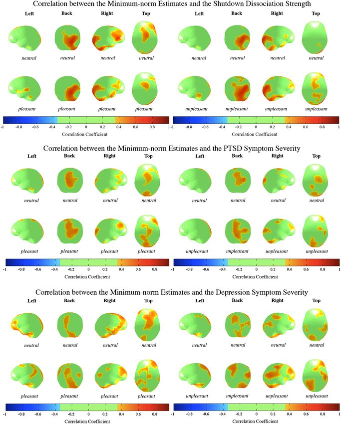
presented in Figure 3. Figure 4 shows significant correla- pleasant pictures. On the behavioural level, PTSD patients
tions for each location of the minimum-norm estimate showed different valence and arousal ratings. They rated
from 60 to 110 ms with the shutdown dissociation score, unpleasant stimuli as more unpleasant compared to the
the PTSD symptom severity and the depression scores. Non-PTSD group. Further, they rated the arousal level of
Due to interrelation of the psychopathological scales, the unpleasant and neutral stimuli as higher compared to the
common variance of the neural brain response was cal- Non-PTSD control group. This finding of different ratings
culated and summarized in Table 2. The highest con-
cordance of brain correlates (67%) was observed for the
unpleasant picture category related to shutdown disso-
ciation and PTSD symptom severity.
Correlates for the time window of interest 228 to 245 ms
within the PTSD group
For both affective conditions, the correlation with the
minimum-norm estimation of the condition across 228
to 245 ms reached significance with the depression score
(pleasant condition: r = .40; unpleasant condition: r = .37,
p < .05), but not with the shutdown dissociation strength
or PTSD symptom severity (all ps > .2). No significant
associations were observed in the neutral condition of
either block.
Discussion
The present study examined processing of affective mater-
ial in a sample of severely traumatized women with PTSD
and varying degrees of dissociation compared to women Figure 3 This figure presents the Pearson correlation coefficient
without PTSD on multiple dimensions (behaviour, heart between the surface minimum-norm estimate in source space
rate, subjective and neural processing). Using a dimen- and the psychopathological scale (circle: Shutdown dissociation;
triangle: Posttraumatic Stress Disorder (PTSD) symptom severity;
sional approach, we assessed the early dynamics of visual
rechtangle: Depression symptom severity) as a function of
processing in response to RSVP with respect to shutdown picture category. The grey horizontal line presents the significance
dissociation, PTSD and depression symptom severity. level indicating significant correlations above the line. The dashed
Despite obvious cultural differences, the IAPS valence line separate the blocks presenting the results of the unpleasant/
ratings of the present Non-PTSD sample are in accord neutral block at the left side and the results of the pleasant/neutral
block at the right side. The black solid line and * indicate the
with the normative ratings of the American sample.
significant difference between the correlation coefficient.
Arousal ratings were higher for unpleasant compared toSchalinski et al. BMC Psychiatry 2014, 14:193 Page 9 of 13 http://www.biomedcentral.com/1471-244X/14/193 Figure 4 Brain maps present the correlation between the minimum-norm estimate (average from 60 to 110 ms) and the shutdown dissociation strength in response to the processing of rapidly presented pictures, PTSD symptom severity and depression severity, projected on schematic cortical surfaces. Brain maps are shown across the picture category and block type for the PTSD group. The brain maps are presented from different perspectives (left, back, right and top view). Correlations between -.3 and .3 were supressed. PTSD = Posttraumatic Stress Disorder. for unpleasant stimuli is in line with a previous study with were not personalized for the traumatic events, 60% of PTSD patients showing exaggerated emotional responding the PTSD sample experienced intrusive memories of [36]. PTSD patients show more extreme ratings (e.g. very their own trauma in response to the stimulation. The negative, very arousing), making more of a distinct cate- observation is consistent with the theory of a fear/trauma gorization rather than graded evaluation of the stimuli. network, i.e., an interconnected network of neural rep- The arousal rating differed between the pleasant and resentations formed through multiple threatening ex- unpleasant stimuli in both PTSD and Non-PTSD con- periences. It encompasses sensory, cognitive, physiological, trols, with unpleasant stimuli rated as more arousing. and emotional experiences and includes the action dispos- Consistent with the arousal ratings, the heart rate data ition related to the experience. When a few representa- confirmed a higher physiological arousal in the unpleas- tions within this network (e.g. the sight of blood or ant/neutral block compared to the pleasant/neutral weapons, cognitions, like “I cannot do anything”) become block for both groups. Although the unpleasant pictures activated, the excitation will begin to spread through this
Schalinski et al. BMC Psychiatry 2014, 14:193                                                                                     Page 10 of 13
http://www.biomedcentral.com/1471-244X/14/193

Table 2 Similarities (r2) of brain correlates across                             severity is related to more severe dissociative responding
conditions and symptom severities                                                [40]. So far, no studies have assessed how cumulative
                             Shutdown           Depression/           PTSD/      trauma affects the temporal pattern of the development
                            dissociation/        shutdown           depression   of trauma-related symptoms, but it has been shown that
                                PTSD            dissociation
                                                                                 dissociative symptoms play a role, even years after the
Unpleasant                       67%                 31%                   33%
                                                                                 traumatic experiences [41].
Neutral (unpleasant/             26%                 26%                   26%      The analysis of the differential effects of the shutdown
neutral block)
                                                                                 dissociation, PTSD and depression symptom severity
Pleasant                         42%                 15%                   28%   addresses the issue of complex psychopathology in this
Neutral (pleasant/               41%                 25%                   28%   sample and shed light upon the influences on emotional
neutral block)
                                                                                 processing. Regardless of the picture category, positive
Note. All p < .0001. Similarities are measured as proportion of variance
explained, i.e. as squared correlation coefficient. PTSD = Posttraumatic
                                                                                 correlations of the minimum-norm estimates with depres-
Stress Disorder.                                                                 sion severity were found at a very early stage of visual pro-
                                                                                 cessing between 60 and 110 ms. Shutdown dissociation
interconnected excitatory network and activation of other                        was more strongly correlated with the processing of un-
trauma-related memory traces will have a low threshold                           pleasant/threatening stimuli compared to the correlates of
see, e.g., [37]. The dominant peri-traumatic response –                          the neutral condition. In contrast, for both levels of the
fear/anger or dissociative responding – will also be more                        pleasant/neutral block, positive associations between shut-
likely to appear. Indeed, those PTSD patients who re-                            down dissociation during testing and minimum-norm
ported more intense shutdown dissociation in their daily                         estimate were observed for the early time window. These
life experienced more of these symptoms also during the                          very early effects of visual processing of salient emotional
emotional visual processing. To overcome the intrusive                           stimuli are consistent with previous findings [15,16]. The
memories, the shutdown of perceptual channels, bodily                            standard account of early processing holds that crude per-
functions and emotions may be rewarding in the short-                            ceptual information can be relayed to important emotion
term, as it interrupts the perception of trauma reminders                        centres of the brain before undergoing more sophisticated
and thus reduces the symptoms and autonomic arousal                              cortical processing [42,43]. Depression severity seems to
[10]. However, in the long-term this response will also                          affect the cortical processing regardless of the picture cat-
disrupt psychosocial functioning and will leave the sur-                         egory. In contrast, the shutdown dissociation differentially
vivor without behavioural control and proper regulation                          modulates the processing of the unpleasant compared to
of emotions [38].                                                                neutral pictures. This is consistent with the theory of a
                                                                                 fear-network, whereby shutdown dissociation is one possi-
Minimum-norm estimates and brain correlates                                      bility for the peri-traumatic response that in the course of
Within the PTSD group, an arousal effect (mean of 60                             multiple traumatic experiences may have become the
to 110 ms) was found, showing higher global field power                          primary mode of responding. From its brain activation
in source space in the pleasant and unpleasant compared                          pattern very early after stimulus onset, we might speculate
to the neutral conditions. This arousal effect was also                          that the shutdown dissociation enhances immediate and
observed in the Non-PTSD control group in the un-                                crude visual processing of threat cues. Cortical as well as
pleasant versus neutral condition, but not for the plea-                         subcortical networks that could be involved in the rela-
sant versus neutral condition. Elbert and co-workers                             tionships are likely to be widespread (compare Figure 4).
[16] also found a very early arousal modulation in a sam-                        Shutdown dissociation and PTSD symptom severity show
ple of PTSD using the RSVP design. The affective modu-                           a 67% overlap of significant correlations for unpleasant
lation in the Non-PTSD sample could represent a higher                           stimuli, which is considerable but does not suggest that
behavioural importance of more arousing threatening                              the two measures are identical. Although one could antici-
stimuli compared to less arousing pleasant stimuli. The                          pate fundamental processing differences between a shut-
present study used a dimensional approach to assess the                          down dissociation response and the hyperarousal that
modulation of emotional processing. Shutdown disso-                              characterizes some PTSD patients, in terms of the peri-
ciation, PTSD symptom severity and depression severity                           traumatic psychophysiological defence response, both
were all inherently associated with each other. The inter-                       scales show similar but not identical modulations of the
relations are described in literature: persistent trauma-                        brain circuits when the traumatized brain is confronted
related dissociation has been shown to be strongly                               with unpleasant/threatening stimuli. Examining the results
associated with PTSD [2,3]. A person that went through                           in further detail, the lower overlap between the brain’s
a greater number of different types of traumatic events                          correlate of shutdown dissociation and PTSD symptom
is more likely to develop PTSD and a co-morbid depres-                           severity for neutral stimuli suggest a qualitatively differen-
sive disorder [39]. At the same time, the PTSD symptom                           tial modulation of the processing for the two clinical
Schalinski et al. BMC Psychiatry 2014, 14:193 Page 11 of 13
http://www.biomedcentral.com/1471-244X/14/193
syndromes. Additionally, the examination of the correla- these three symptom clusters are interrelated but not
tions with the minimum-norm estimates in the unpleasant interchangeable. All three are not only distinct clinical
condition and the standardized difference between PTSD manifestations but also appear as different forms of
symptom severity and shutdown dissociation (the resid- functional brain organisation. Differential effects become
uals) result in non-significant correlations. The overlap visible in time (60-110 ms vs. 228-245 ms), space (cor-
may indicate that shutdown dissociation appears at the relations of regional pattern of activation with symptom
upper end of the posttraumatic stress response, rather severity) and affective arousal. With the dimensional
than being uniformly present in all survivors with PTSD. approach, we could show that in an early time window,
DSM-5 includes, as a subtype, PTSD with prominent dis- affective modulation of cortical response is associated with
sociative symptoms. Our data would argue for a dimen- all three symptoms clusters, in an overlapping but also
sional/severity difference rather than a separate category, partly differential pattern. Regardless of the emotional sa-
as in our sample, high shutdown dissociation is inherently lience, these variables seem to affect the streams of visual
associated with more severe PTSD. The high cortical over- emotional processing. The stronger correlation between
lap in the processing of threatening/unpleasant pictures the shutdown dissociation and the minimum-norm esti-
raises the question of whether dissociation relates to a mate in the unpleasant versus neutral condition indicated
distinct categorical construct or appears as a modification differential processing. In the later time window (228 to
of brain circuitry with increasing symptom severity. A 245 ms), selective arousal modulation associated with
greater exposure to traumatic stressors would increase depressive symptoms could also be observed. The brain
PTSD symptom severity and also make peri-traumatic regions involved cause widespread activity. A high con-
shutdown more likely, which then might replay as dis- cordance of brain correlations was found for shutdown
sociative responding when cued by trauma-related re- dissociation and PTSD severity in the unpleasant con-
minders. A recent study found evidence of a dissociative dition, but this overlap was much lower in the neutral
symptom cluster that is correlated with the core PTSD condition. In sum, these results would support a model in
symptoms and associated with higher PTSD symptom which increasing exposure to traumatic stress, brain pro-
severity as well as more severe co-morbidity pattern [44]. cessing becomes altered on qualitatively different dimen-
It is likely that dissociation, with its ongoing disruption of sions, as captured by symptoms of PTSD, depression and
integrative processes, would play a key role in the severity dissociation. That is, survivors of traumatic stressors
and maintenance of PTSD. present with a set of different functional reorganization of
Depression symptom severity also correlates with a pat- brain activity and hence a comparison of patients with
tern of brain activity, however, there are clear differences and without PTSD may produce quite variable results if
in functional brain activity between depression and the the other dimensions as well as their intensities are not
other two symptom clusters. In contrast to the early ef- considered.
fects of visual processing and depression severity, the later
time window from 228 to 245 ms revealed only significant Additional file
correlations with the minimum-norm estimates and the
depression strength in the high arousing conditions (pleas- Additional file 1: Brain maps present the minimum-norm estimate
ant and unpleasant). These effects seem to be specific for (average from 128 to 143 ms) in the unpleasant condition for the
Non-PTSD control group and the PTSD group. The brain maps on the
the emotionally salient stimuli categories and present lower line show the significance of the group difference between the
differential affective cortical processing. Usually, patients Non-PTSD control and PTSD group. The brain maps are presented from
with depressive disorders show lower cortical modulation different perspectives (left, right, top and back view). Posttraumatic Stress
Disorder.
for affective arousal stimulation [45]. In contrast, the
present results suggest that the depressive brain reacts
Competing interests
more strongly towards arousing content than neutral con- The authors declare that they have no competing interests.
tent. This is in line with the results from another study
that found the complementary modulation, namely hyper- Authors’ contributions
MS & TE developed the study concept. TE and IS contributed to the study
activity to arousing stimuli in patients with depression and design. Data collection and data preprocessing were performed by JM and
comorbid anxiety disorders [46,47]. Further, our results IS. IS developed scripts to analyse the event-related magnetic fields and
suggest that increasing severity of depression in a sample performed the data analysis and interpretation under the supervision of TE.
TE and IS drafted the paper, and JM and MS provided critical revisions.
of patients with PTSD is associated with more pro- All authors approved the final version of the paper for submission.
nounced response to highly arousing stimuli.
Acknowledgements
Conclusion We thank the respondents who participated in the study with great courage
and openness. The project was supported by the Deutsche
Repeated exposure to traumatic stressors may result in Forschungsgemeinschaft (DFG) and the European Refugee Fund. We are
PTSD, shutdown dissociation, and depression, whereby grateful to Heike Riedke, Ursula Lommen, Alexandra Geist, Charlotte SalmenSchalinski et al. BMC Psychiatry 2014, 14:193 Page 12 of 13
http://www.biomedcentral.com/1471-244X/14/193
and Franziska Unholzer for help with data acquisition and logistics and to dimensions and emotion dysregulation in responses to script-driven
Dres. Katalin Dohrmann, Julia Morath, Maria Roth and Roland Weierstall for trauma imagery. J Trauma Stress 2007, 20:713–725. doi:10.1002/jts.20284.
conducting structured interviews. 21. Ray WJ, Odenwald M, Neuner F, Schauer M, Ruf M, Rockstroh B, Elbert T:
Decoupling neural networks from reality: dissociative experiences in
Received: 30 January 2014 Accepted: 3 July 2014 torture victims are reflected in abnormal brain waves in left frontal
Published: 5 July 2014 cortex. Psychol Sci 2006, 17:825–829.
22. Peyk P, Schupp HT, Elbert T, Junghöfer M: Emotional processing in the
visual brain: a MEG analysis. Brain Topogr 2008, 20:205–215. doi:10.1007/
References s10548-008-0052-7.
1. Marx BP, Forsyth JP, Lexington JM: Tonic immobility as an evolved 23. Schupp HT, Stockburger J, Codispoti M, Junghöfer M, Weike AI, Hamm AO:
predator defense: implications for sexual assault survivors. Sci Pract 2008, Stimulus novelty and emotion perception: the near absence of
15:74–90. habituation in the visual cortex. Neuroreport 2006, 17:365–369.
2. Briere J, Scott C, Weathers F: Peritraumatic and persistent dissociation in 24. Bradley MM: Natural selective attention: orienting and emotion.
the presumed etiology of PTSD. Am J Psychiat 2005, 162:2295–2301. Psychophysiology 2009, 46:1–11. doi:10.1111/j.1469-8986.2008.00702.x.
3. Panasetis P, Bryant RA: Peritraumatic versus persistent dissociation in 25. Öhman A, Flykt A, Esteves F: Emotion drives attention: detecting the
acute stress disorder. J Trauma Stress 2003, 16:563–566. snake in the grass. J Exp Psychol Gen 2001, 130:466–478.
4. Werner KB, Griffin MG: Peritraumatic and persistent dissociation as 26. Rockstroh B, Elbert T: Traces of fear in the neural web-
predictors of PTSD symptoms in a female cohort. J Trauma Stress 2012, Magnetoencephalographic responding to arousing pictorial stimuli.
25:401–407. Int J Psychophysiol 2010, 78:14–19. doi:10.1016/j.ijpsycho.2010.01.012.
5. Bremner JD, Southwick S, Brett E, Fontana A, Rosenheck R, Charney DS: 27. Schalinski I, Elbert TR, Schauer M: Cardiac defense in response to
Dissociation and posttraumatic stress disorder in Vietnam combat imminent threat in women with multiple trauma and severe PTSD.
veterans. Am J Psychiat 1992, 149:328–332. Psychophysiology 2013, 50:691–700. doi:10.1111/psyp.12051.
6. Ginzburg K, Koopman C, Butler LD, Palesh O, Kraemer HC, Classen CC, 28. Blake DD, Weathers FW, Nagy LM, Kaloupek DG, Gusman FD, Charney DS,
Spiegel D: Evidence for a dissociative subtype of Post-traumatic stress Keane TM: The development of a Clinician-Administered PTSD Scale.
disorder among help-seeking childhood sexual abuse survivors. J Trauma Stress 1995, 8:75–90.
J Trauma Dissociation 2006, 7:7–27. doi:10.1300/J229v07n02_02. 29. Sheehan DV, Lecrubier Y, Sheehan KH, Amorim P, Janavs J, Weiller E,
7. Murray J, Ehlers A, Mayou RA: Dissociation and post-traumatic stress Dunbar GC: The Mini-International Neuropsychiatric Interview (M.I.N.I.):
disorder: two prospective studies of road traffic accident survivors. The development and validation of a structured diagnostic psychiatric
Br J Psychiatry 2002, 180:363–368. interview for DSM-IV and ICD-10. J Clin Psychiatry 1998, 59:22–33.
8. Feeny NC, Zoellner LA, Fitzgibbons LA, Foa EB: Exploring the roles of 30. Williams JB: A structured interview guide for the Hamilton Depression
emotional numbing, depression, and dissociation in PTSD. J Trauma Stress Rating Scale. Arc Gen Psychiatry 1988, 45:742–747.
2005, 13:489–498. doi:10.1023/A:1007789409330. 31. Lang PJ, Bradley MM, Cuthbert BN: International Affective Picture System
9. van der Hart O, Nijenhuis E, Steele K, Brown D: Trauma-related (IAPS): Affective Ratings of Pictures and Instruction Manual. Technical Report
dissociation: conceptual clarity lost and found. Aust N Z J Psychiatry 2004, A-8. University of Florida, Gainesville, FL.FL: The Center for Research in
38:906–914. Psychophysiology, University of Florida; 2008.
10. Schauer M, Elbert T: Dissociation following traumatic stress: etiology 32. Oldfield RC: The assessment and analysis of handedness: the Edinburgh
and treatment. J Psychol 2010, 218:109–127. doi:10.1027/0044-3409/ inventory. Neuropsychologia 1997, 9:97–113.
a000018. 33. Bradley MM, Lang PJ: Measuring emotion: the self-assessment manikin and
11. Bracha HS: Freeze, flight, fight, fright, faint: adaptationist perspectives on the semantic differential. J Behav Ther Exp Psychiatry 1994, 25:49–59.
the acute stress response spectrum. CNS Spectr 2004, 9:679–685. 34. Hämäläinen MS, Ilmoniemi RJ: Interpreting magnetic fields of the brain:
12. Noyes R, Kletti R: Depersonalization in response to life-threatening Minimum norm estimates. Med Biol Eng Comput 1994, 32:35–42.
danger. Compr Psychiatry 1977, 18:375–384. 35. Rosenthal R: Meta-Analytic Procedures for Social Research (Vol. 6). Newbury
13. Lanius RA, Vermetten E, Loewenstein RJ, Brand B, Schmahl C, Bremner JD, Park, CA: SAGE Publications, Incorporated; 1991.
Spiegel D: Emotion modulation in PTSD: Clinical and neurobiological 36. Wolf EJ, Miller MW, McKinney AE: Emotional processing in PTSD:
evidence for a dissociative subtype. Am J Psychiatry 2010, 167:640–647. heightened negative emotionality to unpleasant photographic
doi:10.1176/appi.ajp.2009.09081168. stimuli. J Nerv Ment Dis 2009, 197:419–426. doi:10.1097/
14. Adenauer H, Pinösch S, Catani C, Gola H, Keil J, Kissler J, Neuner F: Early NMD.0b013e3181a61c68.
processing of threat cues in Posttraumatic Stress disorder—evidence for 37. Schauer M, Neuner F, Elbert T: Narrative Exposure Therapy. 2nd edition.
a cortical vigilance-avoidance reaction. Biol Psychiatry 2010, 68:451–458. Göttingen, Germany: Hogrefe & Huber; 2011.
doi:10.1016/j.biopsych.2010.05.015. 38. Feeny NC, Zoellner LA, Foa EB: Anger, dissociation, and posttraumatic stress
15. Catani C, Adenauer H, Keil J, Aichinger H, Neuner F: Pattern of cortical disorder among female assault victims. J Trauma Stress 2000, 13:89–100.
activation during processing of aversive stimuli in traumatized survivors 39. Neuner F, Schauer M, Karunakara U, Klaschik C, Robert C, Elbert T:
of war and torture. Eur Arch Psychiatry Clin Neurosci 2009, 259:340–351. Psychological trauma and evidence for enhanced vulnerability for PTSD
doi:10.1007/s00406-009-0006-4. through previous trauma among West Nile refugees. BMC Psychiatry 2004,
16. Elbert TR, Schauer M, Ruf M, Weierstall R, Neuner F, Rockstroh B, Junghöfer 4:34. doi:10.1186/1471-244X-4-34.
M: The tortured brain: imaging neutral representation of the traumatic 40. Schalinski I, Elbert T, Schauer M: Female dissociative responding to
tress experiences using RSVP with affective pictorial stimuli. J Psychol extreme sexual violence in a chronic crisis setting: The case of eastern
2011, 219:167–174. doi:10.1027/21512604/a000064. congo. J Trauma Stress 2011, 24:235–238. doi:10.1002/jts.20631.
17. Junghöfer M, Schauer M, Neuner F, Odenwald M, Rockstroh B, Elbert T: 41. Carlson EB, Dalenberg C, McDade-Montez E: Dissociation in posttraumatic
Enhanced fear-network in torture survivors activated by RVSP of stress disorder part I: definitions and review of research. Psychol Trauma
aversive material can be monitored by MEG. Psychophysiology 2003, Theory Res Pract Policy 2012, 4:479–489.
40(Suppl):51. 42. LeDoux JE: Synaptic Self: How our Brains Become Who We Are. New York:
18. Matz K, Junghöfer M, Elbert T, Weber K, Wienbruch C, Rockstroh B: Viking Penquin; 2002.
Adverse experiences in childhood influence brain responses to 43. Pessoa L, Adolphs R: Emotion processing and the amygdala: from a ‘low
emotional stimuli in adult psychiatric patients. Int J Psychophysiol 2010, road’ to ‘many roads’ of evaluating biological significance. Nat Rev Neurosci
75:277–286. doi:10.1016/j.ijpsycho.2009.12.010. 2010, 11:773–782. doi:10.1038/nrn2920.
19. Moratti S, Rubio G, Campo P, Keil A, Ortiz T: Hypofunction of right 44. Steuwe C, Lanius RA, Frewen PA: Evidence for a dissociative subtype of
temporoparietal cortex during emotional arousal in depression. PTSD by latent profile and confirmatory factor analyses in a civilian
Arch Gen Psychiatry 2008, 65:532–541. doi:10.1001/archpsyc.65.5.532. sample. Depress Anxiety 2012, 29:689–700.
20. Hopper JW, Frewen PA, van der Kolk BA, Lanius RA: Neural correlates of 45. Heller W, Nitscke JB: Regional brain activity in emotion: a framework for
reexperiencing, avoidance, and dissociation in PTSD: Symptom understanding cognition in depression. Cogn Emot 1997, 11:637–661.You can also read