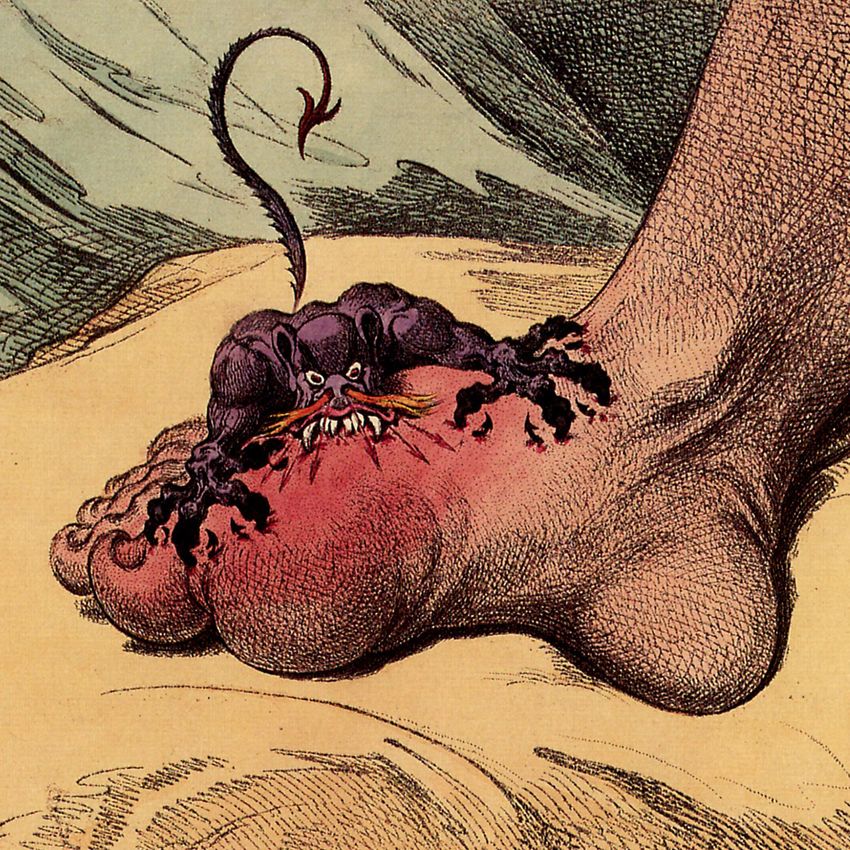
Revisiting the pathogenesis of podagra: why does gout target the foot? - Roddy - JOURNAL OF FOOT AND ANKLE RESEARCH
←
→
Page content transcription
If your browser does not render page correctly, please read the page content below
JOURNAL OF FOOT
        AND ANKLE RESEARCH

Revisiting the pathogenesis of podagra: why does
gout target the foot?
Roddy

                                     Roddy Journal of Foot and Ankle Research 2011, 4:13
                             http://www.jfootankleres.com/content/4/1/13 (13 May 2011)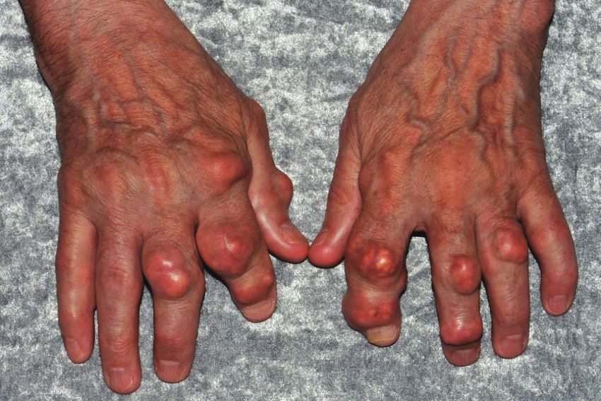
Roddy Journal of Foot and Ankle Research 2011, 4:13
http://www.jfootankleres.com/content/4/1/13
                                                                                                                                               JOURNAL OF FOOT
                                                                                                                                               AND ANKLE RESEARCH

 REVIEW                                                                                                                                         Open Access

Revisiting the pathogenesis of podagra: why does
gout target the foot?
Edward Roddy

  Abstract
  This invited paper provides a summary of a keynote lecture delivered at the 2011 Australasian Podiatry Conference.
  Gout is the most prevalent inflammatory arthropathy. It displays a striking predilection to affect the first
  metatarsophalangeal joint as well as joints within the mid-foot and ankle. A number of factors are known to
  reduce urate solubility and enhance nucleation of monosodium urate crystals including decreased temperature,
  lower pH and physical shock, all of which may be particularly relevant to crystal deposition in the foot. An
  association has also been proposed between monosodium urate crystal deposition and osteoarthritis, which also
  targets the first metatarsophalangeal joint. Cadaveric, clinical and radiographic studies indicate that monosodium
  urate crystals more readily deposit in osteoarthritic cartilage. Transient intra-articular hyperuricaemia and
  precipitation of monosodium urate crystals is thought to follow overnight resolution of synovial effusion within the
  osteoarthritic first metatarsophalangeal joint. The proclivity of gout for the first metatarsophalangeal joint is likely to
  be multi-factorial in origin, arising from the unique combination of the susceptibility of the joint to osteoarthritis
  and other determinants of urate solubility and crystal nucleation such as temperature and minor physical trauma
  which are particularly relevant to the foot.

Background                                                                            dramatically distant from humoral theory, these observa-
Gout is a true crystal deposition disease in which all                                tions concerning the intimate relationship between gout
clinical manifestations are considered to be directly                                 and the foot have been reinforced over the centuries
attributable to the presence of monosodium urate                                      and continue today. This review will consider the ways
(MSU) crystals. It is one of the most prevalent inflam-                               in which gout affects the foot and discuss potential
matory arthropathies with a prevalence of approximately                               mechanisms underlying this relationship.
1.4%, and is the most common inflammatory arthropa-
thy in men [1]. Both the prevalence and incidence of                                  Clinical presentation of gout and involvement of
gout appear to be rising [2]. The primary risk factor for                             the foot
the development of gout is elevation of serum uric acid                               After an often prolonged period of asymptomatic hyper-
(urate) levels, or hyperuricaemia. As uric acid levels rise                           uricaemia, the initial manifestation of gout is usually an
and exceed the physiological saturation threshold of uric                             acute attack of synovitis affecting a single peripheral
acid in body tissues, formation and deposition of MSU                                 joint, most commonly the first metatarsophalangeal
crystals occurs in and around joints.                                                 joint (MTPJ). Other commonly affected joints include
  The propensity of gout for the foot was recognised by                               the mid-tarsal joints, ankles, knees, fingers, wrists and
the ancient Greeks who referred to it as podagra, lit-                                elbows (Figure 1). Such attacks are characterised by sud-
erally “foot-grabber” [3]. The name “gout” derives from                               den onset of excruciating joint pain, typically taking less
humoral theory and the Latin word gutta or “drop”,                                    than 24 hours from symptom onset to reach peak inten-
podagra being thought to arise as a result of the bodily                              sity, with associated joint swelling, overlying erythema
humours falling to the affected body part. Although our                               and exquisite tenderness to touch. Although acute gout
current understanding of the pathogenesis of gout is                                  should be treated rapidly with a non-steroidal anti-
                                                                                      inflammatory drug (NSAID) or colchicine, it usually
Correspondence: e.roddy@cphc.keele.ac.uk                                              resolves completely over a period of two to three weeks
Arthritis Research UK Primary Care Centre, Primary Care Sciences, Keele
University, Keele, UK                                                                 even without treatment. A variable period of time then

                                        © 2011 Roddy; licensee BioMed Central Ltd. This is an Open Access article distributed under the terms of the Creative Commons
                                        Attribution License (http://creativecommons.org/licenses/by/2.0), which permits unrestricted use, distribution, and reproduction in
                                        any medium, provided the original work is properly cited.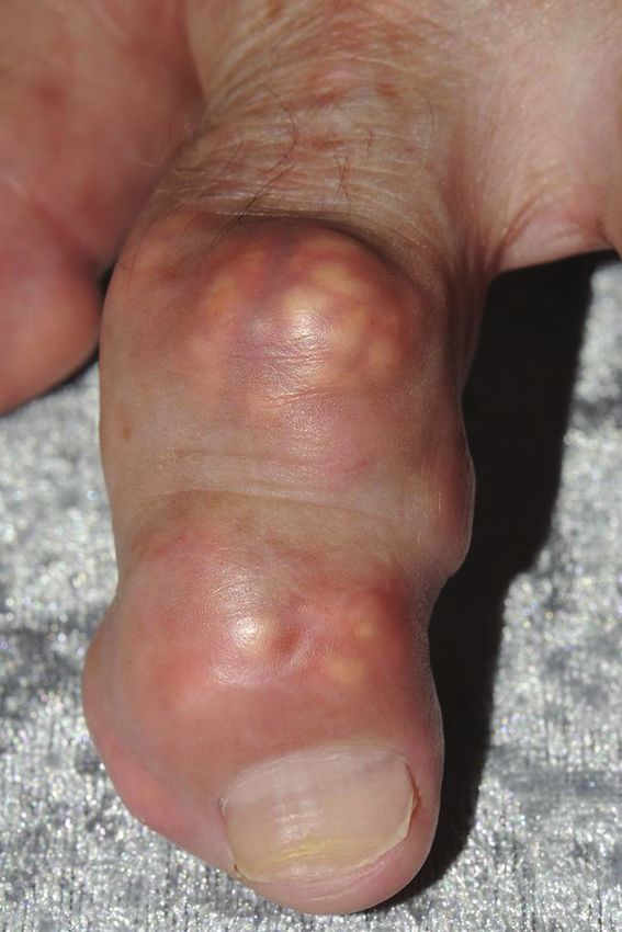
Roddy Journal of Foot and Ankle Research 2011, 4:13                                                                   Page 2 of 6
http://www.jfootankleres.com/content/4/1/13

                                                                    disease in 59-89% [4,6,8-10]. Fewer studies report the
                                                                    frequency of involvement of other joints. However,
                                                                    mid-foot and ankle involvement occurs in 25-50% and
                                                                    18-60% of patients respectively [5,8,9]. In contrast, the
                                                                    upper limb is involved in 13-46% [4,6,8,10] and the
                                                                    finger interphalangeal joints in only 6-25% [5,8,9].
                                                                      Sub-clinical involvement in the foot also appears to be
                                                                    common-place. MSU crystal deposits have been
                                                                    observed in synovial fluid aspirated from first MTPJs
                                                                    that have never been affected by an acute attack of gout
                                                                    [11,12]. Furthermore, a study which examined the first
                                                                    MTPJs of 39 males with gout using high resolution
                                                                    ultrasonography found erosions to be present in 45% of
                                                                    22 first MTPJs that had never been affected by acute
                                                                    gout [13].
                                                                      Gout has a number of chronic manifestations which
                                                                    are easily recognisable as such including tophaceous
                                                                    deposits and a characteristic erosive arthropathy. How-
                                                                    ever, it is also associated with a number of other less
                                                                    specific foot problems. Perhaps not surprisingly given
                                                                    the frequency of first MTPJ involvement, hallux valgus
                                                                    is a common finding. In a community-based case-
                                                                    control study, hallux valgus was found in 41% of gout
                                                                    suffers compared to 25% of age- and gender-matched
                                                                    control subjects (odds ratio (OR) 2.10, 95% confidence
                                                                    interval (CI) 1.39 to 3.18, adjusted for body mass index
                                                                    (BMI) and use of diuretics) [14]. Big toe pain occurring
                                                                    on most days for at least a month within the last year
                                                                    was reported by 16% of those with gout compared to
                                                                    6% of controls (adjusted OR 2.94, 95% CI 1.62 to 5.34).
                                                                    Given the striking predilection of gout for the foot,
                                                                    there has been surprisingly little work examining the
                                                                    influence of gout on foot function, gait and plantar pres-
 Figure 1 Distribution of joints typically affected by gout
                                                                    sure distributions. A recent study compared functional
 (reproduced with the permission of the author and the Royal
 College of General Practitioners: Roddy E, Doherty M. Gout. In:    and biomechanical foot characteristics between 25 patients
 RCGP Guide to MSK Disorders in Primary Care. Ed: Warburton L (in   with chronic gout and 25 age- and gender-matched con-
 press)).                                                           trol subjects with no history of gout [15]. Patients with
                                                                    chronic gout were found to have slower walking velocity,
                                                                    reduced step and stride length, reduced peak plantar pres-
elapses until the patient experiences a further attack (the         sure under the hallux, and higher mid-foot pressure-time
“intercritical period”). With time, attacks may increase            integrals compared to controls. The authors postulate that
in severity and frequency, involve different joint sites,           gait pattern is altered in chronic gout in an attempt to off-
and may become oligo- or polyarticular. Eventually,                 load the first MTPJ thereby reducing pain. Further studies
without treatment, the patient may develop chronic                  are necessary to explore these observations in more detail
tophaceous gout, characterised by chonic pain and stiff-            and examine the contribution of chronic pain in the great
ness, joint damage and erosive arthropathy, and clini-              toe, hallux valgus, obesity and osteoarthritis (OA) to gait
cally evident subcutaneous nodular deposits of MSU                  patterns in patients with gout.
crystals (tophi) which can occur at the toes, Achilles’
tendons, pre-patellar tendons, fingers, olecranon pro-              Factors influencing crystal deposition
cesses, and less commonly, the ears (Figure 2).                     Gout is one of the best understood inflammatory arthro-
  Gout displays a striking tendency to affect the foot, in          pathies. Clinical features can be easily understood and
particular the first MTPJ. The initial attack of gout               interpreted in the context of a clearly elucidated patho-
affects the first MTPJ in 56-78% of patients [4-7] and              genetic process. Specific risk factors such as genetics,
the joint is involved at some point in the course of                dietary factors, co-morbidity and its treatment lead to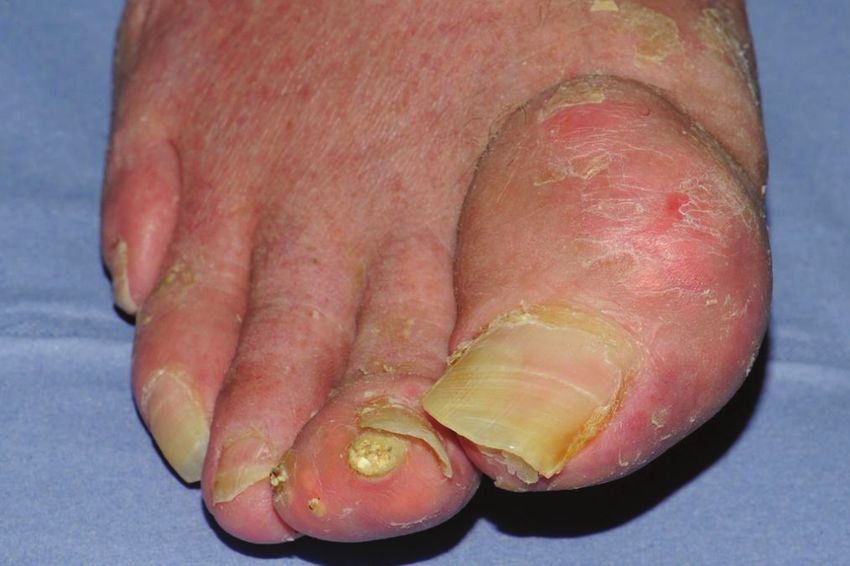
Roddy Journal of Foot and Ankle Research 2011, 4:13                                                                        Page 3 of 6
http://www.jfootankleres.com/content/4/1/13

 Figure 2 Tophaceous gout affecting the right great toe and finger interphalangeal joints. Note the asymmetrical swelling and yellow-
 white discolouration.

hyperuricaemia and subsequently MSU crystal formation               Trauma and pH
occurs [16,17]. Crystals are then shed into the joint and           A further well-recognised clinical feature of gout is the
activate the inflammatory cascade via the NALP3                     tendency of an acute attack to be precipitated by physical
inflammasome [18,19]. Hence, any explanation of why                 trauma such as stubbing the toe or following physical
gout targets the foot must link these pathological pro-             activity. Enhanced MSU crystal nucleation has been
cesses to the specific anatomical, functional, and disease          reported in vitro following mechanical agitation of solu-
characteristics of the foot (Figure 3).                             tions supersaturated with sodium urate [22]. The same
                                                                    authors demonstrated that nucleation is also potentiated
Temperature                                                         by both acidification and addition of calcium ions. Lower-
As described above, gout tends to affect distal peripheral          ing of pH has a direct action on MSU crystal nucleation
joints, not only in the foot but also in the upper limb,            but also enhances nucleation by increasing calcium ion
with central axial joints such as the shoulders, hips and           activity. Whilst their observations concerning mechanical
spine only rarely affected. The solubility of urate                 agitation provide evidence that a physical shock can
decreases with reducing temperature [20,21] enhancing               directly lead to MSU crystal nucleation, the authors
nucleation of MSU crystals, that is, the “birth” of new             hypothesised that local trauma indirectly enhances crystal
crystals. Reduced solubility of urate at lower tempera-             nucleation by lowering synovial pH [22]. Hence, the sus-
tures has therefore been suggested to account for the               ceptibility of the foot to physical trauma might also help
occurrence of gout at cooler distal joints such as the              to explain the predilection of gout for the foot.
foot-ankle complex. However, this theory does not
account for the preference of gout for the first MTPJ               Cartilage damage and osteoarthritis
ahead of the great toe interphalangeal (IP) joint or the            More recently, the deposition of MSU and calcium pyr-
lesser MTPJs.                                                       ophosphate dihydrate (CPPD) crystals in areas of
Roddy Journal of Foot and Ankle Research 2011, 4:13 Page 4 of 6
http://www.jfootankleres.com/content/4/1/13
Lower Hyperuricaemia
temperature
First MTPJ osteoarthritis
Increased calcium • Increased chondroitin sulphate
ion activity concentration
• Degradation of protein-polysaccharide
complexes
• Epitaxial nucleation and growth of MSU
Lower pH crystals on cartilage fragments
MSU crystal formation • Transient increases in synovial fluid
and deposition urate concentration in resolving effusions
Physical
stress/trauma
“Shedding” of crystals
into the joint space
Acute inflammation
“podagra”
Figure 3 Processes enhancing MSU crystal formation and deposition at the first MTPJ.
cartilage damage has been described in a cadaveric study (DIP) joints also affected by OA [25-30]. A Polish hos-
which examined 7855 adult human tali from 4007 pital-based study of 262 patients with gout found an
donors [23]. Crystal deposits, both MSU and CPPD, association of gout and radiographic OA at the first
were an uncommon finding, being present in specimens MTPJs, tarsal joints and knees [31]. A more recent
from only 5% of donors. However, where seen, crystal study of 164 patients with gout recruited from primary
deposits were usually found within or adjacent to a car- care found a very strong association between joints that
tilage lesion. Only 8% of tali with crystal deposits had had previously been the site of an acute attack of gout
no gross evidence of cartilage degeneration. Cartilage and evidence of OA on clinical examination (OR 7.94,
lesions tended to be located at sites of biomechanical 95%CI 6.27 to 10.05, adjusted for age, gender, BMI and
stress such as the articulation of the margin of the tro- diuretic use) [8]. Significant associations were seen
chlea with the tibia or fibula or where apposition with between acute attacks of gout and the presence of clini-
anterior tibial osteophytes was thought to have cal OA at the first MTPJs, mid-foot, knee and finger
occurred. In a separate study, the epitaxial nucleation DIP joints.
and growth of MSU crystals was observed to occur on
fragments of articular cartilage [24]. Thus there appears Why are gout and osteoarthritis associated?
to be a relationship between cartilage lesions and the The observations outlined above that MSU crystals tend
anatomical location of MSU crystal deposition. to deposit at sites of cartilage damage and that clinical
In support of these observations, clinical and radio- and radiographic evidence exists of an association
graphic evidence exists of an association between gout between gout and OA lead to the important question of
and OA. Several case reports and small case series the mechanism by which gout and OA might be asso-
describe the occurrence of acute attacks of gout and/or ciated. There are three possible explanations for this
tophi at first MTPJs and finger distal interphalangeal association.Roddy Journal of Foot and Ankle Research 2011, 4:13 Page 5 of 6 http://www.jfootankleres.com/content/4/1/13 Firstly, does an association exist between the disease Why does gout target the first states of gout and nodal generalised OA? These two metatarsophalangeal joint? conditions share the common risk factor of obesity The studies discussed above provide clear evidence of an [32,33]. In a related study to the primary care study association between MSU crystal deposition and OA. described above [8], generalized nodal OA, defined as Whilst further studies are required to definitively answer the presence of Heberden’s or Bouchard’s nodes on at the questions of direction of association and causality, it least two digits in each hand [34], was no more com- appears that MSU crystals more readily deposit in monplace in subjects with gout than age-and gender- osteoarthritic cartilage and that the presence of OA matched community controls but, as discussed above, influences the distribution of joints affected by gout. hallux valgus and self-reported knee and big toe pain However, OA cannot solely explain the typical distribu- were more frequent in those with gout [14]. Although tion of joints affected by gout, as many joints commonly this case-control study was underpowered, these find- affected by OA such as the knees, finger IP joints, and ings do not suggest that an association exists between hips are less frequently affected by gout than the first the disease states of gout and generalised OA. MTPJ, and other target joints for gout such as the The second and third explanations are related and ankle, wrist and elbow are infrequent sites for primary concern the hypothesis that the association of gout and OA. Is it plausible therefore that the relationship OA occurs at local joint sites and relates to the co-loca- between MSU crystal deposition and OA is of more tion of MSU crystal deposits and cartilage lesions. Speci- relevance for the first MTPJ than other joint sites? fically, they question the direction of this association, The first MTPJ is certainly targeted by OA although namely, does the presence of osteoarthritic cartilage pre- foot OA is under-studied in comparison to other com- dispose to the local formation and deposition of MSU monly affected sites such as the hand and knee. A crystals or do MSU crystals themselves initiate and pro- recent systematic review of population-based epidemio- gress cartilage damage? Evidence to support the deposi- logical studies found that the estimated prevalence of tion of MSU crystals in osteoarthritic cartilage rather radiographic OA at the first MTPJ may be as high as than MSU crystals leading to cartilage damage arises 39% in middle-aged to older adults [40]. Simkin pro- from two sources. Although the primary care study posed a model to explain the clinical observations that described above was cross-sectional, making it difficult acute attacks of gout are commonly precipitated by phy- to infer causality, the strength of the association sical stress and occur overnight, based upon the co- between involvement of gout and OA at individual joint occurrence of gout and OA at the first MTPJ [41]. In sites did not increase with longer duration of gout [8]. this model, a synovial effusion develops in an osteoar- A further insight into the direction of association thritic first MTPJ during the day and subsequently between MSU crystal deposition and OA is provided by resolves when the joint is rested overnight. Synovium is a recent study which examined the relationship between more permeable to water than urate and hence, as the synovial fluid uric acid levels and the radiographic sever- effusion resolves, water leaves the joint more rapidly ity of knee OA [35]. Although synovial fluid uric acid than urate. This results in a transient increase in the was found to correlate with baseline knee OA severity, synovial fluid urate concentration which leads to preci- it was not associated with change in OA severity over 3 pitation of MSU crystals if the saturation threshold of years. These two observations do not suggest that the urate is exceeded. As discussed above, MSU crystal for- association between the occurrence of gout and OA at mation and deposition will be further potentiated in the individual joint sites is due to MSU crystal-initiated osteoarthritic first MTPJ by impaired urate solubility joint damage. Furthermore, certain properties of the and enhanced crystal nucleation arising from factors osteoarthritic joint are thought to influence urate solubi- relating to the anatomical location of the first MTPJ lity and predispose to local MSU crystal disposition [36]. namely lower distal temperature and physical stress Increased concentrations of chondroitin sulphate and [20-22], and those relating to OA namely increased con- degradation of protein-polysaccharide complexes found centrations of chondroitin sulphate, degradation of pro- within articular cartilage have been shown to reduce tein-polysaccharide complexes, and epitaxial MSU urate solubility and lead to the precipitation and growth crystal nucleation and growth on cartilage fragments of MSU crystals [37-39]. However, it is also possible [24,37-39] (Figure 3). that the association between MSU crystal deposition and OA is bi-directional whereby existing osteoarthritic Conclusion change predisposes to local formation and deposition of The striking predilection of gout for the first MTPJ MSU crystals which then initiate further cartilage appears to be multi-factorial in origin and arises from damage. the unique combination of the susceptibility of the joint
Roddy Journal of Foot and Ankle Research 2011, 4:13                                                                                                 Page 6 of 6
http://www.jfootankleres.com/content/4/1/13

to OA and local anatomical considerations of tempera-                              22. Wilcox WR, Khalaf AA: Nucleation of monosodium urate crystals. Ann
                                                                                       Rheum Dis 1975, 34:332-339.
ture, minor physical trauma and biomechanical stress,                              23. Muehleman C, Li J, Aigner T, Rappoport L, Mattson E, Hirschmugl C,
leading to ideal conditions for MSU crystal formation                                  Masuda K, Rosenthal AK: Association between crystals and cartilage
and deposition in predisposed hyperuricaemic indivi-                                   degeneration in the ankle. J Rheumatol 2008, 35:1108-1117.
                                                                                   24. Pascual E, Ordonez S: Orderly arrayed deposit of urate crystals in gout
duals, manifesting as clinical gout.                                                   suggest epitaxial formation. Ann Rheum Dis 1998, 57:255.
                                                                                   25. Simkin PA, Campbell PM, Larson EB: Gout in Heberden’s nodes. Arthritis
                                                                                       Rheum 1983, 26:94-97.
Acknowledgements                                                                   26. O’Dell JR: Gout in Heberden’s nodes. Arthritis Rheum 1983, 26:1413-1414.
The author would like to thank Dr George Peat for helpful comments on              27. Parhami N, Greenstein N, Juozevicius JL: Erosive osteoarthritis and gout:
the manuscript. The author is supported by an Arthritis Research UK Primary            gout in 36 joints. J Rheumatol 1986, 13:469-471.
Care Centre Grant (18139).                                                         28. Lally EV, Zimmermann B, Ho G Jr, Kaplan SR: Urate-mediated inflammation
                                                                                       in nodal osteoarthritis: clinical and roentgenographic correlations.
Competing interests                                                                    Arthritis Rheum 1989, 32:86-90.
The author declares that they have no competing interests.                         29. Foldes K, Petersilge CA, Weisman MH, Resnick D: Nodal osteoarthritis and
                                                                                       gout: a report of four new cases. Skeletal Radiol 1996, 25:421-424.
Received: 21 April 2011 Accepted: 13 May 2011 Published: 13 May 2011               30. Fam AG, Stein J, Rubenstein J: Gouty arthritis in nodal osteoarthritis. J
                                                                                       Rheumatol 1996, 23:684-689.
References                                                                         31. Kawenoki-Minc E, Eyman E, Leo W, Werynska-Przybylska J: Osteoarthrosis
1. Annemans L, Spaepen E, Gaskin M, Bonnemaire M, Malier V, Gilbert T,                 and spondylosis in gouty patients. Analysis of 262 cases of gout.
    Nuki G: Gout in the UK and Germany: prevalence, comorbidities and                  Reumatologia 1974, 12:267-267.
    management in general practice 2000-2005. Ann Rheum Dis 2008,                  32. Choi HK, Atkinson K, Karlson EW, Curhan G: Obesity, weight change,
    67:960-966.                                                                        hypertension, diuretic use, and risk of gout in men: the health
2. Roddy E, Zhang W, Doherty M: The changing epidemiology of gout. Nat                 professionals follow-up study. Arch Intern Med 2005, 165:742-748.
    Clin Pract Rheumatol 2007, 3:443-449.                                          33. Oliveria SA, Felson DT, Cirillo PA, Reed JI, Walker AM: Body weight, body
3. Porter R, Rousseau GS: Gout The Patrician Malady New Haven and London:              mass index, and incident symptomatic osteoarthritis of the hand, hip,
    Yale University Press; 1998.                                                       and knee. Epidemiology 1999, 10:161-166.
4. Puig JG, Michan AD, Jimenez ML, Perez de Ayala C, Mateos FA, Capitan CF,        34. Wright GD, Regan M, Deighton CM, Wallis G, Doherty M: Evidence for
    de Miguel E, Gijon JB: Female gout. Clinical spectrum and uric acid                genetic anticipation in nodal osteoarthritis. Ann Rheum Dis 1998,
    metabolism. Arch Intern Med 1991, 151:726-732.                                     57:524-526.
5. Mijiyawa M: Gout in patients attending the rheumatology unit of Lome            35. Denoble AE, Huffman KM, Stabler TV, Kelly SJ, Hershfield MS, McDaniel GE,
    Hospital. Br J Rheumatol 1995, 34:843-846.                                         Coleman RE, Kraus VB: Uric acid is a danger signal of increasing risk for
6. Lally EV, Ho G Jr, Kaplan SR: The clinical spectrum of gouty arthritis in           osteoarthritis through inflammasome activation. Proc Natl Acad Sci USA
    women. Arch Intern Med 1986, 146:2221-2225.                                        2011, 108:2088-2093.
7. Klemp P, Stansfield SA, Castle B, Robertson MC: Gout is on the increase in      36. Nowatzky J, Howard R, Pillinger MH, Krasnokutsky S: The role of uric acid
    New Zealand. Ann Rheum Dis 1997, 56:22-26.                                         and other crystals in osteoarthritis. Curr Rheumatol Rep 2010, 12:142-148.
8. Roddy E, Zhang W, Doherty M: Are joints affected by gout also affected          37. Laurent TC: Solubility of Sodium Urate in the Presence of Chondroitin-4-
    by osteoarthritis? Ann Rheum Dis 2007, 66:1374-1377.                               Sulphate. Nature 1964, 202:1334.
9. Grahame R, Scott JT: Clinical survey of 354 patients with gout. Ann Rheum       38. Katz WA, Schubert M: The interaction of monosodium urate with
    Dis 1970, 29:461-468.                                                              connective tissue components. J Clin Invest 1970, 49:1783-1789.
10. Hall AP, Barry PE, Dawber TR, McNamara PM: Epidemiology of gout and            39. Burt HM, Dutt YC: Growth of monosodium urate monohydrate crystals:
    hyperuricemia. A long-term population study. Am J Med 1967, 42:27-37.              effect of cartilage and synovial fluid components on in vitro growth
11. Weinberger A, Schumacher HR, Agudelo CA: Urate crystals in                         rates. Ann Rheum Dis 1986, 45:858-864.
    asymptomatic metatarsophalangeal joints. Ann Intern Med 1979, 91:56-57.        40. Trivedi B, Marshall M, Belcher J, Roddy E: A systematic review of
12. Rouault T, Caldwell DS, Holmes EW: Aspiration of the asymptomatic                  radiographic definitions of foot osteoarthritis in population-based
    metatarsophalangeal joint in gout patients and hyperuricemic controls.             studies. Osteoarthritis Cartilage 2010, 18:1027-1035.
    Arthritis Rheum 1982, 25:209-212.                                              41. Simkin PA: The pathogenesis of podagra. Ann Intern Med 1977, 86:230-233.
13. Wright SA, Filippucci E, McVeigh C, Grey A, McCarron M, Grassi W,
    Wright GD, Taggart AJ: High-resolution ultrasonography of the first             doi:10.1186/1757-1146-4-13
    metatarsal phalangeal joint in gout: a controlled study. Ann Rheum Dis          Cite this article as: Roddy: Revisiting the pathogenesis of podagra: why
                                                                                    does gout target the foot? Journal of Foot and Ankle Research 2011 4:13.
    2007, 66:859-864.
14. Roddy E, Zhang W, Doherty M: Gout and nodal osteoarthritis: a case-
    control study. Rheumatology (Oxford) 2008, 47:732-733.
15. Rome K, Survepalli D, Sanders A, Lobo M, McQueen FM, McNair P,
    Dalbeth N: Functional and biomechanical characteristics of foot disease
    in chronic gout: A case-control study. Clin Biomech (Bristol, Avon) 2011,
    26:90-94.
16. Merriman TR, Dalbeth N: The genetic basis of hyperuricaemia and gout.               Submit your next manuscript to BioMed Central
    Joint Bone Spine 2011, 78:35-40.                                                    and take full advantage of:
17. Roddy E, Doherty M: Gout. Epidemiology of gout. Arthritis Res Ther 2010,
    12:223.
                                                                                       • Convenient online submission
18. Martinon F, Petrilli V, Mayor A, Tardivel A, Tschopp J: Gout-associated uric
    acid crystals activate the NALP3 inflammasome. Nature 2006,                        • Thorough peer review
    440:237-241.                                                                       • No space constraints or color figure charges
19. Petrilli V, Martinon F: The inflammasome, autoinflammatory diseases, and
                                                                                       • Immediate publication on acceptance
    gout. Joint Bone Spine 2007, 74:571-576.
20. Kippen I, Klinenberg JR, Weinberger A, Wilcox WR: Factors affecting urate          • Inclusion in PubMed, CAS, Scopus and Google Scholar
    solubility in vitro. Ann Rheum Dis 1974, 33:313-317.                               • Research which is freely available for redistribution
21. Loeb JN: The influence of temperature on the solubility of monosodium
    urate. Arthritis Rheum 1972, 15:189-192.
                                                                                        Submit your manuscript at
                                                                                        www.biomedcentral.com/submit
You can also read