Risk Factors of Influenza-Associated Necrotizing Encephalopathy in Children
←
→
Page content transcription
If your browser does not render page correctly, please read the page content below
Journal of Behavioral and Brain Science, 2021, 11, 193-203
                                                                                                  https://www.scirp.org/journal/jbbs
                                                                                                              ISSN Online: 2160-5874
                                                                                                                ISSN Print: 2160-5866

Risk Factors of Influenza-Associated
Necrotizing Encephalopathy in Children

Guangming Liu1, Sida Yang2, Suyun Li1, Qinglian Chen1, Weiqiang Xiao3, Peiqing Li1*
1
 Pediatric Emergency Department, Guangzhou Women and Children’s Medical Center, Guangzhou Medical University,
Guangzhou, China
2
 Pediatric Neurology Department, Guangzhou Women and Children’s Medical Center, Guangzhou Medical University,
Guangzhou, China
3
 Pediatric Radiology Department, Guangzhou Women and Children’s Medical Center, Guangzhou Medical University,
Guangzhou, China

How to cite this paper: Liu, G.M., Yang,         Abstract
S.D., Li, S.Y., Chen, Q.L., Xiao, W.Q. and Li,
P.Q. (2021) Risk Factors of Influenza-Asso-      Objective: A case-control study of Influenza-Associated Necrotizing En-
ciated Necrotizing Encephalopathy in Chil-       cephalopathy (IANE) in children was conducted to explore the risk factors
dren. Journal of Behavioral and Brain Sci-       for the diagnosis of IANE, and to provide a predictive reference for the diag-
ence, 11, 193-203.
                                                 nosis of IANE. Methods: The children with IANE who received treatment in
https://doi.org/10.4236/jbbs.2021.118015
                                                 our hospital from January 2016 to December 2020 were selected as the study
Received: June 30, 2021                          group, and the children with Influenza-Associated Encephalopathy (IAE) group
Accepted: August 14, 2021                        who received treatment in the same period were selected as the control group.
Published: August 17, 2021                       The blood biochemical, coagulation function and cerebrospinal fluid test re-
                                                 sults of the two groups were analyzed by univariate analysis. Receiver Operat-
Copyright © 2021 by author(s) and
Scientific Research Publishing Inc.              ing Characteristic curve (ROC) analysis was used to determine the optimal
This work is licensed under the Creative         threshold point of each index for the indicators with statistically significant
Commons Attribution International                differences in univariate analysis results, and multivariate Logistic stepwise
License (CC BY 4.0).                             regression analysis was performed according to the optimal threshold points.
http://creativecommons.org/licenses/by/4.0/
                                                 Results: In the IANE group, there were 32 children, including 20 males and
                 Open Access
                                                 12 females, aged 60 (35, 84) months. There were 40 children in IAE group, in-
                                                 cluding 26 males and 14 females, aged 58 (23, 97) months. Univariate results
                                                 showed that serum Lactate Dehydrogenase (LDH), Cerebrospinal Fluid Lac-
                                                 tate Dehydrogenase (CSF LDH) and Cerebrospinal Fluid Protein (CSF PRO)
                                                 in the IANE group were significantly higher than those in the IAE group, and
                                                 the difference between the two groups was statistically significant (P < 0.001).
                                                 The optimal threshold points of blood LDH, CSF LDH and CSF PRO by ROC
                                                 curve analysis were 535 U/L, 67 U/L and 0.49 g/L, respectively. Further Mul-
                                                 tivariate Logistic stepwise regression analysis showed that LDH > 535 U/L
                                                 (OR = 31.264, 95% CI: 5.892 - 165.878, P < 0.001) and CSF PRO > 0.49 g/L
                                                 (OR = 7.695, 95% CI: 1.052 - 56.305, P = 0.044) were independent risk factors
                                                 for IANE. Conclusion: For children with influenza whose neurological symp-

DOI: 10.4236/jbbs.2021.118015           Aug. 17, 2021              193                        Journal of Behavioral and Brain Science
G. M. Liu et al.
toms appear rapidly and persist in the early stages of the disease, blood
LDH > 535 U/L and CSF PRO > 0.49 g/L are independent risk factors for IANE.
Keywords
Children, Influenza-Associated Necrotizing Encephalopathy, Risk Factor
1. Introduction
Influenza occurs frequently in winter and spring in southern China. Most in-
fected children have a fever and respiratory symptoms. In addition to fever and
respiratory symptoms, some children with influenza will have neurological symp-
toms, manifested as convulsions, disturbance of consciousness and coma. Some
cases will die after rapid progress, and the survivors will have severe neurological
sequelae [1] [2] [3]. Nervous system damage caused by influenza is one of the
main causes of influenza death in children, among which IANE is the most se-
rious, with a case fatality rate of up to 30% [4] [5]. However, there is currently a
lack of indicators related to IANE risk factors, which delays the early identifica-
tion of critical cases, leading to poor prognosis and death [6].
This study retrospectively analyzed the blood biochemical, coagulation func-
tion and cerebrospinal fluid results of children with IAE and IANE who were
admitted to our hospital from January 2016 to December 2020 with neurological
symptoms, to explore the possibility of predicting IANE in the early stage of the
disease, so as to detect the children with IANE early, and to closely evaluate and
actively intervene in these children to improve the prognosis. The aim was to re-
duce the nervous system sequelae and mortality.
2. Patients and Methods
2.1. Clinical Data
The gender, age, blood biochemical results, blood coagulation function, cere-
brospinal fluid results, Imaging data and prognosis of children with IAE and
IANE treated in Guangzhou Women and Children’s Medical Center from Janu-
ary 2016 to December 2020 were retrospectively analyzed. This study was approved
by the Ethics Committee of Guangzhou Women and Children Medical Center (NO.
2019-38201). The legal guardians signed the consent forms.
The inclusion criteria were: 1) children ( 7 days after onset; 2) co-infected
with other pathogens; 3) comorbidities like brain trauma, sequelae of viral en-
cephalitis, or metabolic diseases; 4) missing data; or 5) neurological complications
other than IAE or IANE.
IAE was defined as convulsions, acute cognitive impairment, acute disturbance
of consciousness, and coma [7] [8] [9]; without specific biochemistry abnormal,
DOI: 10.4236/jbbs.2021.118015 194 Journal of Behavioral and Brain ScienceG. M. Liu et al.
without or minor imaging changes such as slight cerebral edema [10]. IANE was
defined as acute fever, frequent convulsions, acute disturbance of consciousness,
coma, and multiple organ failure, with a risk of death [11] [12] [13]; biochemis-
try changes could be not specific [10], but imaging shows brain edema and ne-
crosis of thalamus and other deep brain structures [10] [14].
2.2. Methods
Through query the data of children diagnosed with IAE and IANE admitted to
our hospital from January 2016 to December 2020 form structured electronic medi-
cal record system and laboratory information system, the date of gender, age, blood
biochemical results, coagulation function and cerebrospinal fluid results of the
first test after hospitalization, imaging data and prognosis were collected by
clinical and imaging physicians and analyzed by retrospective analysis. The col-
lected clinical data and laboratory examination results were statistically ana-
lyzed.
3. Statistical Analysis
All data were processed using SPSS (V26.0, IBM Corp, Armonk, NY, USA) sta-
tistical software. Enumerative data were expressed by numerical values and per-
centages. Differences between groups were tested by Pearson chi-square (χ2) test
or Fisher’s exact test. Shapiro-Wilk method was used to test the normality of meas-
urement data, and the interval between median and quaternary M (P25, P75)
was used to represent the non-normal distribution, and the differences between
groups were tested by Mann-Whitney U test. Mean ± SD was used for normal
distribution, and t test was used for comparison between groups. P value < 0.05
on both sides was considered statistically significant. ROC curve analysis was
used to calculate the area under the curve and determine the optimal threshold
value. Multivariate logistic stepwise regression analysis was performed on the
variables with statistically significant differences in univariate analysis.
4. Results
4.1. Characteristics of Cases
From January 2016 to December 2020, a total of 72 children with influenza asso-
ciated with neurological complications that meeting inclusion and exclusion
criteria were admitted to our hospital. In the IANE group, there were 32 chil-
dren, including 20 males and 12 females, aged 60 (35, 84) months, and all of
them had an initial infection and persistent neurological symptoms following,
such as Acute Disturbance of Consciousness (ADOC) or seizure occurred within
three days after onset of the disease. There were 40 children in IAE group, in-
cluding 26 males and 14 females, aged 58 (23, 97) months, whose neurological
symptoms were milder and shorter than IANE group. There was no significant
difference in gender ratio and age distribution between the two groups (P > 0.05)
(Table 1).
DOI: 10.4236/jbbs.2021.118015 195 Journal of Behavioral and Brain ScienceG. M. Liu et al.
Table 1. Analysis of patient data and laboratory results of the two groups.
IAE IANE
Variable Χ2/t/U P*
(n = 40) (n = 32)
Male/Female 26/14 20/12 0.048 0.826
Age/Month 58 (23, 97) 60 (35, 84) 437.000 0.545
Blood biochemical
AST/U·L −1
37 (33, 47) 49 (35, 101) 815.000 0.026
ALT/U·L−1 18 (12, 20) 25 (14, 48) 776.500 0.013
CK/U·L−1 174 (115, 279) 239 (159, 356) 440.000 0.036
CK-MB/U·L −1
15 (9, 26) 19 (12, 33) 347.000 0.105
Cr/umol·L −1
32 (24, 38) 41 (23, 49) 1842.000 0.689
Urea/mmol·L−1 3.39 (3.06, 4.40) 3.78 (3.10, 5.30) 655.000 0.164
LDH/U·L−1 336 (218, 493) 567 (369, 916) 109.500G. M. Liu et al.
Figure 1. Brain MRI findings of a child in the IANE group. Note: (A) Symmetrical
multilayer concentric circles of bilateral thalamic lesions (black arrows) on axial T2WI
show slightly higher signal in the center, slightly lower signal in the middle, and
patchlike slightly higher signal in the periphery with blurred edges. Hyperintensity is
seen in the left ventricular voiceover lesion (white arrow). (B) Axial T1WI bilateral
thalamic lesions (black arrow) show slightly higher signal in the center, slightly lower
signal in the middle, and patchlike slightly higher signal in the periphery with blurred
edges. The voiceover lesion (white arrow) of the left ventricle shows low signal. (C)
Axial T2-FLAIR bilateral thalamic lesions (black arrow) showed slightly higher signal
in the center, slightly higher signal in the center, slightly lower signal in the middle,
and patchlike slightly higher signal in the peripheral part. Slight hyperintensity in the
left ventricular voiceover lesion (white arrow); (D) Axial enhancement Bilateral thalamic
lesions (black arrows) on T1WI show no enhancement in the center and periphery,
and ring enhancement in the middle. Small patches of enhancement are seen at the
margin of the peripheral mass of the left ventricle (white arrow).
4.3. Laboratory Results and Prognosis
The blood biochemical results, coagulation function, Cerebrospinal Fluid (CSF)
results and prognosis of 40 IAE patients and 32 IANE patients were statistically
analyzed (Table 1). In terms of blood biochemical results, Aspartate Transami-
nase (AST) of IAE group and IANE group were 37 (33, 47) U/L and 49 (35, 101)
U/L, Alanine Transaminase (ALT) were 18 (12, 20) U/L and 25 (14, 48) U/L,
Creatinine Kinase (CK) were 174 (115, 279) U/L and 239 (159, 356) U/L, Crea-
tine Kinase MB (CK-MB) were 15 (9, 26) U/L and 19 (12, 33) U/L, Creatinine
DOI: 10.4236/jbbs.2021.118015 197 Journal of Behavioral and Brain ScienceG. M. Liu et al.
(Cr) were 32 (24, 38) umol/L and 41 (23, 49) umol /L, Urea were 3.39 (3.06, 4.40)
mmol/L and 3.78 (3.10, 5.30) mmol/L, LDH were 336 (218, 493) U/L and 567
(369, 916) U/L. In terms of blood coagulation function, Prothrombin Time (PT)
of IAE group and IANE group were 14.66 ± 1.08 s and 16.79 ± 5.21 s, Thrombin
Time (TT) were 17.47 ± 1.76 s and 19.58 ± 4.29 s, Activated Partial Thromboplastin
Time (APTT) were 45.97 ± 14.39 s and 49.45 ± 10.82 s, fibrinogen (FIB) was 3.05
± 0.88 g/L and 2.63 ± 0.92 g/L. In terms of CSF results, CSF leukocyte levels in
IAE group and IANE group were 3 (1, 4) ×106/L and 4 (1, 6) ×106/L, cerebrospi-
nal fluid glucose (CSF GLU) were 3.44 (3.20, 3.67) mmol/L and 3.71 (3.17, 4.27)
mmol/L, CSF LDH was 21 (16, 24) U/L and 76 (24, 145) U/L, CSF PRO was 0.28
(0.21, 0.39) g/L and 0.65 (0.43, 0.98) g/L. In terms of prognosis, the sequelae of
IAE and IANE group were 14 (35%) and 15 (53%), death were 0 (0%) and 10
(mortality rate was 31%).
The results showed that there were no statistical differences in CK-MB, Cr,
Urea, PT, TT, APTT, FIB, CSF leukocyte, CSF GLU and sequelae between the
two groups (P > 0.05). Comparing with IAE group, the differences of AST, ALT
and CK in IANE group were statistical (P < 0.05), and that in IANE group was
higher than that in IAE group. Blood LDH, CSF LDH and CSF PRO had statis-
tically significant differences (P < 0.001), and the IANE group was significantly
higher than the IAE group. There was no statistically difference in sequelae be-
tween the two groups (P > 0.05), and the difference in mortality was statistical
difference (P < 0.05).
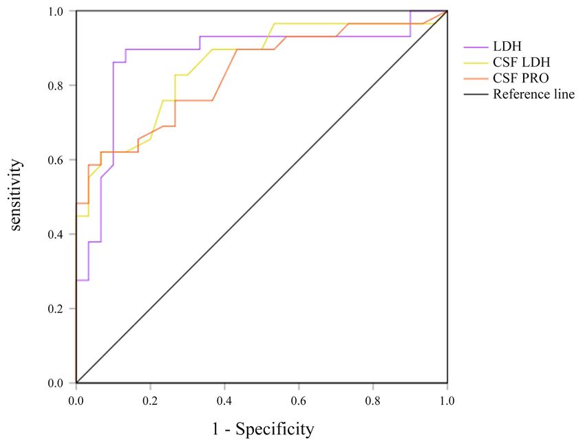
4.4. ROC Curve Analysis
Since LDH, CSF LDH and CSF PRO of the IANE group were significantly higher
than those of the IAE group, ROC curve analysis was performed on the three in-
dicators to determine the area under the curve, the optimal threshold point, sen-
sitivity and specificity of each indicator (Figure 2). The area under curve (AUC)
of blood LDH, CSF LDH and CSF PRO were 0.876, 0.853 and 0.831, respectively
(Table 2). The optimal threshold points of blood LDH, CFS LDH and CSF PRO
were 535 U/L, 67 U/L and 0.49 g/L, respectively. The sensitivity and specificity of
LDH > 535 U/L for predicting IANE were 89.7% and 86.5% respectively. The
sensitivity and specificity of CSF LDH > 67 U/L for predicting IANE were 82.8%
and 73.3% respectively. The sensitivity and specificity of CSF Pro > 0.49 g/L for
predicting IANE was 73.7% and 76.6% respectively.
4.5. Logistic Regression Analysis
Logistic stepwise regression analysis was conducted on the three variables with
statistically significant difference results from univariate analysis according to
the optimal threshold point determined by ROC analysis, two variables were in-
cluded in the final regression model (Table 3). The analysis results showed that
the OR value of blood LDH > 535 U/L was 31.264, 95% CI: 5.892 - 165.878, P <
0.001. The OR value of CSF PRO > 0.49 g/L was 7.695, 95% CI: 1.052 - 56.305, P
= 0.044. Both of the two variables were independent risk factors for IANE.
DOI: 10.4236/jbbs.2021.118015 198 Journal of Behavioral and Brain ScienceG. M. Liu et al.
Figure 2. The ROC curve of LDH, CSF LDH and CSF PRO to predict IANE.
Table 2. Efficiency of predicting IANE by various indicators.
Laboratory AUC Sensitivity Specificity Cut off
LDH 0.876 89.7 86.5 535
CSF LDH 0.853 82.8 73.3 67
CSF PRO 0.831 73.7 76.6 0.49
Note: IANE: Influenza-associated necrotizing encephalopathy; LDH: lactate dehydrogenase; CSF LDH:
cerebrospinal fluid lactate dehydrogenase; CSF PRO: Cerebrospinal fluid protein.
Table 3. Risk factors for IANE by multivariate analysis.
95% CI
Variables β SE P OR
Lower Upper
LDH > 535 U/L 3.442 0.851 0.49 g/L 2.041 1.015 0.044 7.695 1.052 56.305
Constant −8.593 2.157G. M. Liu et al.
demiological survey in Japan showed that the proportion of nervous system
damage in children under 18 years old accounted for 74% and the case fatality
rate was 9% [20]. As the same as ANE causing by a variety of causes [21], the
mortality rate of IANE is about 30% [9], which was close to our findings (see
Table 1).
Flu infections ANE progress rapidly, sequela and high mortality rate, early
diagnosis and intervention is the key. The current IAE and IANE diagnosis were
on the basis of the clinical manifestations, evaluations of the nervous system, and
the brain imaging examinations including CT and MRI. The Cranial imaging of
IAE were without or minor imaging changes such as slight cerebral edema, the
Cranial imaging of IANE were brain edema and necrosis of thalamus and other
deep brain structures. As other studies showed, our study found the symmetry of
thalamus and other brain structures lesions, particularly in the brainstem, basal
ganglia, white matter around ventricle and cerebellum in IANE group. However,
due to the limitations of illness, examination conditions and caregivers’ wishes,
neuroimaging examination cannot be carried out in all children, and simple and
feasible risk factors related indicators for the occurrence of IANE are needed
clinically.
In this study, the data of 40 cases of IAE and 32 cases of IANE who were ad-
mitted to our hospital from January 2016 to December 2020 with influenza in-
fection and nervous system symptoms were retrospective analyzed. The results
showed that there were statistically differences in ALT, AST and CK between the
two groups (P < 0.05), and the IANE group was higher than the IAE group. This
is consistent with previous literature reports that children with IANE have vary-
ing degrees of elevated AST, ALT and CK, which may be related to the fact that
children with IANE are more prone to liver function and muscle injury [22]
[23]. There were statistically significant differences in serum LDH, CSF LDH
and CSF PRO between the two groups (P < 0.001), and the IANE group was sig-
nificantly higher than the IAE group. LDH in serum and cerebrospinal fluid of
the IANE group was significantly higher than that of the IAE group, the reason
being that LDH could be over expressed in cell necrosis [24] [25] [26]. The as-
sessment of LDH in serum and cerebrospinal fluid is helpful for the assessment
of peripheral and brain cell necrosis. The increase of CSF protein in the IANE
group was associated with brain cell necrosis and injury, which was consistent
with previous literature that CSF protein in the IANE group was greater than 0.4
g [13] [27].
ROC curve analysis of serum LDH, CSF LDH and CSF PRO showed that the
areas under the curve of LDH, CSF LDH and CSF PRO were 0.876, 0.853 and
0.831, respectively. The optimal threshold points of LDH, CSF LDH and CSF PRO
were 535 U/L, 67 U/L and 0.49 g/L, respectively. The sensitivity and specificity of
blood LDH > 535 U/L, CSF LDH > 67U/L, CSF PRO > 0.49 g/L were 89.7%, 82.8%,
73.7% and 86.5%, 73.3%, 76.6%, respectively. All three indexes had good sensi-
tivity and specificity in predicting IANE. Logistic stepwise regression analysis
DOI: 10.4236/jbbs.2021.118015 200 Journal of Behavioral and Brain ScienceG. M. Liu et al.
was conducted for the three indicators according to the optimal threshold points
determined by ROC analysis, and the results showed that two variables were in-
cluded in the final regression model. The analysis results indicated that LDH >
535 U/L predicted the risk of IANE 31.264 times as much as LDH ≤ 535 U/L
(OR = 31.264, 95% CI: 5.892 - 165.878), CSF PRO > 0.49 g/L predicted the risk
of IANE 7.695 times as much as CSF PRO ≤ 0.49 g/L (OR = 7.695, 95% CI: 1.052
- 56.305), both of which are independent risk factors for IANE and have high
predictive value for IANE.
6. Conclusion
For children with influenza whose neurological symptoms appear rapidly and
persist in the early stages of the disease, blood LDH > 535 U/L and CSF PRO >
0.49 g/L are independent risk factors for IANE, high vigilance, close evaluation
and active intervention should be taken to reduce sequelae and mortality and
improve the prognosis of these patients.
Conflicts of Interest
The authors declare no conflicts of interest regarding the publication of this pa-
per.
References
[1] Mizuguchi, M., Ichiyama, T., Imataka, G., Okumura, A., Goto, T., Sakuma, H.,
Takanashi, J.I., Murayama, K., Yamagata, T., Yamanouchi, H., et al. (2021) Guide-
lines for the Diagnosis and Treatment of Acute Encephalopathy in Childhood.
Brain & Development, 43, 2-31. https://doi.org/10.1016/j.braindev.2020.08.001
[2] Welk, A., Schmeh, I., Knuf, M., Groendahl, B., Goebel, J., Staatz, G., Gawehn, J. and
Gehring, S. (2016) Acute Encephalopathy in Children Associated with Influenza A:
A Retrospective Case Series. Klinische Pädiatrie, 228, 280-281.
https://doi.org/10.1055/s-0042-111686
[3] Chen, L.W., Teng, C.K., Tsai, Y.S., Wang, J.N., Tu, Y.F., Shen, C.F. and Liu, C.C.
(2018) Influenza-Associated Neurological Complications during 2014-2017 in Tai-
wan. Brain & Development, 40, 799-806.
https://doi.org/10.1016/j.braindev.2018.05.019
[4] Akins, P.T., Belko, J., Uyeki, T.M., Axelrod, Y., Lee, K.K. and Silverthorn, J. (2010)
H1N1 Encephalitis with Malignant Edema and Review of Neurologic Complications
from Influenza. Neurocritical Care, 13, 396-406.
https://doi.org/10.1007/s12028-010-9436-0
[5] Britton, P.N., Dale, R.C., Blyth, C.C., Macartney, K., Crawford, N.W., Marshall, H.,
Clark, J.E., Elliott, E.J., Webster, R.I., Cheng, A.C., et al. (2017) Influenza-Associated
Encephalitis/Encephalopathy Identified by the Australian Childhood Encephalitis
Study 2013-2015. The Pediatric Infectious Disease Journal, 36, 1021-1026.
https://doi.org/10.1097/INF.0000000000001650
[6] Ekstrand, J.J. (2012) Neurologic Complications of Influenza. Seminars in Pediatric
Neurology, 19, 96-100. https://doi.org/10.1016/j.spen.2012.02.004
[7] Sugaya, N. (2002) Influenza-Associated Encephalopathy in Japan. Seminars in Pe-
diatric Infectious Diseases, 13, 79-84. https://doi.org/10.1053/spid.2002.122993
DOI: 10.4236/jbbs.2021.118015 201 Journal of Behavioral and Brain ScienceG. M. Liu et al.
[8] Shiomi, M. (2011) Pathogenesis of Acute Encephalitis and Acute Encephalopathy.
Nihon Rinsho Japanese Journal of Clinical Medicine, 69, 399-408.
[9] Togashi, T., Matsuzono, Y., Narita, M. and Morishima, T. (2004) Influenza-Associated
Acute Encephalopathy in Japanese Children in 1994-2002. Virus Research, 103, 75-78.
https://doi.org/10.1016/j.virusres.2004.02.016
[10] Chen, Q., Li, P., Li, S., Xiao, W., Yang, S. and Lu, H. (2020) Brain Complications with
Influenza Infection in Children. Journal of Behavioral and Brain Science, 10, 129-152.
https://doi.org/10.4236/jbbs.2020.103008
[11] Howard, A., Uyeki, T.M. and Fergie, J. (2018) Influenza-Associated Acute Ne-
crotizing Encephalopathy in Siblings. Journal of the Pediatric Infectious Diseases
Society, 7, e172-e177. https://doi.org/10.1093/jpids/piy033
[12] Weitkamp, J.H., Spring, M.D., Brogan, T., Moses, H., Bloch, K.C. and Wright, P.F.
(2004) Influenza A Virus-Associated Acute Necrotizing Encephalopathy in the United
States. The Pediatric Infectious Disease Journal, 23, 259-263.
https://doi.org/10.1097/01.inf.0000115631.99896.41
[13] Mizuguchi, M., Abe, J., Mikkaichi, K., Noma, S., Yoshida, K., Yamanaka, T. and
Kamoshita, S. (1995) Acute Necrotising Encephalopathy of Childhood: A New Syn-
drome Presenting with Multifocal, Symmetric Brain Lesions. Journal of Neurology,
Neurosurgery, and Psychiatry, 58, 555-561. https://doi.org/10.1136/jnnp.58.5.555
[14] Kirat, N., De Cauwer, H., Ceulemans, B., Vanneste, D. and Rossi, A. (2018) Influ-
enza-Associated Encephalopathy with Extensive Reversible Restricted Diffusion
within the White Matter. Acta Neurologica Belgica, 118, 553-555.
https://doi.org/10.1007/s13760-018-1004-y
[15] Azziz Baumgartner, E., Dao, C.N., Nasreen, S., Bhuiyan, M.U., Mah, E.M.S., Al
Mamun, A., Sharker, M.A., Zaman, R.U., Cheng, P.Y., Klimov, A.I., et al. (2012) Sea-
sonality, Timing, and Climate Drivers of Influenza Activity Worldwide. The Journal
of Infectious Diseases, 206, 838-846. https://doi.org/10.1093/infdis/jis467
[16] Bloom-Feshbach, K., Alonso, W.J., Charu, V., Tamerius, J., Simonsen, L., Miller,
M.A. and Viboud, C. (2013) Latitudinal Variations in Seasonal Activity of Influenza
and Respiratory Syncytial Virus (RSV): A Global Comparative Review. PLoS ONE,
8, e54445. https://doi.org/10.1371/journal.pone.0054445
[17] Zou, J., Yang, H., Cui, H., Shu, Y., Xu, P., Xu, C. and Chen, T. (2013) Geographic
Divisions and Modeling of Virological Data on Seasonal Influenza in the Chinese
Mainland during the 2006-2009 Monitoring Years. PLoS ONE, 8, e58434.
https://doi.org/10.1371/journal.pone.0058434
[18] Fraaij, P.L. and Heikkinen, T. (2011) Seasonal Influenza: The Burden of Disease in
Children. Vaccine, 29, 7524-7528. https://doi.org/10.1016/j.vaccine.2011.08.010
[19] Cowling, B.J., Perera, R.A., Fang, V.J., Chan, K.H., Wai, W., So, H.C., Chu, D.K.,
Wong, J.Y., Shiu, E.Y., Ng, S., et al. (2014) Incidence of Influenza Virus Infections
in Children in Hong Kong in a 3-Year Randomized Placebo-Controlled Vaccine Study,
2009-2012. Clinical Infectious Diseases: An Official Publication of the Infectious Dis-
eases Society of America, 59, 517-524. https://doi.org/10.1093/cid/ciu356
[20] Okuno, H., Yahata, Y., Tanaka-Taya, K., Arai, S., Satoh, H., Morino, S., Shimada,
T., Sunagawa, T., Uyeki, T.M. and Oishi, K. (2018) Characteristics and Outcomes of
Influenza-Associated Encephalopathy Cases among Children and Adults in Japan,
2010-2015. Clinical Infectious Diseases: An Official Publication of the Infectious
Diseases Society of America, 66, 1831-1837. https://doi.org/10.1093/cid/cix1126
[21] Wu, X., Wu, W., Pan, W., Wu, L., Liu, K. and Zhang, H.L. (2015) Acute Necrotizing
Encephalopathy: An Underrecognized Clinicoradiologic Disorder. Mediators Inflamm,
DOI: 10.4236/jbbs.2021.118015 202 Journal of Behavioral and Brain ScienceG. M. Liu et al.
2015, Article ID: 792578. https://doi.org/10.1155/2015/792578
[22] Lee, Y.J., Hwang, S.K. and Kwon, S. (2019) Acute Necrotizing Encephalopathy in
Children: A Long Way to Go. Journal of Korean Medical Science, 34, Article ID:
e143. https://doi.org/10.3346/jkms.2019.34.e143
[23] Trujillo-Gomez, J. and Cabrera-Hemer, D.N. (2019) Acute Necrotizing Encephalo-
pathy. Revue Neurologique, 69, 349-350. https://doi.org/10.33588/rn.6908.2019113
[24] Huang, C.F., Liu, S.H. and Lin-Shiau, S.Y. (2012) Pyrrolidine Dithiocarbamate Aug-
ments Hg (2+)-Mediated Induction of Macrophage Cell Death via Oxidative Stress-
Induced Apoptosis and Necrosis Signaling Pathways. Toxicology Letters, 214, 33-45.
https://doi.org/10.1016/j.toxlet.2012.08.006
[25] Franke, R.P., Fuhrmann, R., Mrowietz, C., Rickert, D., Hiebl, B. and Jung, F. (2010)
Reduced Diagnostic Value of Lactate Dehydrogenase (LDH) in the Presence of Ra-
diographic Contrast Media. Clinical Hemorheology and Microcirculation, 45, 123-130.
https://doi.org/10.3233/CH-2010-1290
[26] Tran, T.T., Groben, P. and Pisetsky, D.S. (2008) The Release of DNA into the Plas-
ma of Mice following Hepatic Cell Death by Apoptosis and Necrosis. Biomarkers,
13, 184-200. https://doi.org/10.1080/13547500701791719
[27] Wang, H.S. (1995) Acute Necrotising Encephalopathy of Childhood Presenting with
Multifocal, Symmetric Brain Lesions Occurring Outside Japan. Journal of Neurolo-
gy, Neurosurgery, and Psychiatry, 59, 661. https://doi.org/10.1136/jnnp.59.6.661
DOI: 10.4236/jbbs.2021.118015 203 Journal of Behavioral and Brain ScienceYou can also read