SARS-COV-2 GASTROINTESTINAL INFECTION CAUSING HEMORRHAGIC COLITIS: IMPLICATIONS FOR DETECTION AND TRANSMISSION OF COVID-19 DISEASE
←
→
Page content transcription
If your browser does not render page correctly, please read the page content below
SARS-CoV-2 Gastrointestinal Infection Causing Hemorrhagic Colitis:
Implications for Detection and Transmission of COVID-19 Disease

Alexandre Carvalho, MD, MPH1; Rana Alqusairi, MD1; Anna Adams, DO1; Michelle Paul, BS2;
Neelay Kothari, MD1,3; Stevany Peters, MD1,4; and Anthony T. DeBenedet, MD, MSc1,4

1
  Department of Internal Medicine, St. Joseph Mercy Health System, Ann Arbor, MI
2
  Eastern Michigan University, College of Health and Human Services, Ypsilanti, MI
3
  Division of Infectious Disease, St. Joseph Mercy Health System, Ann Arbor, MI
4
  Huron Gastroenterology Associates, Ypsilanti, MI

Correspondence: Anthony T. DeBenedet, MD, MSc, Huron Gastroenterology Associates, 5300
Elliott Dr., Ypsilanti, MI 48197; Tel: (734) 434-6262; E-mail: debenedeta@hurongastro.com

INTRODUCTION
        The betacoronavirus, SARS-CoV-2, that is responsible for COVID-19 disease and that
was first described in Wuhan, China in late 2019, has swiftly made its way around our world,
resulting in excess of 30,000 deaths to date (1). Efforts to recognize SARS-CoV-2 infection have
focused on respiratory symptoms such as cough and shortness of breath (2, 3). Currently, the
Centers for Disease Control and Prevention (CDC) criteria for identifying persons under
investigation (PUI) for SARS-CoV-2 infection in the United States comprise respiratory
symptoms and/or fever only (5).
         Recent reports from China have described concomitant digestive symptoms, such as
nausea, vomiting, diarrhea, and abdominal pain, in patients with confirmed SARS-CoV-2
pulmonary infection (6, 7, 8, 9, 10) and the presence of SARS-CoV-2 RNA in fecal samples (9,
11). However, it remains unclear whether these digestive symptoms were causally related to
SARS-CoV-2 gastrointestinal infection. Because the main goals of the care in these cases were
to treat the pulmonary disease and limit healthcare worker exposure, a comprehensive evaluation
of the gastrointestinal system to implicate the virus and rule out alternative etiologies was not
undertaken.
         We present a case of SARS-CoV-2 gastrointestinal infection causing acute hemorrhagic
colitis and signaling COVID-19 disease which endoscopy confirmed colonic injury and helped
exclude other etiologies of disease. We believe that this observation has important implications
for the ongoing management and prevention of COVID-19 disease.
CASE REPORT
        A 71-year-old woman with a history of hypertension, depression, and chronic back pain,
had returned to the United States in early March 2020 following a 10-day trip to Egypt which
included a 4-day cruise on the Nile River. On her last day in Egypt, she developed diffuse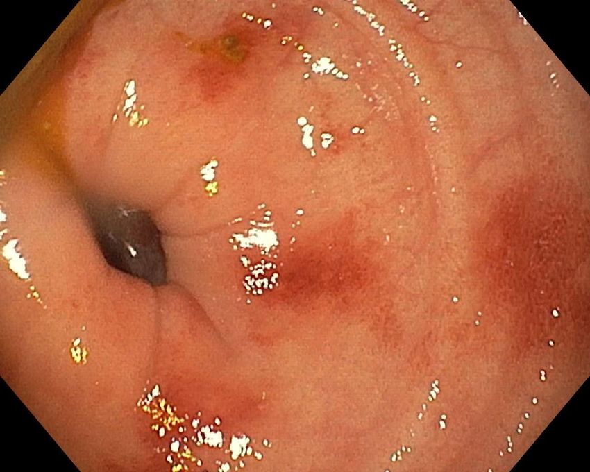
abdominal pain and non-bloody diarrhea. The next day, while traveling back to the United
States, her diarrhea became bloody. Over the next 4 days she experienced nausea, vomiting,
anorexia, diffuse abdominal pain and distention, and 10-20 bloody bowel movements daily.
         She presented to our emergency department 5 days following the onset of her symptoms.
Physical examination revealed a temperature of 36.4°C (97.6°F), blood pressure of 140/81 mm
Hg, pulse of 98 beats per minute, respiratory rate of 18 breaths per minute, and oxygen saturation
of 99% on ambient air. Lung auscultation was normal. Abdominal examination demonstrated
normal bowel sounds and diffuse tenderness to palpation, but no signs of peritonitis. Red blood,
mixed with loose stool, was present in her bedside commode. On further questioning, she denied
fever, cough, shortness of breath, sore throat, or any other symptoms. She also denied a personal
and family history of gastrointestinal disease and had undergone a normal screening colonoscopy
one month prior. She denied antibiotic, antidiarrheal, and non-steroidal anti-inflammatory use,
food allergies, lactose intolerance, alcohol abuse, smoking, and drug use. Her medications did
include llisinopril, as well as desvenlafaxine, amlodipine, and as-needed morphine for chronic
back pain. She reported having been vaccinated against Hepatitis A, B, and typhoid before her
trip to Egypt.
       Laboratory evaluation was notable for an elevated white blood cell count of 24.4 K/μL,
with 20.8 K/μL neutrophils and normal lymphocyte and eosinophil distributions, a normal
hemoglobin, and slightly elevated creatinine at 1.31 mg/dL (baseline 0.90 mg/dL). CT scan of
her abdomen and pelvis with intravenous contrast showed severe colonic inflammation that was
most pronounced in the ascending, transverse, and descending colon, but was also apparent in
the sigmoid colon (Figure 1). There was also a small, right pleural effusion.
        Given the presumptive diagnosis of traveler’s diarrhea with dysentery, empiric
ceftriaxone, azithromycin, and metronidazole were initiated intravenously. Before administration
of antimicrobials, a fecal sample was obtained and was negative for fecal leukocytes, stool
culture (Campylobacter, Salmonella, Shigella, Shiga-toxin producing E. coli, and Yersinia), ova
and parasites, and Clostridium difficile toxin (GDH Antigen-toxin screen). The next day, another
fecal sample was negative for Entamoeba histolytica antigen and Giardia antigen. Of note, later
in the hospitalization (hospital day 7), fecal molecular testing (FilmArray; BioFire Diagnostics,
Salt Lake City, UT) was also negative for bacterial, viral, and parasitic pathogens. HIV 1, 2
antibodies and Legionella urine antigen were also negative.
        Over the next 3 days, the patient’s abdominal pain and bloody diarrhea persisted despite
antimicrobial support. Given a concern for inflammatory bowel disease, a C-reactive protein on
hospital day 3 was 11.6 mg/dL. Also on hospital day 3, the patient learned that someone in her
travel group had been diagnosed with SARS-CoV-2 pulmonary infection. The patient was then
immediately moved to a negative-pressure room and SARS-CoV-2 precautions were instituted.
On hospital day 4, nine days following the onset of her digestive symptoms, the patient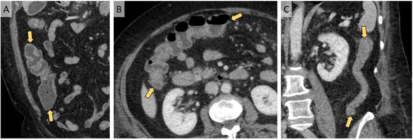
developed a cough; nasopharyngeal swabs were sent for comprehensive viral detection,
including SARS-CoV-2 RNA (Quest Diagnostics).
       Given the patient’s elevated C-reactive protein and persistent abdominal pain and bloody
diarrhea, a flexible sigmoidoscopy was performed on hospital day 4 to evaluate for evidence of
inflammatory bowel disease or ischemic colitis. Endoscopic evaluation to 40 cm from the anal
verge revealed patchy areas of focal erythema without ulceration in the descending colon,
sigmoid colon, and rectum (Figure 2). Histological examination of the colon and rectal biopsies
by hematoxylin and eosin stain under light microscopy showed slight expansion of the lamina
propria by edema with normal cellularity and intact crypts. No virocytes or protozoa were seen.
There were no microscopic changes to indicate the presence of classic infectious colitis,
ischemia, or inflammatory bowel disease.
        On the evening of hospital day 4, the patient’s nasopharyngeal swab for comprehensive
respiratory viral panel returned positive for rhinovirus and herpes simplex virus 1. Her SARS-
CoV-2 RNA was also positive by reverse-transcriptase-polymerase-chain-reaction (rRT-PCR).
Over the next several days, the patient’s abdominal pain and bloody diarrhea persisted, and a
sore throat developed. On hospital day 7, a SARS-CoV-2 rRT-PCR performed on a fecal sample
using the swab and viral transport media from a SARS-CoV-2 nasopharyngeal testing kit was
also positive.
       On hospital day 8, the patient’s respiratory status worsened and her oxygen saturation
declined to 91% on ambient air. She was given two 400 mg doses of hydroxychloroquine
followed by 200 mg twice daily. Within the next 48 hours, she had improvement in her
abdominal pain and bloody diarrhea. Her respiratory symptoms did not evolve further, but she
did require 5 L of oxygen via nasal cannula for several days. CT scan of the chest and CT
angiogram of the abdomen and pelvis performed on hospital day 10 showed multifocal
pneumonia consistent with pulmonary COVID-19 disease as well as resolution of colonic
inflammation (Figure 3). There was no evidence of vascular compromise.
        Over the next 12 days, the patient’s respiratory status gradually improved and she was
weaned off oxygen supplementation. Her digestive symptoms also improved. The patient was
discharged on hospital day 20 in good health, off all antimicrobials. Unfortunately, at the time of
the writing of this report, the patient has been readmitted with mental status changes that are
currently being evaluated.
DISCUSSION
        There has been a growing appreciation of the importance of digestive symptoms (nausea,
vomiting, anorexia, non-bloody diarrhea, and abdominal pain) in the spectrum of COVID-19
disease. Presumed gastrointestinal manifestations have been reported in anywhere from 3-50% of
patients with concomitant SARS-CoV-2 pulmonary infection (6, 7, 8, 12). SARS-CoV-2 RNA
has been found in fecal samples from patients with COVID-19 pulmonary disease, and initial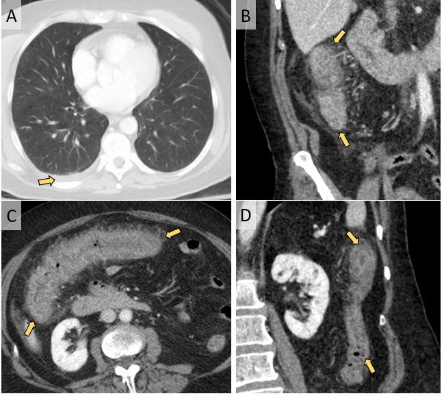
case series have noted that 3-10% of patients who are eventually found to have SARS-CoV-2
pulmonary infection initially presented with isolated digestive symptoms (6, 8). What has been
more difficult to establish is whether SARS-CoV-2 infection is directly responsible for the
digestive symptoms. Since the focus of care in most hospitalized patients is the respiratory
illness, and endoscopy—as a possible virus-aerosolizing procedure—is employed judiciously,
diagnostic studies to implicate the virus in gastrointestinal pathology and to exclude other
etiologies are generally not undertaken.
         Because our patient presented with bloody diarrhea, which has not previously been
described as a manifestation of COVID-19, and our index of suspicion in early March 2020 was
low, our patient did undergo a comprehensive evaluation. This strongly suggested that SARS-
CoV-2 gastrointestinal infection was responsible for her acute hemorrhagic colitis. We
demonstrated that SARS-CoV-2 RNA was present in our patient’s feces, and the endoscopic
findings of coloproctopathy in her descending colon, sigmoid colon, and rectum confirmed
colonic injury and pointed toward an infectious process. We were also able to eliminate, to the
greatest extent possible, other potential etiologies of hemorrhagic colitis—such as alternative
infections, inflammatory bowel disease, and ischemic colitis—through lab testing, radiological
imaging, and colon and rectal biopsies. Although fecal molecular testing was performed after the
initiation of antimicrobials, it is well described that even in the treated patient, fecal molecular
testing will remain positive for up to several weeks (13). This, combined with the fact that our
patient did not improve with standard antimicrobial therapy, makes a multi-infection scenario
unlikely.
         Interestingly, although our patient had endoscopic evidence of coloproctopathy and
colonic thickening on CT, her sigmoid colon and rectal biopsies were histologically
unremarkable. There is currently no commercially-available assay in the United States to test
tissue for the presence of SARS-CoV-2 RNA, so we were not able to do this. However, such
normal histologic findings are in line with the 2003 SARS-CoV experience wherein, under light
microscopy, small intestinal and colonic specimens of patients with confirmed SARS-CoV
gastrointestinal infection showed normal architecture, without evidence of villous atrophy,
inflammatory infiltrates, or virocytes (14). We also did not have access to electron microscopy;
in the 2003 SARS-CoV experience, viral particles were seen by electron microscopy in the small
intestinal and colonic epithelial cells (15).
         Related, there was also a disconnect between the degree of colonic inflammation seen on
initial CT scan and the endoscopically-observed coloproctopathy seen on flexible
sigmoidoscopy. We suspect this is because the CT scan was performed three days before the
flexible sigmoidoscopy, and thus some healing was likely already taking place in at least the left
colon. Moreover, on CT scan, the colonic inflammation was most pronounced in the ascending
and transverse colon, with the left colon not too far behind. Our patient’s continued bloody
diarrhea post-flexible sigmoidoscopy was likely from resolving mucosal damage in the
ascending and transverse colon that was not observed on flexible sigmoidoscopy.
It has become established that the target viral receptor for SARS-CoV-2 is angiotensin-
converting enzyme 2 (ACE2) (9, 15, 16). This receptor is highly expressed on type II alveolar
cells, esophagus epithelial cells, and both small intestine and colonic cells, among other cell
types (9, 17, 18, 19). Additionally, immunofluorescence analysis has shown that the ACE2
receptor is abundantly expressed in also gastric and rectal epithelia (9). These data suggest that
SARS-CoV-2 may gain entry into and potentially damage gastrointestinal host cells, causing the
array of digestive symptoms that are currently being observed.
Our patient was taking lisinopril 40mg daily as part of her regimen for hypertension.
There have been some reports suggesting that patients treated with ACE-inhibitors and
angiotensin-receptor blockers may theoretically have increased numbers of ACE2 receptors,
making them more prone to infection with SARS-CoV-2 and perhaps higher risk for severe
COVID-19 disease (20). It is certainly plausible this applied to our patient. Our patient did
clinically improve with hydroxychloroquine administration, and there have been some reports
suggesting a possible benefit (21, 22). We are unsure whether this was truly a therapeutic effect
or coincidental. More research is certainly needed in regards to the clinical efficacy of
hydroxychloroquine in the treatment of SARS-CoV-2 infection.
From a transmission perspective, oral and respiratory droplets are well described as the
major mode of transmission of SARS-CoV-2 viral particles. However, live SARS-CoV-2 virus
has also been isolated from fecal samples and viral particles have been detected in the feces even
after resolution of respiratory symptoms, suggesting the potential for fecal-oral transmission
beyond the symptomatic period (9, 23). When our patient was admitted, she did not meet CDC
guidelines at the time for PUI for SARS-CoV-2 infection as she was afebrile, had no respiratory
symptoms, and had not travelled to China, Italy, Iran, or South Korea. We were unfortunately not
aware of the Washington Post article that ran 3 days before her presentation, reporting a cluster
of SARS-CoV-2 cases associated with Nile River cruises (24). Awareness of the gastrointestinal
manifestations of SARS-CoV-2 may have increased our index of suspicion and encouraged us to
institute SARS-CoV-2 precautions on arrival, avoiding the exposure and subsequent quarantine
of 72 healthcare workers, including many of us.
To our knowledge, this is the first report of SARS-CoV-2 gastrointestinal infection
causing hemorrhagic colitis in which colonic injury was demonstrated endoscopically and other
etiologies were excluded. This case adds to the body of evidence implicating the gastrointestinal
tract in the clinical expression and transmission of SARS-CoV-2 infection. On this basis, we
believe it is important to institute SARS-CoV-2 precautions in patients who present with either
respiratory or digestive symptoms. We also encourage the rapid development and deployment of
fecal testing kits for SARS-CoV-2 RNA and encourage institutions to use their nasopharyngeal
kits for fecal testing in the interim.
On March 29, 2020, New York City healthcare professionals made the recommendation
that anyone presenting to New York City hospitals (even without respiratory or digestivesymptoms) be considered SARS-CoV-2 positive and appropriate safeguards taken (25). We have not reached this level universally in our country, yet. But this emerging disease will continue to evolve, and so must we. For the maxim “when you hear hoofbeats, think horses not zebras” works well, unless you are on a safari—or in the middle of a pandemic. REFERENCES 1. World Health Organization Director-General’s opening remarks at COVID-19 media briefing. World Health Organization, February 2020 (https://www.who.int/dg/speeches/detail/who- director-general-s-opening-remarks-at-the-media-briefing-on-covid-19---24-february-2020). 2. Zhu N, Zhang D, Wang W, et al. A Novel Coronavirus from Patients with Pneumonia in China, 2019. N Engl J Med 2020; 382:727-733. 3. Coronavirus Disease Testing (COVID-19): Symptoms & Testing. Centers for Disease Control and Prevention, 2020 (https://www.cdc.gov/coronavirus/2019-ncov/symptoms- testing/symptoms.html). 4. Bajema KL, Oster AM, McGovern OL, et al. Persons Evaluated for 2019 Novel Coronavirus—United States, January 2020. (https://www.who.int/dg/speeches/detail/who- director-general-s-opening-remarks-at-the-media-briefing-on-covid-19---24-february- 2020).MMWR Morb Mortal Wkly Rep 2020; 69:166-70. 5. Centers for Disease Control and Prevention. https://www.cdc.gov/coronavirus/2019- ncov/hcp/clinical-criteria.html. 6. Wang D, Hu B, Hu C, et al. Clinical Characteristics of 138 Hospitalized Patients with 2019 Novel Coronavirus-Infected Pneumonia in Wuhan, China. JAMA 2020; 323:1061-9. 7. Huang C, Wang Y, Li X, et al. Clinical Features of Patients Infected with 2019 Novel Coronavirus in Wuhan, China. Lancet 2020; 395: 497-506. 8. Pan L, Mu M, Ren HG, et al. Clinical characteristics of COVID-19 patients with digestive symptoms in Hubei, China: a descriptive, cross-sectional, multicenter study. American Journal of Gastroenterology, preprint. 2020 March 18 https://journals.lww.com/ajg/Documents/COVID_Digestive_Symptoms_AJG_Preproof.pdf?PRI D=AJG_PR_031820. 9. Xiao F, Tang M, et al. Evidence for gastrointestinal infection of SARS-Cov-2. Gastroenterology. 2020 Mar 3; https://doi.org/10.1053/j.gastro.2020.02.055. 10. Pan L, Mi M, Pengcheng Y, et al. Clinical Characteristics of COVID-19 patients with digestive symptoms in Hubei, China: A descriptive, cross-sectional, multicenter study. American Journal of Gastroenterology, preprint. 2020 March 30 https://journals.lww.com/ajg/Documents/COVID_Digestive_Symptoms_AJG_Preproof.pdf
11. Lijuan C, Lou J, Bai Y et al. COVID-19 Disease with Positive Fecal and Negative
Pharyngeal and Sputum Viral Tests. American Journal of Gastroenterology, preprint.
https://journals.lww.com/ajg/Citation/publishahead/COVID_19_Disease_With_Positive_Fecal_a
nd_Negative.99371.aspx.
12. Zhang J-J, Dong X, Cao Y-Y, et al. Clinical characteristics of 140 patients infected with
SARS-CoV-2 in Wuhan, China. Allergy 2020;00:1-12.
13. Riddle MS, DuPont HL, Connor BA. ACG Clinical Practice Guideline: Diagnosis,
Treatment, and Prevention of Acute Diarrheal Infections in Adults. American Journal of
Gastroenterology. 111: 602-622.
14. Leung WK, To KF, Chan PK, et al. Enteric involvement of severe acute respiratory
syndrome-associated coronavirus infection. Gastroenterology. 2003; 125:1011-7.
15. Li W, Moore M, Vasilieva N, et al. Angiotensin-converting enzyme 2 is a functional receptor
for the SARS coronavirus. Nature 2003; 426:450-4.
16. Hamming I, Timens W, Bulthuis MLC, Lely AT, Navis GJ, Van Goor H. Tissue distribution
of ACE2 protein, the functional receptor for SARS coronavirus. A first step in understanding
SARS pathogenesis. J Pathol 2004;203:631-7.
17. Zou X, Chen K, Zou J, et al. Single-cell RNA-seq data analysis on the receptor ACE2
expression reveals the potential risk of different human organs vulnerable to 2019-nCoV
infection. Front Med, 2020. [Epub ahead of print] doi: 10.1007/s11684-020-0754-0.
18. Zhao Y, et al. Single-cell RNA expression profiling of ACE2, the putative receptor of Wuhan
2019-nCov. Preprint pending peer review.
https://www.biorxiv.org/content/10.1101/2020.01.26.919985v1(2020).
19. Zhang, H. et al. The digestive system is a potential route of 2019-nCov infection: a
bioinformatics analysis based on single-cell transcriptomes. March 2020. Preprint pending peer
review https://www.biorxiv.org/content/10.1101/2020.01.30.927806v1(2020).
20. James H Diaz. Hypothesis: Angiotensin-converting enzyme inhibitors and Angiotensin
receptor blockers may increase the risk of severe COVID-19. Journal of Travel Medicine, 2020;
DOI: 10.1093/jtm/taaa041.
21. Chen Z, Hu J, Zhang Z, et al. Efficacy of Hydroxycholoroquine in patients with COVID-19:
Results of a Randomized Clinical Trial. Preprint 22 March 2020. MedRxIv, BMJ/Yale.
https://doi.org/10.1101/2020.03.22.20040758.
22. Liu J., Ruiyan C., Mingyue X et al. Hydroxychloroquine, a less toxic derivative of
chloroquine, is effective in inhibiting SARS-CoV-2 infection in vitro. Cell Discovery. Volume 6,
Article number: 16 (2020).
23. Wang W, Xu Y, Gao R, et al. Detection of SARS-CoV-2 in Different Types of Clinical
Specimens. JAMA. Published online March 11, 2020. doi:10.1001/jama.2020.3786.
24. Washingon Post report about Nile River cruises and SARS-CoV-2 infection.
https://www.washingtonpost.com/world/middle_east/new-coronavirus-cluster-linked-to-nile-
river-cruise-ship-popular-with-tourists/2020/03/06/33c79b24-5fae-11ea-ac50-
18701e14e06d_story.html.
25. Gross S, Robbin D, Greenwald D, et al. Preparation in the Big Apple: New York City, A
New Epicenter of the COVID-19 Pandemic. American Journal of Gastroenterology, preprint. 29
March 2020 https://journals.lww.com/ajg/Documents/COVID_NYC_AJG_Preproof.pdf.

Acknowledgements: We gratefully acknowledge the following individuals for their
contributions to the clinical care of our patient and/or this report: Michelle Robida, MD; Igor
Shkolnik, MD; Holly Murphy, MD, MPH; Joseph Tworek, MD; Zeeshaan Bhatti, MD; Shawna
Newsome, BSN, RN; Katherine Madaleno, BSN, RN; Christopher McCall, BSN, RN; Bradley
A. Connor, MD; and B. Joseph Elmunzer, MD, MSc

Guarantor of the article: Anthony T. DeBenedet, MD, MSc
Specific author contributions: Alexadre Carvalho, MD, MPH: Report concept, acquisition of
data, analysis and interpretation of data, drafting and finalizing the manuscript. Rana Alqusairi,
MD: Report concept, acquisition of data, analysis and interpretation of data, drafting and
finalizing the manuscript. Anna Adams, DO: Report concept, acquisition of data, analysis and
interpretation of data, drafting and finalizing the manuscript. Michelle Paul, BS: Report concept,
acquisition of data, analysis and interpretation of data, drafting and finalizing the manuscript.
Neelay Kothari, MD: Acquisition of data, intellectual manuscript revision. Stevany Peters, MD:
Acquisition of data, intellectual manuscript revision. Anthony T. DeBenedet, MD, MSc: Report
concept, acquisition of data, analysis and interpretation of data, drafting and finalizing the
manuscript.
Financial support: None to report.
Potential competing interests: All authors state that they have no conflict of interest.
Figure 1. Initial CT scan of the abdomen and pelvis in the emergency room. (A): Axial CT
image of the lower thorax shows no airspace disease in the lungs. A small, right pleural effusion
is present (arrow). (B, C, D): Intravenous contrast-enhanced CT scan of the abdomen and pelvis
in the coronal (B & D) and axial (C) planes shows severe inflammation of the ascending colon
(B), transverse colon (C), and descending colon (D), characterized by circumferential wall
thickening, mural hyperenhancement, mesenteric hypervascularity, and pericolic fat stranding
(arrows).
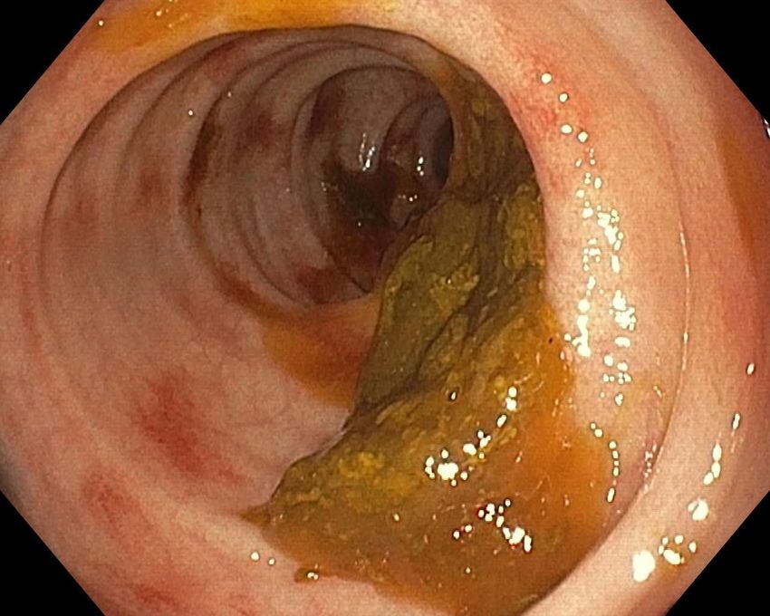
Figure 2. Descending colon and sigmoid colon on flexible sigmoidoscopic examination. The
descending colon, sigmoid colon, and rectum contained patchy areas of focal erythema (arrows).

Figure 3: CT angiogram of the abdomen and pelvis on hospital day 9. Intravenous contrast-
enhanced CT scan of the abdomen and pelvis in the coronal (A & C) and axial (B) planes shows
resolution of the prior inflammation involving the ascending colon (A), transverse colon (B), and
descending colon (C). Specifically, bowel wall thickening, mural hyperenhancement, mesenteric
hypervascularity, and pericolic fat stranding (arrows) have resolved.
You can also read