POTENTIAL ANTIGENIC CROSS-REACTIVITY BETWEEN SEVERE ACUTE RESPIRATORY SYNDROME CORONAVIRUS 2 (SARS-COV-2) AND DENGUE VIRUSES - OXFORD UNIVERSITY PRESS
←
→
Page content transcription
If your browser does not render page correctly, please read the page content below
Clinical Infectious Diseases
    MAJOR ARTICLE

Potential Antigenic Cross-reactivity Between Severe Acute
Respiratory Syndrome Coronavirus 2 (SARS-CoV-2) and
Dengue Viruses
Yaniv Lustig,1 Shlomit Keler,2,3 Rachel Kolodny,4 Nir Ben-Tal,5 Danit Atias-Varon,2 Ekaterina Shlush,6 Motti Gerlic,7 Ariel Munitz,7 Ram Doolman,8,9

                                                                                                                                                                                                 Downloaded from https://academic.oup.com/cid/advance-article/doi/10.1093/cid/ciaa1207/5892809 by guest on 29 December 2020
Keren Asraf,8 Liran I. Shlush,10,a and Asaf Vivante2,3,11,a
1
 Central Virology Laboratory, Ministry of Health, Chaim Sheba Medical Center, Ramat Gan, Israel, 2Department of Pediatrics B, Edmond and Lily Safra Children’s Hospital, Sheba Medical Center,
Ramat-Gan, Israel, 3Sackler School of Medicine, Tel Aviv University, Tel Aviv, Israel, 4Department of Computer Science, University of Haifa, Mount Carmel, Israel, 5Department of Biochemistry
and Molecular Biochemistry, George S. Wise Faculty of Life Sciences, Tel Aviv University, Tel-Aviv, Israel, 6IVF Unit Department of Obstetric and Gynecology Galilee Medical Center, Naharia,
Israel, 7Department of Clinical Microbiology and Immunology, Sackler School of Medicine, Tel Aviv University, Tel Aviv, Israel, 8The Dworman Automated-Mega Laboratory, Sheba Medical Center,
Tel-Hashomer, Ramat-Gan, Israel, 9The Mina and Everard Goodman Faculty of Life Sciences, Bar-Ilan University, Ramat-Gan, Israel, 10Department of Immunology, Weizmann Institute of Science,
Rehovot, Israel, and 11Talpiot Medical Leadership Program, Sheba Medical Center, Tel-Hashomer, Israel

   Background. Coronavirus disease 2019 (COVID-19) and dengue fever are difficult to distinguish given shared clinical and lab-
oratory features. Failing to consider COVID-19 due to false-positive dengue serology can have serious implications. We aimed to
assess this possible cross-reactivity.
   Methods. We analyzed clinical data and serum samples from 55 individuals with severe acute respiratory syndrome corona-
virus 2 (SARS-CoV-2) infection. To assess dengue serology status, we used dengue-specific antibodies by means of lateral-flow rapid
test, as well as enzyme-linked immunosorbent assay (ELISA). Additionally, we tested SARS-CoV-2 serology status in patients with
dengue and performed in-silico protein structural analysis to identify epitope similarities.
   Results. Using the dengue lateral-flow rapid test we detected 12 positive cases out of the 55 (21.8%) COVID-19 patients
versus zero positive cases in a control group of 70 healthy individuals (P = 2.5E−5). This includes 9 cases of positive immu-
noglobulin M (IgM), 2 cases of positive immunoglobulin G (IgG), and 1 case of positive IgM as well as IgG antibodies. ELISA
testing for dengue was positive in 2 additional subjects using envelope protein directed antibodies. Out of 95 samples obtained
from patients diagnosed with dengue before September 2019, SARS-CoV-2 serology targeting the S protein was positive/
equivocal in 21 (22%) (16 IgA, 5 IgG) versus 4 positives/equivocal in 102 controls (4%) (P = 1.6E−4). Subsequent in-silico
analysis revealed possible similarities between SARS-CoV-2 epitopes in the HR2 domain of the spike protein and the dengue
envelope protein.
   Conclusions. Our findings support possible cross-reactivity between dengue virus and SARS-CoV-2, which can lead to false-
positive dengue serology among COVID-19 patients and vice versa. This can have serious consequences for both patient care and
public health.
   Keywords. COVID-19; dengue; West Nile.

Coronavirus disease 2019 (COVID-19), the illness caused by                                              Symptoms of COVID-19 can also be nonspecific and be mis-
the recently identified severe acute respiratory syndrome co-                                        diagnosed. For instance, both dengue fever and COVID-19 are
ronavirus 2 (SARS-CoV-2), has rapidly spread worldwide. As                                           difficult to distinguish as they share similar clinical and labo-
of 1 August 2020, over 10 million patients have been reported                                        ratory features [3–5]. Moreover, in 2 cases from Singapore, it
globally (https://covid19.who.int/). The symptoms of SARS-                                           has been reported that rapid serological testing for dengue can
CoV-2 infection vary widely, from asymptomatic disease to                                            show false-positive results among COVID-19 patients [6]. This
multisystem organ failure [1, 2].                                                                    clinical diagnostic challenge is particularly important in regions
                                                                                                     around the world with dual outbreak of both COVID-19 and
                                                                                                     dengue [7, 8].
   Received 24 June 2020; editorial decision 7 August 2020; accepted 13 August 2020; published
online August 14, 2020.
                                                                                                        The global burden of dengue has grown dramatically
   a
    L. I. S. and A. V. contributed equally to this work.                                             in recent decades as half of the world’s population lives in
   Correspondence: A. Vivante, Pediatric Department B, Edmond and Lily Safra Children’s
Hospital, Chaim Sheba Medical Center, Tel Hashomer and the Sackler Faculty of Medicine, Tel
                                                                                                     dengue-endemic areas [9]. Approximately 100–400 million
Aviv University, Tel Aviv, Israel (Asaf.Vivante@Sheba.health.gov.il).                                cases occur each year (https://www.who.int/en/news-room/
Clinical Infectious Diseases®  2020;XX(XX):1–6                                                       fact-sheets/detail/dengue-and-severe-dengue). Laboratory
© The Author(s) 2020. Published by Oxford University Press for the Infectious Diseases Society
of America. All rights reserved. For permissions, e-mail: journals.permissions@oup.com.
                                                                                                     diagnosis of dengue is established directly by detection of
DOI: 10.1093/cid/ciaa1207                                                                            viral components in serum or indirectly by serologic means.

                                                                                                 SARS-CoV-2 and Dengue Cross-reactivity • cid 2020:XX (XX XXXX) • 1
During the febrile phase detection of the virus-expressed sol- ELISA (anti-SARS-CoV-2 ELISA IgG, Euroimmun, Germany)
uble nonstructural protein 1 (NS1) or dengue-specific immu- performed according to the manufacturer’s recommendation
noglobulin M (IgM) antibodies by means of enzyme-linked [16, 17]. (2) Mount Sinai Hospital Clinical Laboratory COVID-
immunosorbent assay (ELISA) or the lateral-flow rapid test 19 ELISA Antibody Test [18], which was modified accordingly:
is sufficient for a confirmatory diagnosis [9]. Lateral-flow a 96 well microtiter Polysorb plate (Nunc, Thermo, Denmark)
based tests are the most prevalent diagnostic method espe- coated overnight with 1 μg/mL of Receptor Binding Domain
cially in dengue endemic countries because they do not re- (RBD) antigen for detection of IgG and 2 μg/mL for detection of
quire special knowledge or equipment and are not affected IgA antibodies was blocked with 5% skimmed milk at 25º C for
by high ambient tropical temperatures or humidity [10, 11]. 60 minutes, and human serum samples (diluted 1:100 with 3%
Given that a possible cross-reactivity between dengue virus skimmed milk) were added to antigen coated wells. The plate
Downloaded from https://academic.oup.com/cid/advance-article/doi/10.1093/cid/ciaa1207/5892809 by guest on 29 December 2020
and SARS-CoV-2 could lead to false-positive results for both was incubated at 25º C for 120 minutes, washed, and goat anti-
diseases, we aimed to assess the magnitude of such possible- human IgG horseradish peroxidase (HRP) conjugate (Jackson
cross reactivity in a large cohort of individuals who were ImmunoResearch, Philadelphia, PA, USA) (diluted 1:15000) or
known to have either SAR-CoV-2 or dengue virus. anti human IgA HRP conjugate (Abcam) was added to each well
for 60 minutes after addition of tetramethylbenzidine (TMB)
METHODS substrate and stop solution (1M HCl) the OD of each well was
measured at 450 nm. ELISA index value above or equal to 1.1
Study Participants
was considered positive. SARS-CoV-2 RBD IgM was detected
We included in the study clinical data and serum samples
by a electrochemiluminescence serological test as described
obtained from individuals with proven SARS-CoV-2 infection
(medRxiv preprint doi: https://doi.org/10.1101/2020.06.28.201
who were tested for COVID-19 in the Israeli Ministry of Health
41838).
Central Virology Laboratory. The assay is based on the World
Health Organization standard. It targets the SARS-CoV-2 en-
velope (E), N and RdRp genes [12]. Diagnosis was based on Bioinformatics
Protein Structure Analysis
nasopharyngeal swabs, which were tested for SARS-CoV-2
We hypothesized that cross-reactivity between SARS-CoV-2
by reverse transcription polymerase chain reaction. Only
and dengue virus could be the result of similarities in the
laboratory-confirmed cases were included. The institutional re-
structures of outer proteins of these 2 viruses. Such similar-
view board of Sheba Medical Center approved the study and
ities could potentially cause cross-reactivity if the chains that
waived the requirement for informed consent on the basis of
appear in the outer surface of the virus are exposed to the im-
preserving participants’ anonymity.
mune system and trigger the expansion of similar antibodies.
Serological Assays Accordingly, we identified the outer chains of selected
Serology Testing to Detect Dengue, West Nile Virus, and SARS-CoV-2 flaviviruses and of SARS-CoV-2 and compared their struc-
Antibodies tures (Supplementary Table 1S). Cross-reactivity between the
Antibodies against dengue virus were detected by the following different flaviviruses is well established [19]; accordingly we
commercially available immunoassays: lateral-flow rapid diag- used this information as a positive control to test whether
nostic test (RDT), which detects antibodies against the envelope such cross-reactivity could be observed also at the epitope
protein (RDT; SD BIOLINE Dengue IgG/IgM WB (Abbott, similarity level. We used the 0–1 template modeling (TM)-
Chicago, IL, USA)) and shown to be highly specific [13, 14] and score scale to measure similarity: TM-score < 0.30 is random
by ELISA against the envelope ((Panbio® Dengue IgG and IgM structural similarity, whereas 0.5 < TM-score implies the
Indirect ELISA (Abbott, Chicago, IL, USA)) and nonstructural structures have the same fold (ie, are similar in shape) [20];
(NS)-1 (anti dengue NS1 type 1–4 IgG, Euroimmun, Germany) here, we use the threshold of 0.45 to identify similar struc-
proteins. Detection of IgM or immunoglobulin G (IgG) anti- tures. TM-score reports were normalized by both aligned
bodies against West Nile virus (WNV) in serum samples was chains; we use the maximum among these scores. To study
performed using ELISA (WNV IgM capture DxSelect ELISA and all similar protein chains in these viruses, we organize the
IgG DxSelect ELISA kits by Focus Diagnostics Inc., Cypress, CA, data as a network and use Cytoscape [21] to visualize it.
USA). Samples were tested in all the immunoassays according to
the manufacturer’s instructions. WNV neutralization was per- Statistical Analysis
formed as described [15] using a WNV strain (S42H) isolated in Descriptive statistics were used to summarize the data; results are
Israel in 2015. A dilution equal to 1:10 or above was considered reported as means and standard deviations (SD), as appropriate.
neutralizing. SARS-CoV-2 immunglobulin A (IgA) and IgG Categorical variables were summarized as counts and percent-
antibodies were detected by 2 assays: (1) A semi-quantitative ages. P values were calculated using the 2-tailed Fisher exact test.
2 • cid 2020:XX (XX XXXX) • Lustig et alRESULTS samples from a control group of 70 healthy individuals, which
Clinical Findings
we obtained prior to September 2019. In this cohort the dengue
During the period from 17 March through 13 April, we identi- rapid test was negative in all samples (P = 2.5E−5). Subsequently,
fied 55 patients with confirmed COVID-19 infection. The dem- we assessed dengue virus serostatus using ELISA, which detects
ographic and clinical characteristics of the patients are shown antibodies directed against dengue E protein. This analysis
in Supplementary Table 2S. The mean (±SD) age of the patients demonstrated that out of the 12 COVID-19 patients who were
was 45 ± 20 years (range, 14–84); 53% were men. The mean du- positive using the dengue rapid test, only 1 had a weak posi-
ration of symptoms before COVID-19 diagnosis was 7 ± 8 days. tive IgG result and none were positive for ELISA IgM (Table 1).
Thirty-one patients were hospitalized (56%), and 24 were man- In addition, dengue IgM and IgG E based ELISA were positive
aged as outpatients (44%). Full clinical and laboratory details of in 1 and 5 patients, respectively, out of the 43 COVID-19 pa-
Downloaded from https://academic.oup.com/cid/advance-article/doi/10.1093/cid/ciaa1207/5892809 by guest on 29 December 2020
the former group are shown in Supplementary Table 2S. None tients who were negative using the initial dengue rapid test.
of the patients had recently traveled to a dengue virus endemic Dengue has known cross-reactivity with the E protein of WNV.
region. Additionally, WNV seropositivity in the Israeli population is es-
timated at approximately 11% [22]. Therefore, we tested these
Dengue Virus Rapid Test Serology Was Positive in 12 out of 55 Patients IgG dengue E positive samples for WNV E based IgG and IgM
(22%) With SARS-CoV-2 Infection ELISA and subsequently performed WNV neutralization. Out
We first tested the study cohort for IgG and IgM antibodies using of the 5 dengue positive samples, 3 were positive for WNV E
the rapid dengue virus diagnostic test (SD BIOLINE Dengue). IgG. These samples were also positive in WNV-neutralization,
We found 9 patients positive for IgM, 3 positive for IgG, and 1 further providing evidence that these dengue fever positive
positive for both IgM and IgG out of the 55 individuals with ELISA samples represented antigenic cross-reactivity with past
confirmed COVID-19 (Table 1). In addition, we tested serum infection with WNV. Because dengue is not endemic in Israel,
Table 1. Serology Characteristics of 14 Patients With Coronavirus Disease 2019 (COVID-19) and False-Positive Dengue Serology
Electro
chemilumine
Dengue scence
Positive Dengue NS1- Euroimmun SARS- SARS-CoV-2 Mount-Sinai
Index COVID-19 PCR to Dengue Rapid E-ELISA ELISA WNV E-ELISA West-Nile CoV-2 ELISA ELISA ELISA
Case Sample Collec- Neutraliza-
No. tion (days) IgM IgG IgM IgG IgG IgM IgG tion Assay d IgA IgG IgM IgA IgG
3a 4 Positive Neg Neg Neg Neg Neg Positiveit is highly unlikely that these COVID-19 patients with posi- spike protein and several chains in the envelope protein of both
tive E IgG dengue and negative E IgG WNV were previously dengue (4CBF, 4UIF, 5A1Z, etc) and Zika viruses (6JFI, 5YGH,
infected with dengue. Nevertheless, these samples were tested etc). The similarity between SARS-CoV-2 6LVN chains and
negative with a NS1 based ELISA, which detects antibodies WNV were not with chains of the envelope protein but rather
against dengue NS1 and therefore efficiently evaluate past ex- with chains of the NS1 protein (4O6C) of WNV (Figure 1B).
posure to dengue virus [23]. Overall, these results suggest Few other SARS-CoV-2 chains from the spike protein (6LXT,
that cross-reactivity between ELISA dengue E IgG and SARS- 6VSB, 6VXX etc) demonstrated high similarity with chains
CoV-2 may also exist (Table 1). Testing the 14 samples from from the envelope protein of both Zika and dengue viruses, al-
dengue cross-reactive COVID-19 patients with 2 ELISAs and though such similarity could not be apparent with chains from
an electrochemiluminescence serological test for IgA, IgM and WNV (Figure 1B). Altogether, we identified structure simi-
Downloaded from https://academic.oup.com/cid/advance-article/doi/10.1093/cid/ciaa1207/5892809 by guest on 29 December 2020
IgG demonstrated that all but 1 sample had equivocal or posi- larities between chains of the SARS-CoV-2 spike protein and
tive antibodies against SARS-CoV-2. chains of the envelope protein of both Zika and dengue but not
with WNV. Such similarities could potentially explain the cross
SARS-CoV-2 Serology Status Among Patients With Dengue Fever reactivity between SARS-CoV-2 and dengue virus.
To test the possibility of false-positive (FP) SARS-CoV-2 se-
rology in patients diagnosed with dengue fever, we assessed DISCUSSION
SARS-CoV-2 serology status in serum samples obtained prior
to September 2019 from patients with clinical and serological Here we provide lines of evidence to support cross-reactivity
diagnosis of acute (IgM) and past (IgG) dengue fever. Using between dengue and SARS-CoV-2, which can lead to false-
the Euroimmun ELISA, which detects antibodies against the S positive dengue serology among patients with COVID-19.
protein, we found 21 out of 95 samples to be positive or equiv- Failing to consider COVID-19 infection because of a positive
ocal for SARS-CoV-2 IgG or IgA antibodies. This is signifi- dengue result can have serious consequences for both patient
cantly higher FP rates as compared with rates detected in 102 care as well as for public health policies.
healthy controls (4% vs 22%, P = 1.6E−4) and in prior studies Dengue fever and COVID-19 are difficult to distinguish be-
[16, 17] (Supplementary Table 3S). We next examined if the cause they share clinical and laboratory features [3–5]. Yan G
cross-reactivity observed was a general or more specific phe- et al recently described 2 cases that were wrongly diagnosed as
nomenon by testing samples from acute (IgM) and past (IgG) dengue but later confirmed to be COVID-19 [5]. In addition, it
dengue fever patients with an ELISA that detects antibodies has been recently reported in Brazil and other regions that both
against the RBD antigen. Results (Supplementary Table 3S) viruses spread simultaneously as copandemics [7, 24]. In these
show higher, although not statistically significant, FP rates for regions, diagnostic misclassification can be highly problematic.
samples obtained from past dengue fever patients as compared Cross-reactivity between SARS-CoV-2 and dengue viruses may
with healthy controls. To further explore such cross-reactivity interfere with accurate clinical diagnosis and treatment, under-
between SARS-CoV-2 and dengue virus, we hypothesized that standing of undelaying pathomechanisms, and possible devel-
although they belong to different families of viruses they might opment of vaccine.
share structural similarities. We cannot exclude the possibility that the phenomenon seen
in this study is assay-specific false positive and not true antigenic
In-silico Protein Structure Analysis Reveals Possible Similarity Between cross-reactivity. Our results demonstrating cross-reactivity of
SARS-CoV-2 Spike Protein and Dengue Envelope Protein samples from COVID-19 patients on the dengue RDT as well
Because sequence similarity could not explain the observed as samples from dengue patients with the Euroimmun ELISA in
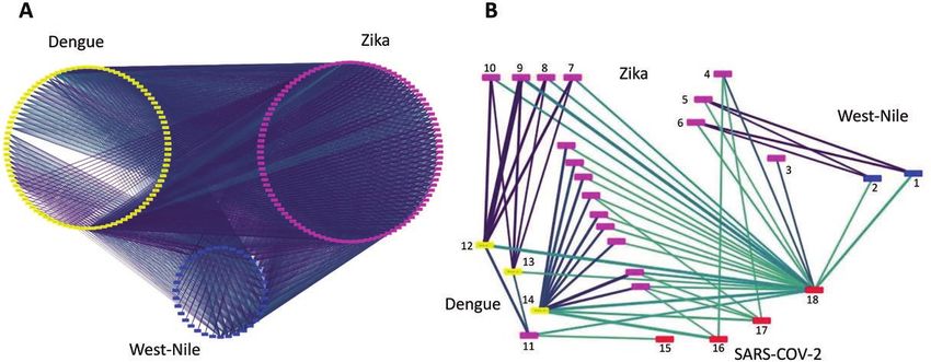
cross-reactivity (Supplementary Figure 1S), we hypothesized addition to our in-silico protein structure analysis suggests that
that structure similarity might expose regions in the outer pro- at least some specific antigenic cross-reactivity exists. However,
tein chains that can explain the immune cross-reactivity. We the results of the dengue ELISA and SARS-CoV-2 Mount Sinai
focused our analysis on the comparison of folds of chains and RBD-ELISA demonstrating only minimal cross-reactivity sug-
structures that are known to be in the outer surface of a protein, gests that this cross-reactivity is not due to a significant anti-
because it is estimated that antibodies identify epitopes that genic similarity. It is possible to speculate that the existence of
are in specific folds. Based on these assumptions, we propose minor structural similarities between dengue and SARS-CoV-2
that structure similarity might correlate with positivity rate. may result in selected epitopes identity. As a consequence only
Indeed, very high similarity (TM score 0.75–1.0) was found be- assays detecting antibodies targeting these epitopes will show
tween many chains of the different flaviviruses (Supplementary differential cross reactivity. Nevertheless, both the RDT and the
Figure 2S, Figure 1A). The same analysis between flaviviruses SARS-CoV-2 ELISA are widely used worldwide, and therefore
and SARS-CoV2 identified high similarity between the SARS- the issue of cross-reactivity with these assays have huge implica-
COV-2 6LVN chains, which are part of the HR2 domain of the tions. One interesting example is the RDT dengue by SD bioline
4 • cid 2020:XX (XX XXXX) • Lustig et alDownloaded from https://academic.oup.com/cid/advance-article/doi/10.1093/cid/ciaa1207/5892809 by guest on 29 December 2020 Figure 1. Structural similarities between flaviviruses and SARS-CoV-2. Nodes represent outer protein chains, colored based on their virus. Edges connect chains in two viruses that are structurally similar (max TM-score > 0.45) and colored based on the TM-score, with darker edges representing more similar structures. A, Extensive structural similarity among the outer proteins of the 3 flaviviruses. To compare the structural similarities between coronavirus and the 3 flaviviruses, we first cluster the protein chains: 2 chains are grouped into the same cluster if they belong to the same virus, their sequences are >90% identical, and the structures of the aligned residues are essentially the same (
and Eleanore Reznik Family Cancer Research Fund, Steven B. Rubenstein                10. Phommasone K, Sengvilaipaseuth O, de Lamballerie X, et al. Temperature and the
Research Fund for Leukemia and Other Blood Disorders, the Rising Tide                    field stability of a dengue rapid diagnostic test in the tropics. Am J Trop Med Hyg
Foundation, and the Applebaum Foundation. R. K. and N. B.-T. acknowl-                    2015; 93:33–9.
                                                                                     11. Blessmann J, Winkelmann Y, Keoviengkhone L, et al. Assessment of diagnostic
edge the support of ISF grant 450/16 of the Israeli Science Foundation (ISF).
                                                                                         and analytic performance of the SD Bioline Dengue Duo test for dengue virus
N. B.-T.’s research is supported in part by the Abraham E. Kazan Chair in
                                                                                         (DENV) infections in an endemic area (Savannakhet province, Lao People’s
Structural Biology, Tel Aviv University.                                                 Democratic Republic). PLoS One 2020; 15:e0230337.
   Potential conflicts of interest. A. M. and M. G. filed a patent application       12. Corman VM, Landt O, Kaiser M, et al. Detection of 2019 novel coronavirus
regarding antibodies that were presented in this study. They report grants               (2019-nCoV) by real-time RT-PCR. Euro Surveill 2020; 25:2000045.
from Biological Industries for development of the electrochemiluminescense           13. Lee H, Ryu JH, Park HS, et al. Comparison of six commercial diagnostic tests for
COVId-19 IgM. All other authors report no potential conflicts. All authors               the detection of dengue virus non-structural-1 antigen and IgM/IgG antibodies.
have submitted the ICMJE Form for Disclosure of Potential Conflicts of                   Ann Lab Med 2019; 39:566–71.
Interest. Conflicts that the editors consider relevant to the content of the         14. Matusali G, Colavita F, Carletti F, et al. Performance of rapid tests in the manage-
                                                                                         ment of dengue fever imported cases in Lazio, Italy 2014–2019. Int J Infect Dis
manuscript have been disclosed.

                                                                                                                                                                                  Downloaded from https://academic.oup.com/cid/advance-article/doi/10.1093/cid/ciaa1207/5892809 by guest on 29 December 2020
                                                                                         2020; 99:193–8.
                                                                                     15. Lustig Y, Mannasse B, Koren R, et al. Superiority of West Nile virus RNA detection
References                                                                               in whole blood for diagnosis of acute infection. J Clin Microbiol 2016; 54:2294–7.
 1. Berlin DA, Gulick RM, Martinez FJ. Severe Covid-19. N Engl J Med 2020. doi:      16. Tang MS, Hock KG, Logsdon NM, et al. Clinical performance of the Roche SARS-
    10.1056/NEJMcp2009575.                                                               CoV-2 serologic assay. Clin Chem 2020; 66:1107–9.
 2. Gandhi RT, Lynch JB, Del Rio C. Mild or moderate COVID-19. N Engl J Med          17. Krüttgen A, Cornelissen CG, Dreher M, Hornef M, Imöhl M, Kleines M.
    2020. doi: 10.1056/NEJMcp2009249.                                                    Comparison of four new commercial serologic assays for determination of SARS-
 3. Nunthavichitra S, Prapaso S, Luvira V, Muangnoicharoen S, Leaungwutiwong P,          CoV-2 IgG. J Clin Virol 2020; 128:104394.
    Piyaphanee W. Case report: COVID-19 presenting as acute undifferen-              18. Amanat F, Stadlbauer D, Strohmeier S, et al. A serological assay to detect SARS-
    tiated febrile illness, a tropical world threat. Am J Trop Med Hyg 2020;             CoV-2 seroconversion in humans. Nat Med 2020; 26:1033–6.
    103:83–5.                                                                        19. Rathore APS, St John AL. Cross-reactive immunity among flaviviruses. Front
 4. Janah H, Zinebi A, Elbenaye J. Atypical erythema multiforme palmar plaques le-       Immunol 2020; 11:334.
    sions due to SARS-CoV-2. J Eur Acad Dermatol Venereol 2020; 34:e373–5.           20. Zhang Y, Skolnick J. TM-align: a protein structure alignment algorithm based on
 5. Bokhari S, Mahmood F, Bokhari S. Case report: diagnosis of novel coronavirus         the TM-score. Nucleic Acids Res 2005; 33:2302–9.
    disease (COVID-19) versus tropical diseases in Pakistan. Am J Trop Med Hyg       21. Shannon P, Markiel A, Ozier O, et al. Cytoscape: a software environment for
    2020; 103:77–8.                                                                      integrated models of biomolecular interaction networks. Genome Res 2003;
 6. Yan G, Lee CK, Lam LTM, et al. Covert COVID-19 and false-positive dengue se-         13:2498–504.
    rology in Singapore. Lancet Infect Dis 2020; 20:536.                             22. Bassal R, Shohat T, Kaufman Z, et al. The seroprevalence of West Nile virus in
 7. Magalhaes T, Chalegre KDM, Braga C, Foy BD. The endless challenges of                Israel: a nationwide cross-sectional study. PLoS One 2017; 12:e0179774.
    arboviral diseases in Brazil. Trop Med Infect Dis 2020; 5:75.                    23. Nascimento EJM, George JK, Velasco M, et al. Development of an anti-dengue
 8. Lam LTM, Chua YX, Tan DHY. Roles and challenges of primary care physicians           NS1 IgG ELISA to evaluate exposure to dengue virus. J Virol Methods 2018;
    facing a dual outbreak of COVID-19 and dengue in Singapore. Fam Pract 2020;          257:48–57.
    37:578–9.                                                                        24. Navarro JC, Arrivillaga-Henríquez J, Salazar-Loor J, Rodriguez-Morales AJ. COVID-
 9. Simmons CP, Farrar JJ, Vinh Chau N, Wills B. Dengue. N Engl J Med 2012;              19 and dengue, co-epidemics in Ecuador and other countries in Latin America:
    366:1423–32.                                                                         pushing strained health care systems over the edge. Travel Med Infect Dis 2020:101656.

6 • cid 2020:XX (XX XXXX) • Lustig et al
You can also read