THE USE OF ENDOSCOPIC DIAGNOSIS IN DOGS WITH UPPER RESPIRATORY DISEASES WITH RESPECT TO THE LOCALISATION OF PATHOGENS AND THE SUBSEQUENT THERAPY ...
←
→
Page content transcription
If your browser does not render page correctly, please read the page content below
DOI: 10.2478/fv-2021-0010

                              FOLIA VETERINARIA, 65, 1: 75—83, 2021

           THE USE OF ENDOSCOPIC DIAGNOSIS IN DOGS WITH UPPER
          RESPIRATORY DISEASES WITH RESPECT TO THE LOCALISATION
                OF PATHOGENS AND THE SUBSEQUENT THERAPY

                                              Bajtoš, M., Kožár, M.

             University of Veterinary Medicine and Pharmacy in Košice, Komenského 73, 041 81, Košice
                                                     Slovakia

                                                  martin.kozar@uvlf.sk

ABSTRACT                                                      amination that involved the collection of samples. The
                                                              samples were examined microbiologically, tested for
    Bacterial diseases of the upper respiratory tract ac-     antibiotic resistance, and a targeted therapy was imple-
companied with various degrees of clinical signs are          mented. The endoscopic and complex microbiological
relatively frequent in a small animal clinical practice.      diagnosis enabled: more effective management of the
The clinical signs are usually mild, mostly connect-          bacterial infection; shortening of the therapy; and a con-
ed with clinical manifestation of nasal discharge, mild       valescence period that reduced the risk of the develop-
dyspnoea, sneezing, and coughing; however, in some            ment, or the spreading of resistant bacteria.
cases they may convert to a chronic stage with serious
systemic manifestations. The course and development               Key words: bacterial infection; laryngotracheitis;
of complications depends on the etiological agent and         rhinitis; samples cultivation and endoscopy visualis-
the success or failure of the subsequent therapy. An ac-      ation; upper respiratory diseases
curate diagnosis is of the upmost importance in order
to develop an appropriate therapeutic plan for disease
management. The present study focused on: endoscopic          INTRODUCTION
visualisation of the upper respiratory tract of the affect-
ed animals; localisation of pathological changes, and the         Infectious diseases of the upper canine respiratory tract
targeted collection of the samples. This clinical study in-   constitute a significant worldwide health issue encoun-
volved 26 patients with long-term signs of the affected       tered in a veterinary clinical practice. The affected group
airways that progressed to chronic stages after the fail-     includes senior dogs and also young dogs with reduced
ure of the prescribed therapy. Each patient was clinically    immunity, development disorders and chronic degenera-
examined, sedated and subjected to an endoscopic ex-          tive changes. Both acute and chronic infections of the up-

                                                                                                                        75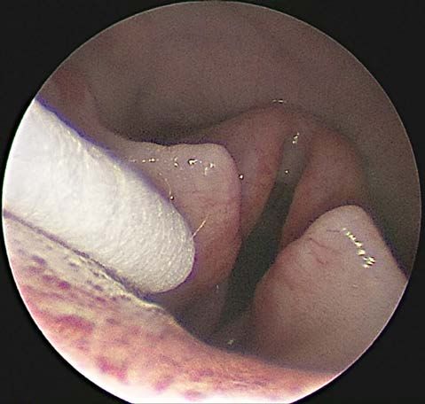
per respiratory airways frequently lead to a wide range of infiltration of the inflammatory process into the surround-
clinical signs. The most frequent are: mucopurulent nasal ing structures and the development of sinusitis maxillaris,
discharge, sniffling, as well as coughing associated with the frontalis and infraorbitalis [11]. A non-specific infectious
excitation of the patient and dyspnoea that can also be re- rhinitis is very rare in dogs. Other factors participating in
lated to diseases of other organ systems [9]. the development of inflammation of the upper respiratory
The etiology of the development of respiratory diseases tract include nasal trauma, allergy, or the penetration of a
is frequently associated with various causative agents, such foreign object [15].
as bacteria, viruses, fungi, parasites and other pathogens. Laryngotracheitis (Fig. 1) is another disease charac-
The respiratory tract is constantly exposed to these infec- terised by the inflammation of the upper respiratory tract.
tious agents either by aerogenic or haematogenic pathways. The etiology of this disease is polyfactorial. The pathogens
The pathogenic invasion is arrested by physical, chemical that most frequently cause this disease include: bacteria
and immunological mechanisms that include mucin, mu- (Bordetella bronchiseptica, Mycoplasma cynos, and Strep-
cocilliary clearance and various inherent antimicrobial tococcus equi subsp. zooepidemicus), and viruses (canine
factors [15]. The impairment of the protective mechanisms parainfluenza virus CAV-2, canine adenovirus CAV-1,
caused, for example, by immunosuppression, stress, tox- canine influenza virus, and canine respiratory virus). The
ins, environment and infectious agents, frequently results primary way of pathogen transfer involves aerosol and the
in the development of chronic inflammatory processes af- clinical signs appear about 3—10 days after the exposure
fecting individual compartments of the respiratory tract. to the pathogens. Additional sources of infection are: in-
In the majority of cases, rhinitis as a disease of the up- adequate sanitation of surfaces, contaminated examination
per respiratory tract is frequently overlooked during the instruments, endotracheal tubes, and toys [3].
primary diagnosis. From the etiological point of view, rhi- The correct diagnosis plays an important role in the
nitis is most frequently induced by the propagation of viral complex of prognosis, selection of therapy and its effec-
infections. However, secondary bacterial contaminations tiveness. Endoscopic imaging diagnosis is a targeted di-
may occur in long term inflammation or chronic diseas- agnostic method. The advantages of this method are: the
es. In such conditions connected with untreated cases, in- visualisation of the internal structures of the nasal and
flammation from rhinitis may spread to the surrounding frontal cavities, larynx, pharynx, trachea and bronchi, the
areas. The subsequent progressive chronic rhinitis involves identification of anatomic abnormalities, and also the lo-
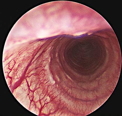
Fig. 1. Laryngotracheitis Fig. 2. Swab collecting in the area of larynx
76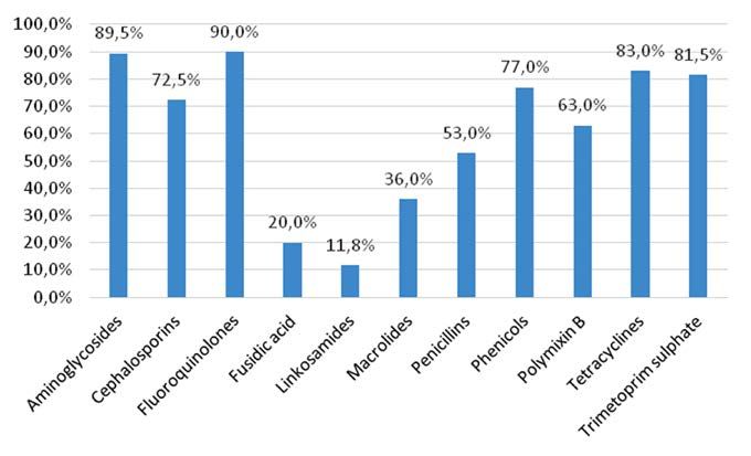
calisation of foreign bodies [7]. The possibilities of endo-      of bacterial culture examinations and the development of
scopic surgical interventions of congenital abnormalities,        the targeted therapeutic plan, based on the susceptibility of
such as: stenotic nostrils, lengthening of the soft palate,       pathogens to the selected antimicrobial preparations.
stricture of nasopharyngeal compartments and tracheal
stenosis which assist in: the therapy management complex,
improvement of prognosis and the course and duration of           MATERIALS AND METHODS
the respiratory disease.
     Endoscopic visualisation is the most suitable diagnos-           This study was carried out on patients (n = 26) exhib-
tic method capable of revealing the sites of pathological         iting long-term clinical signs, such as: coughing, difficult
changes and the parallel collecting of samples for cytolog-       breathing, asphyxia, nasal discharge, hyperaemia and de-
ical and microbiological examinations. One of the most            pigmentation in the nostril zone. In 18 patients the disease
frequently used ways of the collection of samples from the        was localised only in the upper airways, while 8 of them
suspect regions of the respiratory apparatus involves swab-       showed signs of a disease spreading into the tracheal zone.
bing under direct endoscopic control [14]. Mucosal biopsy         All patients were diagnosed and treated at the Small animal
is indicated with the presence of various pathological neo-       clinic of the University of Veterinary Medicine and Phar-
plasms, as well as suspected mycotic diseases. The most ef-       macy (UVMP) in Košice. A rigid type of endoscope (Karl
fective way of sampling is the nasal sinuses, laryngeal swab      Storz, Germany) 2 mm in diameter and 15 cm in length
(Fig. 2), or bronchoalveolar lavage (BAL). During lavage,         was used for the examination of the nasal cavity. The tra-
the appropriate volume of saline at body temperature will         chea and bronchi were examined with a lavage channel en-
entrap sputum, exudate and surface cells revealing the po-        doscope, 2.7 mm in diameter and 25 cm in length.
tential for the development of respiratory infections [2].            The group of patients with a disease localised in the
     The suspicion of the presence of a bacterial infection is    nasal cavity included the following breeds: Maltese dogs
frequently formulated during the primary clinical exami-          (n = 4), Komondors (n = 2), Dachshunds (n = 2), mon-
nation. When detecting signs, such as mucopurulent nasal          grels (n = 2), Pitbull (n = 1), Hungarian Vizsla (n = 1),
discharge, fever, lethargy, inappetence, the best option ac-      Chihuahua (n = 1), and German short-haired pointer
cording to the International Society for Companion Ani-           (n = 1). The mean age of the patients was 6 years. After
mal Infectious Diseases (ISCAID) is the empirical admin-          the targeted endoscopic visualisation of the pathological
istration of doxycycline for 7—10 days as the first line of       changes sampled from the nasal cavities of the dogs, they
antibiotic effectiveness against Mycoplasma spp. and Bor-         were obtained by swabbing of the nasal mucosa in 14 cases
detella bronchiseptica. However, much more effective is the       (74 %): Maltese dogs (n = 4), Dachshunds (n = 2), Komon-
therapy of respiratory infections of bacterial origin based       dors (n = 2), mongrels (n = 2), Yorkshire terriers (n = 2),
on the determination of antibiotic susceptibility of isolated     Pitbull (n = 1), and Hungarian Vizsla (n = 1). Nasopharyn-
strains. For example, in the presence of secondary bacterial      geal swabs were obtained from 4 dogs (21 %): Yorkshire
agents such as Pasteurella spp. and Streptococcus spp., the       terriers (n = 2), German short-haired pointer (n = 1), and
administration of amoxicillin appears to be the most ade-         Chihuahua (n = 1). The sample obtained from a 9 years old
quate treatment. For the treatment of infection with Staph-       Yorkshire terrier male consisted of a swab and the lavage of
ylococcus spp. it is recommended to use amoxicillin and           sinuses with saline (in 1 case—5 %).
clavulanic acid since such treatment is not effective against         The group of patients with clinical signs involving the
Mycoplasma spp. and Bordetella bronchiseptica [10].               trachea consisted of: Yorkshire terriers (n = 4), Dachs-
     The high prevalence of microbial contamination of the        hunds (n = 2), Maltese dog (n = 1), and mongrel (n = 1).
upper airways and increasing resistance to antimicrobials         Their mean age was 7 years. Samples from these dogs were
indicate the necessity of the utilisation of the most accu-       obtained in the same way, namely by swabbing the tracheal
rate diagnosis and targeted medicinal therapy. The aim of         mucosa.
our clinical study was by means of the targeted endoscopic            All patients were subjected to a basic clinical examina-
visualisation of pathological changes, to point out the in-       tion which included: sampling of venous blood; evaluation
creased effectiveness of the collection of samples, reliability   of a roentgenogram of the thoracic cavity in lateral projec-

                                                                                                                            77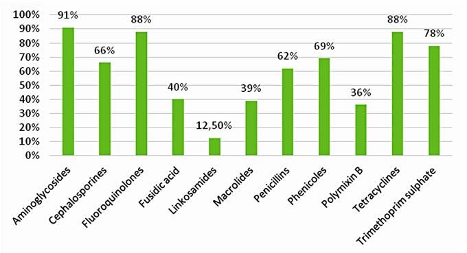
tion; and auscultation. Before each targeted endoscopic ex-      RESULTS
amination, the animals were sedated utilizing a combina-
tion of three anaesthetics administered to the patients in-          The importance of endoscopic visualisation of the res-
travenously, i. e. (Cepetor 1 mg.ml–1 inj. solution, CP-Phar-    piratory tract for the diagnosis of acute and chronic dis-
ma, Germany at a dose of 0.015 mg.kg–1 body weight), di-         eases was confirmed. This examination revealed a broad
azepam (Apaurin 5 mg.ml–1 inj. solution, KRKA, Slovenia          range of pathological changes in the majority of the pa-
at a dose of 0.3 mg.kg–1 b. w.), and propofol (Propofol 10       tients. These pathologic manifestations ranged from mu-
mg.ml–1 inj. suspension, Fresenius Kabi, Germany at a dose       cous clumps adhered to the mucous membrane of the res-
of 3 mg.kg–1 b. w.).                                             piratory airways, through typical macroscopic hyperaemic
    For the primary bacterial culture of the samples and         manifestations of inflammation, up to anatomical changes
isolation of microorganisms we used a non-selective cul-         observed most frequently in the conchae, nasopharynx
ture medium [Columbia agar (HiMedia, India) enhanced             and trachea.
with 5 % sheep blood]. In parallel, as another non-selective         The results of the microbiological examinations of the
medium suitable also for propagation of pigment forming          samples collected in this study revealed the prevalence of
bacteria (such as Pseudomonas aeruginosa and Staphylo-           bacterial strains involved in diseases of the upper respira-
coccus aureus), we used meat-peptone agar (Nutrient agar         tory tract. The pathogens found in the highest abundance
No. 2, HiMedia, India). On the basis of the character of         in the nasal cavity and nasopharynx area were: Enterococ-
growth and haemolytic activity of the isolated bacteria, the     cus faecalis (15.6 %), Staphylococcus aureus (12.5 %), Staph-
colonies were inoculated to the specific selective Endo agar     ylococcus pseudintermedius (9.4 %), Escherichia coli (9.4 %),
(HiMedia, India) with the focus on enterobacteria, includ-       Bacillus spp. (6.3 %), Pseudomonas aeruginosa (6.3 %) and
ing E. coli, Klebsiella and Proteus, and on to a diagnostic      others (Table 1). In the samples from the trachea, Escher-
medium Baird-Parker agar (HiMedia, India) for identifica-        ichia coli (20 %), Enterobacter ludwigii (13 %), haemolytic-
tion of S. aureus. The basic diagnosis included microscopic      Escherichia coli (13 %) and others (Table 2) were found in
examination of the native preparations and Gram-stained          the highest abundance.
preparations.                                                        In addition to the microbiological cultivation of the
    Commercial biochemical tests were used for addition-         samples, antibiograms were prepared illustrating the effec-
al, more accurate identifications. The basic tests used in       tiveness of various groups of antibiotics used in veterinary
our study included catalase (3 % hydrogen peroxide) and          practice. The determination of antibiotic resistance of indi-
oxidase test (OxiTest, Erba Lachema, Czech Republic).            vidual pathogens provided the necessary information for
For the species specification we used EnteroTest 24, Nefer-      the initiation of targeted individual antibiotic therapy. The
mTest 24, StaphyTest 24, and Streptotest 24 (Erba Lachema,       results of the antibiotic resistance of contaminants of the na-
Czech Republic). The results of the tests were evaluated by      sal cavity and trachea isolated in our study may be seen in
means of software TNW ProAuto 7.0.                               Figures 3 and 4. The effectiveness of individual groups of an-
    The identified bacterial strains were subsequently sub-      tibiotics used in the treatment of common infections of the
jected to tests of susceptibility/resistance to antibiotics by   upper respiratory tract in descending sequence were as fol-
means of a modern automatized instrument VITEK® 2 in-            lows: fluoroquinolones (90.0 %), aminoglycosides (89.5 %),
tended for the determination of the minimum inhibition           tetracyclines (83.0 %), trimetoprim-sulphate (81.5 %),
concentrations of antibiotics. The results of the tests were     phenicols (77.0 %), cephalosporins (72.5 %), polymyxin B
interpreted in agreement with the criteria determined by         (63.0 %), penicillins (53.0 %), macrolides (36.0 %), fusidic
the European Committee on Antimicrobial Susceptibility           acid (20.0 %), and lincosamides (11.8 %), see Figure 5.
Testing (EUCAST). The complex microbiological diag-
nosis was carried out in specialised clinical microbiology
laboratories.

78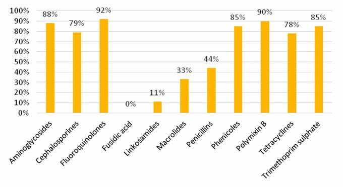
Table 1. Bacterial species found in the nasal cavity                           Table 2. Bacterial species found in the trachea

         Bacterial species               Number of findings       %                  Bacterial species             Number of findings         %

                                                                                Escherichia coli                            3               20.0
Enterococcus faecalis                             5              15.6

Staphylococcus aureus                             4              12.5           Enterobacter ludwigii                       2               13.3

Escherichia coli                                  3               9.4           Haemolytical E. coli                        2               13.3

Staphylococcus pseudintermedius                   3               9.4           Enterobacter cloacae                        1                6.7
Bacillus spp.                                     2               6.3
                                                                                Enterobacter spp.                           1                6.7
Pseudomonas aeruginosa                            2               6.3
                                                                                Beta haemolytic streptococci                1                6.7
Acinetobacter baumanii                            1               3.1
                                                                                Klebsiella pneumoniae                       1                6.7
Acinetobacter pittii                              1               3.1

Acinobacter spp.                                  1               3.1           Pasteurella multocida                       1                6.7

Aeromonas caviae                                  1               3.1           Pasteurella stomatis                        1                6.7

Klebsiella pneumoniae                             1               3.1           Proteus mirabilis                           1                6.7
Neisseria zoodegmatis                             1               3.1
                                                                                Streptococcus viridans                      1                6.7
Pantoea agglomerans                               1               3.1

Pasteurella canis                                 1               3.1

Pasteurella dagmatis                              1               3.1

Pasteurella stomatis                              1               3.1

Staphylococcus epidermidis                        1               3.1

Staphylococcus intermedius                        1               3.1

Staphylococcus epidermidis                        1               3.1

                        Table 3. The effect of antimicrobial drugs used in the UVMP hospital on bacteria isolated in the study

                             Acinetobacter      Pasteurella     Staphylococci      Streptococci         Bacillus
                                                                                                                   Enterobacteriacae      E. coli
 Active ingredient                spp.              spp.             spp.              spp.               spp.
                                                                                                                         (n = 4)          (n = 8)
                                 (n = 3)           (n=5)            (n = 9)           (n = 2)            (n = 2)

 Benzyl penicillin                 –                  0%              28 %              0%                 –            100 %             100 %

 Cephalexin                      100 %                0%              14 %              0%                 –            100 %             29 %

 Enrofloxacin                     0%                  0%              13 %              0%                 –             0%               25 %

 Amoxicillin
                                 100 %                0%              25 %              0%                 –            100 %             25 %
 clavulanic acid

 Amoxicillin
                                 100 %                0%              25 %              0%                 –            100 %             25 %
 clavulanic acid

 Gentamycin                       0%                  0%                0%            100 %                –             0%               13 %

                                                                                                                                                    79
Fig. 3. The effect of antibiotics against bacterial species found in the nasal cavity
Fig. 4. The effect of antibiotics against bacterial species found in the trachea
Fig. 5. The effectiveness of antibiotics used in the treatment of common infections of the upper respiratory tract
80DISCUSSION uate specific sites. Each procedure and normal appearance
like cytology and culture results from each region will en-
Numerous infectious agents are responsible for causing hance the diagnostic success [5].
primary or secondary respiratory diseases in dogs. Clinical By means of endoscopic diagnosis and direct collection
signs may vary from mild dyspnoea, sneezing, and cough- of samples from the nasal cavity (n = 18), nasopharynx
ing to severe pneumonia with systemic manifestations. De- (n = 4) and trachea (n = 8) of 26 animal patients, microbial
pending on the etiological agent, the gross and microscop- cultures allowed us to identify 27 bacterial species in a total
ic changes observed during these infections can be rather number of 47 bacterial findings. The most frequently isolat-
unspecific or have highly characteristic patterns [15]. ed Gram-negative bacteria were: Escherichia coli (17.0 %),
There are many diagnostic tools available on the mar- Pasteurella spp. (10.6 %), members of the family—Entero-
ket that can be used for the identification of respiratory bacteriacae (10.6 %), Acinetobacter spp. (6.4 %), Klebsiel-
agents. The development of molecular diagnostic tools al- la pneumoniae (4.2 %), Pseudomonas aeruginosa (4.2 %),
lows the rapid identification of a wide variety of pathogens Proteus mirabilis (2.1 %), Aeromonas caviae (2.1 %), and
and the establishment of more accurate treatments [16]. Neisseria zoodegmatis (2.1 %). Of the Gram-positive bac-
Most common bacterial cultures from the nasal cavity can teria, we detected most frequently: Staphylococcus aureus
be performed from specimens obtained by nasal swabbing. (8.5 %), Staphylococcus pseudintermedius (6.4 %), Strep-
However nasal lavage specimens gave a higher sensitivi- tococcus spp. (6.4 %), Staphylococcus spp. (4.2 %) and Ba-
ty for bacterial growth than tissue biopsy specimens and cillus spp. (4.2 %). Our results resemble those published
swabs [8]. In comparison with dogs, bacterial culture re- by A d a s z e k et al. [1], showing relatively frequent in-
sults can be difficult or impossible to interpret as bacteria volvement of the pathogenic bacteria E. coli, Klebsiella spp.
can also be cultured from the nasal cavity of healthy cats. and Staphylococcus spp. in the respiratory tract infections
However multidrug‐resistant bacteria can colonize and be in the dogs.
grown from the nasal passages in the absence of infection In the relevant study conducted by D a o d u et al.
[10]. Bacterial cultures from the respiratory tract can be [6], the authors isolated 222 bacterial species. The most
performed from specimens obtained from oropharyngeal frequently identified species were: Staphylococcus spp.
swabs, tracheal wash, or bronchoalveolar lavage (BAL). (21.7 %), E. coli (18.5 %), Proteus spp. (17.1 %), Acineto-
Collection of a tracheal wash or bronchoalveolar lavage bacter spp. (9.0 %), Pseudomonas spp. (6.8 %) and Strep-
specimen is indicated in dogs with more severe clinical tococcus spp. (5.4 %). The results of culture studies of
signs or evidence of pneumonia [13]. Some clinicians rec- samples from 90 nasal swabs published by A y o d h y a
ommend quantitative or semiquantitative bacterial cultures et al. [2] revealed various types of bacteria in mild cases:
of BAL fluid, and it has been reported that counts greater E. coli (63.33 %), Klebsiella spp. (30 %) and mixed infec-
than 104 colony forming units (CFU.ml–1) (or grown from tions (E. coli and Klebsiella spp.) (6.67 %), in 19, 9 and
primary culture) represent true infection whereas less than 2 dogs (out of 30 samples), respectively. Similarly, in the
103 CFU.ml–1 (or grown from subculture) represent con- case of moderate respiratory diseases, samples revealed
tamination [12]. the presence of various bacteria: E. coli (30 %), Klebsiella
The treatment of upper respiratory diseases is sympto- spp. (33.33 %), Streptococcus spp. (30 %) and mixed infec-
matic; however, due to the common occurrence of second- tions (Klebsiella spp. and Streptococcus spp.) (6.67 %), in 9,
ary infections with a broad spectrum of bacteria, antibiotic 10, 9 and 2 dogs (out of 30 samples), respectively. In the
treatment is the first therapeutic approach. Antimicrobial case of severe respiratory diseases, various bacteria were
agents should be selected based on culture and sensitivity identified: E. coli (6.67 %), Klebsiella spp. (3.33 %), Strepto-
tests of airway specimens collected by bronchoscopy [14]. coccus spp. (46.67 %), Staphylococci spp. (40 %) and mixed
Endoscopy is a valuable diagnostic approach to the upper infection (E. coli and Streptococcus spp.) (3.33 %) in 2, 1,
and lower respiratory tract, because it allows direct visual- 14, 12 dogs and 1 dog (out of 30 samples), respectively. The
isation and sample collection. However, each anatomical study conducted by C h a r k r a b a r t h i [4] showed that
region may require a range of specialized technical equip- Klebsiella pneumoniae and E. coli spp. were the common
ment and varying levels of experience to access and eval- bacteria involved in respiratory infectious diseases.
81The information related to microbial contamination REFERENCES
of the upper respiratory tract obtained in our study were
subsequently used for the determination of antibiotic re- 1. Adaszek, L., Gorna, M., Zietek, J., Kutrzuba, J., Win-
sistance of the isolates against 11 groups of antibiotics. iarczyk, S., 2009: Bacterial nosocomial infections in dogs
The effectiveness in descending sequence was as follows: and cats. Zycie Weterynaryjne, 84, 805—808.
aminoglycosides (89.8 %), fluoroquinolones (89.5 %), 2. Ayodhya, S., Rao, D. T., Reddy, Y. N., Sundar, N. S., Kumar,
tetracyclines (84.2 %), trimetoprim-sulphate (80.7 %), V. G., 2013: Isolation and characterization of bacteria from
phenicols (77,0 %), cephalosporins (71.2 %), polymyx- canine respiratory diseases in and around Hyderabad city,
in B (67,8 %), penicillins (55,0 %), macrolides (35.6 %), Andhra Pradesh, India. Vet. World, 6, 9, 601‒604. DOI: 10.
fusidic acid (16.6 %) and lincosamides (11.6 %). In the 5455/vetworld.2013.601-604.
study published by D a d o u et al. [6] the effectiveness 3. Carey, S. A., 2019: Canine Infectious Respiratory Disease
of fluoroquinolones on Gram-positive and Gram-negative Complex: Host, Pathogen, and Environmental Interactions.
bacteria ranged from 78.4 % to 93.3 %. The effectiveness Re
trieved from https://www.michvma.org/resources/Docu-
of cephalosporines against Gram-positive bacteria reached ments/MVC/2018%20Proceedings/carey_05.pdf.
31.7 % while the effect of penicillins (e. g. amoxicillin), 4. Charkrabarthi, A., 2009: Textbook of Clinical Veterinary
as the first line antibiotics, was paradoxically rather low. Medicine. 3rd edn., Ludhiana, Kalayani Publishers, Punjub,
It reached 15.0 % against Gram-positive and 37.7 % against India, 327—372.
Gram-negative strains. 5. Creevy, K. E., 2009: Airway evaluation and flexible endo-
scopic procedures in dogs and cats: laryngoscopy, transtra-
cheal wash, tracheobronchoscopy, and bronchoalveolar lav-
CONCLUSIONS age. Vet. Clin. North Am. Small Anim. Pract., 39, 5, 869—880.
DOI: 10.1016/j.cvsm.2009.05.001.
Chronic and long-term diseases of the upper respirato- 6. Daodu, O. B., Amosun, E. A., Oluwayelu, D. O., 2017:
ry tract require correct and targeted diagnosis. Direct vis- Antibiotic resistance profiling and microbiota of the upper
ualisation of the respiratory airways is capable of ensuring respiratory tract of apparently healthy dogs in Ibadan, South
precise sampling, accurate diagnosis and the development west Nigeria. Afr. J. Infect. Dis., 11, 1, 1—11. DOI: 10.4314/
of the subsequent rational therapeutic plan. Direct targeted ajid.v11i1.4533.
collection of samples is advantageous with respect to the 7. Elie, M., Sabo, M., 2006: Basics in canine and feline rhinos-
elimination of the complex multifactorial influences and copy. Clin. Tech. Small Anim. Pract., 21, 2, 73—77. DOI: 10.
contribution to the therapy effectiveness. Microbiologi- 1053/j.ctsap.2005.12.011.
cal culture methods focused on pathogenic bacteria con- 8. Johnson, L. R., Kass, P. H., 2009: Effect of sample collection
stitute an inseparable part of the diagnosis of respiratory methodology on nasal culture results in cats. J. Feline Med.
diseases especially with respect to prevention of increasing Surg., 11, 645—649. DOI: 10.1016/j.jfms.2008.12.004.
antibiotic resistance and incidence of chronic conditions. 9. Kuehn, N. F., 2013: Tracheobronchitis in Small Animals. Re-
Although the financial demands of endoscopic diagnostic trieved in Sept. 2020, from https://www.msdvetmanual.com/
approach are moderately increased, it has been confirmed respiratory-system/respiratory-diseases-of-small-animals/
that the results obtained in this way are reliable. In prac- tracheobronchitis -in-small-animals.
tice it may result in reduced administration of therapeu- 10. Lappin, M. R., Blondeau, J., Boothe, D., Breitschwerdt,
tically effective medications, more effective treatment and E. B., Guardabassi, L., Lloyd, D. H., et al., 2017: Antimicro-
prevention of an increase in antibiotic resistance and thus, bial use guidelines for treatment of respiratory tract disease
also reduced load on the patient. in dogs and cats: Antimicrobial Guidelines Working Group of
the International Society for Companion Animal Infectious
Diseases. J. Vet. Intern. Med., 31, 2, 279—294. DOI: 10.1111/
jvim.14627.
8211. Levkut, M., Švický, E., Lenhardt, Ľ., Ševčíková, Z., Reva- 15. Vieson, M. D., Pineyro, P., LeRoith, T., 2012: A review of
jová, V., Herich, R., 2007: Special Veterinary Pathology (In the pathology and treatment of canine respiratory infections.
Slovak). Vol. I. University of Veterinary Medicine and Phar- Vet. Med., 3, 25—39. DOI: 10.2147/VMRR.S25021.
macy in Košice, Slovakia, 239 pp. 16. Windsor, R. C., Johnson, L. R., Sykes, J. E., Drazenovich,
12. Padrid P., 2000: Pulmonary diagnostics. Vet. Clin. North Am. T. L., Leutenegger, C. M., De Cock, H. E. V., 2006: Molecular
Small Anim. Pract., 30, 6, 1187—1206. DOI: 10.1016/s0195- detection of microbes in nasal tissue of dogs with idiopathic
5616(00)06008-3. lymphoplasmacytic rhinitis. J. Vet. Intern. Med., 20, 2, 250—256.
13. Reagan, K. L., Sykes, J. E., 2020: Canine infectious respira- DOI: 10.1892/0891-6640(2006)20[250:MDOMIN]2.0.CO;2
tory disease. Vet. Clin. North Am. Small Anim. Pract., 50, 2,
405—418. DOI: 10.1016/j.cvsm.2019.10.009. Received January 4, 2020
14. Sumner, C. M., Rozanski, E. A., Sharp, C. R., Shaw, S. P., Accepted February 22, 2021
2011: The use of deep oral swabs as a surrogate for transoral
tracheal wash to obtain bacterial cultures in dogs with pneu-
monia. J. Vet. Emerg. Crit. Care, 21, 5, 515—520. DOI: 10.
1111/j.1476-4431.2011. 00670.x.
83You can also read