Patient-Informed Organ Dose Estimation in Clinical CT: Implementation and Effective Dose Assessment in 1048 Clinical Patients - DukeSpace
←
→
Page content transcription
If your browser does not render page correctly, please read the page content below
Patient-Informed Organ Dose Estimation in Clinical CT:
Implementation and Effective Dose Assessment in 1048
Clinical Patients
Wanyi Fu, MS1,2,3, Francesco Ria, PhD3,4, William Paul Segars, PhD1,3,5,6, Kingshuk Roy Choudhury, PhD3,
Joshua M. Wilson, PhD 4,5, Anuj J. Kapadia, PhD1,3,5,7, Ehsan Samei, PhD1,2,3,4,5,6,7
Downloaded from www.ajronline.org by DUMC Univ Med Ctr on 01/25/21 from IP address 152.3.102.254. Copyright ARRS. For personal use only; all rights reserved
Multispecialty Articles · Original Research
Keywords
OBJECTIVE. The purpose of this study is to comprehensively implement a pa-
CT, effective dose, patient-specific organ
dose, radiation burden characterization tient-informed organ dose monitoring framework for clinical CT and compare the effec-
tive dose (ED) according to the patient-informed organ dose with ED according to the
Submitted: Oct 23, 2019 dose-length product (DLP) in 1048 patients.
Revision requested: Dec 9, 2019 MATERIALS AND METHODS. Organ doses for a given examination are computed
Revision received: Mar 9, 2020 by matching the topogram to a computational phantom from a library of anthropomor-
Accepted: Apr 29, 2020 phic phantoms and scaling the fixed tube current dose coefficients by the examination
volume CT dose index (CTDIvol) and the tube-current modulation using a previously val-
The authors declare that they have no idated convolution-based technique. In this study, the library was expanded to 58 adult,
disclosures relevant to the subject matter of 56 pediatric, five pregnant, and 12 International Commission on Radiological Protection
this article.
(ICRP) reference models, and the technique was extended to include multiple protocols,
Based on a presentation at the Radiological a bias correction, and uncertainty estimates. The method was implemented in a clin-
Society of North America 2018 annual ical monitoring system to estimate organ dose and organ dose–based ED for 647 ab-
meeting, Chicago, IL. domen-pelvis and 401 chest examinations, which were compared with DLP-based ED
using a t test.
Supported in part by a grant from NIH (R01 RESULTS. For the majority of the organs, the maximum errors in organ dose estima-
EB001838). The technology has been tion were 18% and 8%, averaged across all protocols, without and with bias correction,
nonexclusively licensed by Duke University respectively. For the patient examinations, DLP-based ED was significantly different from
to GE and Imalogix. Those entities were not organ dose–based ED by as much as 190.9% and 234.7% for chest and abdomen-pelvis
involved in the development of the content scans, respectively (mean, 9.0% and 24.3%). The differences were statistically significant
of this article.
(p < .001) and exhibited overestimation for larger-sized patients and underestimation for
smaller-sized patients.
CONCLUSION. A patient-informed organ dose estimation framework was compre-
hensively implemented applicable to clinical imaging of adult, pediatric, and pregnant
patients. Compared with organ dose–based ED, DLP-based ED may overestimate effec-
tive dose for larger-sized patients and underestimate it for smaller-sized patients.
Because of the widespread utility of CT in clinical diagnosis, its usage has increased
steadily over the last decade. From recent estimates, about 82 million CT scans were per-
formed in 2018 in the United States alone [1]. With the rise in the number of scans, it is ben-
eficial to practice consistency and perform safety and quality control within and across in-
stitutions toward the general goal of increasing patient population safety by improving
CT risk assessment and the design of effective optimization actions. Of particular interest
is the estimation of radiation dose in clinical practice, which has prompted hospitals and
clinics to record CT radiation dose from clinical examinations [2–4]. Most CT dose record-
ings are made according to system outputs and not patient dose. Yet radiation dose is an
individualized burden to each patient, dependent on that patient’s body attributes and
the specific imaging protocol used. Individual patient dose is the attribute that neces-
sitates the monitoring of radiation dose in the first place. Thus, radiation management
would benefit from recording patient dose in an individualized, patient-specific manner
Fu et al. accounting for age, size, and varying radiosensitivities of human tissues [5].
Patient-Informed Organ Dose Estimation in CT
Multispecialty Articles
Department of Radiology, Duke University, 2424 Erwin Rd, Ste 302, Durham, NC 27705. Address correspondence to
1
Original Research W. Fu (wanyi.fu@duke.edu).
Department of Electrical and Computer Engineering, Duke University, Durham, NC.
2
Fu W, Ria F, Segars WP, et al.
Carl E. Ravin Advanced Imaging Laboratories, Duke University, Durham, NC.
3
doi.org/10.2214/AJR.19.22482 4
Clinical Imaging Physics Group, Duke University Health System, Durham, NC.
AJR 2021; 216:1–11 Medical Physics Graduate Program, Duke University, Durham, NC.
5
ISSN-L 0361–803X/21/2163–1 6
Department of Biomedical Engineering, Duke University, Durham, NC.
© American Roentgen Ray Society Department of Physics, Duke University, Durham, NC.
7
AJR:216, March 2021 www.ajronline.org | 1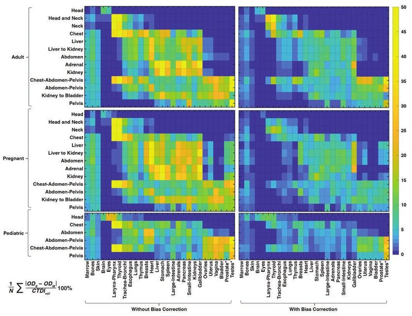
Fu et al.
In making CT radiation dose records individualized, patient-spe- models with 10 gestational ages of 3, 6, 8, 10, 15, 20, 25, 30, 35,
cific organ dose and the corresponding organ dose–derived effec- and 38 weeks [9, 20–22]. The frontal views of phantom renderings
tive dose (EDOD) have been regarded as the reference standard [6]. are shown in Figure 2A. As described in separate publications for
Estimating these quantities, however, requires explicit modeling of establishing the XCAT library, the adult and pediatric phantoms
patient anatomy and CT scan geometry and protocols [7–9]. These used in this study were created according to clinical CT images.
Downloaded from www.ajronline.org by DUMC Univ Med Ctr on 01/25/21 from IP address 152.3.102.254. Copyright ARRS. For personal use only; all rights reserved
tasks are both labor intensive and computationally expensive, which Major organs were segmented using a semiautomatic process,
limits their feasibility and practicality in clinical implementation. As and the remaining organs were added to the phantoms by de-
an alternative, scanner-derived exposure outputs, volume CT dose forming template models according to segmented organs from
index (CTDIvol), and dose-length product (DLP) that are recorded for the National Library of Medicine Visible Human Project images
each examination have been used to approximate effective dose ac- [9, 20, 21, 23]. The ICRP reference phantoms used were made ac-
cording to DLP (EDDLP) using DLP-to–effective dose (ED) conversion cording to the template models, with the body and organ masses
coefficients (k factors) [10–14]. However, the k factors are estimat- scaled according to ICRP 89 [22].
ed with a sexless, ageless, uniform phantom and do not represent Specifically developed for this study, pregnant phantoms were
the varied attributes and organ distributions seen in patient popu- formed according to five maternal models selected from exist-
lations. Consequently, these estimates may not represent the radi- ing adult female phantoms, representing different obesity class-
ation burden to the patient with sufficient accuracy and precision. es (50th percentile height and weight; and body mass index [BMI],
To make the estimation of the radiation burden more specif- measured in weight in kilograms divided by the square of height
ic to the patient, albeit not fully patient-specific, several studies in meters: underweight, 18.2; overweight, 28.6; class I obesity, 30.7;
have introduced patient-informed approaches, in which antici- and class II obesity, 35.5). The fetus models were generated by seg-
pated anatomic attributes of the patient are used to derive the menting organs and major structures from imaging data (CT, MRI,
patient organ dose estimates [15–18]. These methods are some- and histologic data). The maternal models were deformed to com-
times referred to as patient specific, even though the exact spe- bine the fetus models and represent various gestational ages. The
cifics of the patient are not modeled. As such, they are rather pa- BMI and age of the XCAT models are shown in Figure 2B.
tient-informed because they rely on accessible patient attributes In the framework for organ dose estimation, the topogram of
(e.g., patient diameter, sex) along with anthropomorphic models each patient was used to match to one phantom from the XCAT
of the anatomy to inform the organ dose calculation. The compu- library according to anatomic landmark heights. Anatomic land-
tationally expensive modeling of the anatomy and radiation field marks were automatically detected using a commercial software
is performed ahead of time and then ascribed to the patients, (DoseWatch, GE Healthcare) and included top and bottom of the
making the method practical for clinical applications. However, head, shoulders, lungs, and pelvis. These landmarks determined
these methods have not yet been applied across the diversity of patients’ anatomic heights that were used as the criteria to find
clinical protocols. the closest matched phantom. This matching method thus ap-
In this study, we aimed to develop and implement a comprehen- proximated the patient anatomy to an anthropomorphic phan-
sive patient-informed organ dose estimation framework for clinical tom with similar organ longitudinal distribution. The matching
CT dose monitoring with enhanced accuracy, patient-cohort cov- for pregnant patients was according to the longitudinal distribu-
erage, and protocol representation. To show its utility, patient-in- tion and considering any of the modeled gestational ages.
formed organ doses were calculated in two large CT clinical pop- When modeling exposure, the matched phantom was used to in-
ulations, and the results were used to determine the discrepancy dicate patient’s organ longitudinal distribution. The patient width
between ED values derived from organ dose and DLP estimates. was accommodated separately modeling with patient water equiva-
lent diameter. The water equivalent diameter was automatically de-
Materials and Methods rived from the medium slice of the patient CT images (DoseWatch).
This retrospective study was performed in compliance with Modeling the exposure—The patient-informed organ doses were
HIPAA and was determined to be exempt from Duke University estimated accounting for the patient anatomy and the tube cur-
institutional review board requirements. rent modulation incorporating the CTDIvol-to–organ dose conver-
We describe the framework implementation and then its appli- sion coefficients (h factors) [16, 18, 19, 24, 25]. The h factor library
cation to clinical CT images. The implementation includes creating was created a priori for 13 adult protocols (chest, abdomen-pel-
necessary libraries and programs. Once implemented, the frame- vis, chest-abdomen-pelvis, abdomen, pelvis, liver, kidneys, kid-
work is applied to clinical CT images automatically. The workflow ney-to-bladder, adrenal, liver-to-kidneys, head, head-neck, neck)
of the framework on clinical images is illustrated in Figure 1. and six pediatric protocols (chest, abdomen-pelvis, chest-abdo-
men-pelvis, abdomen, pelvis, head) under constant tube current.
Framework Implementation For each XCAT phantom and protocol, the h factors were simulat-
Modeling the anatomy—The study deployed an established pa- ed by a previously validated Monte Carlo simulation package (PE-
tient-informed organ dose estimation method in which the anat- NELOPE, version 2006, Universitat de Barcelona) [7, 8, 26, 27]. For
omy for a given patient is modeled by matching it to a compu- pregnant models, although a fetus at 3 and 6 weeks was created
tational phantom from a library of anthropomorphic phantoms in our initial models, their volumes were too small to obtain credi-
[19]. For this study, the library included extended cardiac-torso ble dose values. Their doses were thus represented by the dose to
(XCAT) phantoms: 58 existing adult models, 56 existing pediat- the uterus. For a fetus older than 6 weeks, the fetus models includ-
ric models, 14 existing International Commission on Radiolog- ed fetus bone structure and the rest as the fetus body and were
ical Protection (ICRP) reference models, and five new pregnant considered as organs in the dose calculation. The h factors calcula-
2 AJR:216, March 2021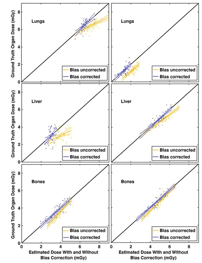
Patient-Informed Organ Dose Estimation in CT
TABLE 1: Summary of Simulated CT Techniques for Bias and Uncertainty Estimation
Cohort Protocols Tube Potential (kVp) Bowtie Filter Pitch Collimation (mm)
Adulta
Body Chest, abdomen-pelvis, chest-abdomen-pelvis, abdomen, 120 Large 1.375 40
Downloaded from www.ajronline.org by DUMC Univ Med Ctr on 01/25/21 from IP address 152.3.102.254. Copyright ARRS. For personal use only; all rights reserved
pelvis, liver, kidneys, kidney-to-bladder, adrenal,
liver-to-kidneys
Neuralgic Head, head-neck, neck 120 Small 1 20
Pediatric
Body Chest, abdomen-pelvis, chest-abdomen-pelvis, abdomen, 120 Small 1.375 40
pelvis
Neuralgic Head 120 Small 1 20
Note—All CT techniques were conducted on a LightSpeed VCT (GE Healthcare) system.
a
Adult cohort includes pregnant women.
tion used the normalized fetal-dose ratio method of Felmlee et al. and organ. Specifically, a linear fit was applied to ODC versus
[28]. The h factors were calculated for fetuses at various gestation- ODM for each protocol, cohort (each gestational age for preg-
al ages and in maternal models with various sizes. Overall, with- nant phantoms), and organ as
in each protocol, the h factors were modeled as a function of the
XCAT scanned region diameter applied to each new patient. For ODM = β0ODC + β1 + ε, (1)
the pregnant phantoms, the h factors were applied to the patients
according to both diameter and gestational ages. where β0 and β1 are fitting coefficients and ε is the fitting error.
The radiation field under modulated tube current was mod- The fits were used to provide bias-corrected, protocol-specific or-
eled as a function of the convolution of the tube current profile gan dose values (ODCB,p, where p denotes a patient) and 95% CI as
and a dose spread function using a previously developed tech-
nique [19]. The dose spread function was simulated by the Mon- ODCB,p = β0ODC,p + β1 (2)
te Carlo method on cylindric phantoms across diameters of 8–50
cm [19]. For each patient, the dose spread function correspond- and
ing to the patient diameter was used. The dose ratio was calcu-
lated as the ratio of dose spread function convolved with mod-
1 (ODC,p – ODC)
2
ulated tube current versus the dose spread function convolved CI = tα RMSE 1 + + (3)
,N N σ 2
with the mean tube current value with the same scan length [19]. 2 OD C
The dose field for each organ (CTDIorgan) was determined accord-
ing to the dose ratio with respect to organ distribution from the where β0 hat and β1 hat are the estimated coefficients obtained
matched XCAT phantom and the examination CTDIvol [19]. The from fitting model (Equation 1) to the data. ODC,p is the organ dose
examination CTDIvol accommodates radiation specification from estimated for a new patient, N is the number of phantoms, the ex-
various scanners and CT techniques. The organ dose was calcu- pression of t is the t value with N – 2 degrees of freedom, RMSE is
lated as a function of the CTDIorgan and h factor [19]. the root mean square error between ODC and ODM for all phantoms
Bias and uncertainties—Organ doses estimated by the pro- of the protocol and cohort, and ODC with the overbar and corre-
posed method showed bias and uncertainties compared with sponding σ2 are the mean and variance of ODC, respectively.
those simulated using the Monte Carlo method (the reference Framework accuracy—To quantify the overall organ dose esti-
standard) [19]. To improve the accuracy of the estimated organ mation accuracy within this XCAT library, the mean difference be-
doses, we developed a secondary correction and uncertainty tween the estimated dose using the leave-one-out method (ei-
estimation strategy using simulated CT scans with known or- ther ODC or ODCB) and ODM was calculated for each protocol.
gan dose ground truth. Simulated CT scans were conducted for
each XCAT phantom and protocol using typical clinical CT ex- Clinical Study
amination protocols. The CT techniques are shown in Table 1. Patients and scan protocols—The framework was tested across
All examinations simulated tube current modulation accord- 1048 patients (mean age, 58 years; range, 18–89 years) who un-
ing to Li et al. [29]. The organ dose estimated using the pro- derwent abdomen-pelvis scans with contrast enhancement (647
posed method (ODC) was obtained in a leave-one-out meth- examinations) or chest scans without contrast enhancement
od in which each XCAT phantom was treated as a new patient, (401 examinations) from January 5, 2017, to August 29, 2018, at
matched to another XCAT to obtain the ODC from the exposure Duke University. Cases included 524 women (mean age, 57 years;
model as proposed. The ground truth organ dose (ODM) was range, 18–88 years) and 524 men (mean age, 59 years; range, 18–
estimated using a validated Monte Carlo simulation package 89 years). All image data were acquired using a commercial scan-
[17]. The statistics of ODC and ODM were used to generate linear ner (Discovery CT750 HD, GE Healthcare) with tube current mod-
models that provided organ doses with bias correction (ODCB) ulation, a tube voltage of 120 kV, and pitch values of 1.53 for chest
and CIs on new clinical patients within each cohort, protocol, and 0.98 or 1.53 for abdomen-pelvis scans.
AJR:216, March 2021 3
Fu et al.
Effective dose estimation and data analysis—For each examina- chest scans, the lung-to-liver and lung-to-bone dose differences
tion, the ED was calculated using two methods evaluated in this were 36.7% ± 9.6% and 52.7% ± 5.4% of the CTDIvol, respectively,
study according to the patient-informed organ dose estimated averaged across all chest examinations. For abdomen-pelvis scans,
using the proposed method as the liver-to-lung and liver-to-bone dose differences were 67.0% ±
22.5% and 26.8% ± 12.3% of CTDIvol, respectively, averaged across
ΣOD
Downloaded from www.ajronline.org by DUMC Univ Med Ctr on 01/25/21 from IP address 152.3.102.254. Copyright ARRS. For personal use only; all rights reserved
EDOD,p = BC,p
wo, (4) all abdomen-pelvis examinations.
o
Figure 6 shows the ED as a function of water equivalent diame-
where p denotes a patient and wo is the tissue weighting coeffi- ter for the 401 chest and 647 abdomen-pelvis scans. EDDLP was, on
cients defined by ICRP Publication 103 [30]; and according to DLP as average, greater than EDOD by 9.0% (p < .001, range from –18.1%
to 190.9%) and 24.3% (p < .001, range from –28.4% to 234.7%) for
EDDLP,p = ΣDLP
r
k ,
r,p r,p
(5) chest and abdomen-pelvis scans, respectively. For water equiv-
alent diameter ranges of less than 25, 25–30, 30–35, and greater
where DLPr,p is the DLP for region r (e.g., chest, abdomen-pelvis); than 35 cm, the percentage differences between EDDLP and EDOD
and kr is the DLP-to-ED conversion coefficients for region r de- were –6.3% ± 21.8% (p < .001), 8.8% ± 21.2% (p < .001), 31.6% ±
fined by ICRP Publication 102 [6]. The mean difference between 34.9% (p < .001), and 38.3% ± 11.0% (p = .04) for chest, and –13.3%
EDOD,p and EDDLP,p was calculated for each protocol and evaluated ± 14.3% (p < .001), 4.4% ± 18.1% (p < .001), 31.3% ± 25.1% (p <
using a t test (Matlab, version 2018a, Mathworks) in terms of the .001), and 61.9% ± 32.2% (p < .001) for abdomen-pelvis protocols.
difference across all patients and across different-sized groups On group average, EDDLP was larger than EDOD for larger patients
(grouped according to the water equivalent diameter). Although (water equivalent diameter, > 25 cm) and smaller than EDOD for
some other investigators have used the ICRP reference phantom smaller patients (water equivalent diameter, < 25 cm).
for the purpose of overall radiation exposure estimation [30], this
study calculated the EDOD,p for each patient using a virtual anthro- Discussion
pomorphic phantom matched to the patient. In the wake of accreditation and regulatory program require-
ments, an increasing number of hospitals and clinics are record-
Results ing radiation dose from CT examinations. When combined with
Extended Cardiac-Torso Phantom Validation image quality or other diagnostic performance measurements,
Figure 3 shows ODM versus ODC and ODCB and their corre- they can be used to manage exposure and optimize imaging pro-
sponding linear model for sample protocols. The ODC was cor- tocols. To facilitate this management, it is best to use metrics that
related with ODM for organs inside, on the periphery, outside, and are most relevant to the actual burden on a patient. Current clini-
distributed (R, 0.78 ± 0.24 [SD]; 0.62 ± 0.28; 0.34 ± 0.33; 0.63 ± 0.31, cal practice typically relies on scanner-derived dose indexes such
respectively), and averaged across all protocols, cohorts, and or- as CTDIvol and DLP, which are more of a reflection of the expo-
gans within each location group according to Sahbaee et al. [17]. sure output of the machine than the actual radiation burden to
The linear model with bias correction obtained unified the slope the patient. Patient-specific approaches require time-consum-
and reduced the intercept. ing and labor-intensive anatomic modeling of each patient and
Figure 4 shows the difference between ODM versus ODC and computationally expensive Monte Carlo simulations, which al-
ODCB for each organ and protocol averaged across all of the XCAT though accurate, are challenging in clinical applications. In this
phantoms within each cohort. When accounting for all the or- study, we implemented a comprehensive patient-informed or-
gans, the protocol-averaged maximum error was 53% and 18%, gan dose estimation framework. The patient information is taken
without and with bias correction, respectively. The correspond- into account and precalculated anatomy and exposure datasets
ing errors within each cohort were 53% (adult), 56% (pregnant), are used to provide clinical dose estimation in real time and with
and 45% (pediatric) without bias correction, and 21% (adult), high accuracy. This system may assist in exposure standardiza-
13% (pregnant), and 17% (pediatric) with bias correction. When tion and comparison within and across health care providers and
accounting for the majority of organs (excluding 15% of outli- potentially protocol optimization when combined with diagnos-
er organs with the largest errors), the protocol-averaged max- tic image quality–related measurements.
imum errors were 18% and 8%, without and with bias correc- To facilitate exposure management, ED according to organ
tion, respectively. The corresponding errors within each cohort doses and tissue weighting coefficients is a relevant quantity, ac-
were 17% (adult), 18% (pregnant), and 17% (pediatric) without counting for the actual radiation burden to the patient. Organ
bias correction, and 9% (adult), 7% (pregnant), and 8% (pediat- doses determined in a patient-relevant manner, however, are usu-
ric) with bias correction. As shown, for the majority of organs, ally not known. In such cases, ED according to DLP is commonly
including fetus as an organ, the dose estimation showed rea- used. The difference between the two quantities has previously
sonable errors, and the error reduced with bias correction. The been noted for a limited number of protocols and reference phan-
bias correction was more effective for organs with larger errors. toms under fixed tube current condition [12–14]. In this study, the
comparison was performed under a widely used modulated tube
Clinical Study current condition in a clinical population. The ED from patient-in-
Figure 5 shows the liver, lung, and bone-surface organ doses as formed organ dose showed a substantial difference from those
a function of examination mean CTDIvol for the 401 chest and 647 estimated using the DLP method. This difference can be as large
abdomen-pelvis scans. The plots indicate that for each examina- as –28.4% to 234.7%, with the differences larger for larger-sized
tion mean CTDIvol, different organs have diverse dose values. For patients. This suggests that care should be exercised when using
4 AJR:216, March 2021
Patient-Informed Organ Dose Estimation in CT
EDDLP to inform clinical decisions, because overestimating doses size-specific conversion factors [33–35]. However, future studies
may lead to unnecessary concern and result in insufficient expo- can extend our approach to include the comparison with other
sures, and vice versa. patient-informed radiation metrics. Finally, the clinical study was
There is an increase in the ED with increased patient size, which limited to the adult population without any confirmed cases of
agrees with previous studies reporting the ED according to pa- pregnancy. Future work will include pediatric populations.
Downloaded from www.ajronline.org by DUMC Univ Med Ctr on 01/25/21 from IP address 152.3.102.254. Copyright ARRS. For personal use only; all rights reserved
tient-specific organ doses [15, 31]. Our clinical study showed that In conclusion, in this study we comprehensively implement-
EDDLP is overestimated for larger patients and underestimated for ed and showed the feasibility of a patient-informed organ dose
smaller patients. This trend agrees with a clinical study on abdo- monitoring system in the clinical setting. Compared with scan-
men-pelvis examinations with tube current modulation by Ha- ner-derived dose indexes such as CTDIvol or DLP, the system pro-
ji-Momenian et al. [14], who explained that this trend was a result vides a more accurate estimate of organ dose by taking into ac-
of using size-generic k factors [31]. In a previous work deriving count variation of tissue radiation sensitivity and heterogeneity
patient-specific k factors using patient-specific organ dose, the in the radiation field, especially under tube current modulation.
patient-specific k factors were shown to decrease with increased Of notable innovation, the framework provides for estimation of
patient size [15, 31]. Moreover, Haji-Momenian et al. [14] reported fetal dose under variable habitus and gestational conditions.
that EDDLP agreed with EDOD for patients with a diameter of 28 cm,
matching the diameter of phantoms used to derive the gener- References
ic k factors commonly used for EDDLP calculation [32]. Our study 1. IMV Medical Information Division. 2018 CT Market Outlook Report. IMV, 2018
confirmed that there is a better agreement between EDDLP and 2. McCollough CH, Chen GH, Kalender W, et al. Achieving routine submil-
EDOD for patients with water equivalent diameter ranging from lisievert CT scanning: report from the summit on management of radiation
25 to 30 cm, with EDDLP being underestimated for patients smaller dose in CT. Radiology 2012; 264:567–580
than 25 cm and overestimated for patients larger than 30 cm. This 3. Brenner DJ, Hall EJ. Computed tomography: an increasing source of radia-
is likely a result of the fact that in larger-sized patients, remain- tion exposure. N Engl J Med 2007; 357:2277–2284
der tissue consists of more volume, and this radiation sensitivity 4. Schauer DA, Linton OW. NCRP Report No. 160, ionizing radiation exposure
distribution change is not taken into account in EDDLP. As shown, of the population of the United States, medical exposure: are we doing less
the DLP-based method may likely overestimate the dose for larg- with more, and is there a role for health physicists? Health Phys 2009; 97:1–5
er-sized patients and underestimate for the smaller patients. The 5. Jaffe TA, Yoshizumi TT, Toncheva G, et al. Radiation dose for body CT proto-
underestimation for dose can negatively affect risk assessment cols: variability of scanners at one institution. AJR 2009; 193:1141–1147
(for pediatric patients), whereas overestimation can compromise 6. Valentin J; International Commission on Radiological Protection. Manag-
needed quality for image protocols. Therefore, only a robust and ing patient dose in multi-detector computed tomography (MDCT): ICRP
consistent dose estimate for both cohorts can ensure the evalua- publication 102. Ann ICRP 2007; 37:1–79, iii
tion and the design of optimal radiologic procedures. 7. Li X, Samei E, Segars WP, et al. Patient-specific radiation dose and cancer
There are a few limitations in this study. First, the organ dose risk estimation in CT. Part I. Development and validation of a Monte Carlo
estimation system accuracy was only validated for one scanner. program. Med Phys 2011; 38:397–407
Different hospitals may use different scan protocols. Although it 8. Li X, Samei E, Segars WP, et al. Patient-specific radiation dose and cancer risk
was not performed in the current study, the same method can estimation in CT. Part II. Application to patients. Med Phys 2011; 38:408–419
be extended to other scanners and manufacturers. The differenc- 9. Segars WP, Bond J, Frush J, et al. Population of anatomically variable 4D
es in dose among scanners and CT techniques have been shown XCAT adult phantoms for imaging research and optimization. Med Phys
to be largely normalized by using the CTDIvol; thus, when imple- 2013; 40:043701
menting the framework we used quantities normalized by the 10. Christner JA, Kofler JM, McCollough CH. Estimating effective dose for CT
CTDIvol [16, 25]. In the application, the examination CTDIvol is used using dose-length product compared with using organ doses: conse-
as a parameter to address the radiation field under different scan- quences of adopting International Commission on Radiological Protection
ners and CT techniques with tube current modulation separate- publication 103 or dual-energy scanning. AJR 2010; 194:881–889
ly incorporated in the calculation. Second, the XCAT dataset was 11. McCollough CH, Christner JA, Kofler JM. How effective is effective dose as a
used both to create and validate the system. This was done be- predictor of radiation risk? AJR 2010; 194:890–896
cause it is time and resource expensive to create accurate compu- 12. Newman B, Ganguly A, Kim JE, Robinson T. Comparison of different meth-
tational phantoms. In future work we will examine a completely ods of calculating CT radiation effective dose in children. AJR 2012;
different dataset to validate the overall system. Furthermore, the 199:[web]W232–W239
framework implementation is portable to other phantom librar- 13. Brady SL, Mirro AE, Moore BM, Kaufman RA. How to appropriately calculate
ies. The users may use alternative libraries either publicly avail- effective dose for CT using either size-specific dose estimates or dose-
able or through licensing. Third, in the clinical study the ED did length product. AJR 2015; 204:953–958
not have a corresponding ground truth to address the accuracy 14. Haji-Momenian S, Ellenbogen A, Khati N, et al. Comparing dose-length
of EDOD and EDDLP. However, compared with EDDLP, EDOD consid- product-based and Monte Carlo simulation organ-based calculations of
ers heterogeneity in both the tissue radiation sensitivity and tube effective dose in 16- and 64-MDCT examinations using automatic tube
current modulation radiation field, which in theory is more accu- current modulation. AJR 2018; 210:583–592
rate and relevant to the patient dose. Fourth, the study focused 15. Ding A, Mille MM, Liu T, Caracappa PF, Xu XG. Extension of RPI-adult male
on organ dose–based ED estimation on clinical images and did and female computational phantoms to obese patients and a Monte Carlo
not investigate other alternative patient-informed metrics such study of the effect on CT imaging dose. Phys Med Biol 2012; 57:2441–2459
as size-specific dose estimate and the dose estimation using 16. Turner AC, Zhang D, Khatonabadi M, et al. The feasibility of patient size-cor-
AJR:216, March 2021 5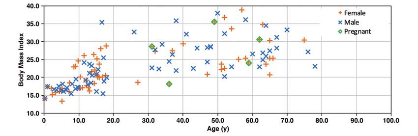
Fu et al.
rected, scanner-independent organ dose estimates for abdominal CT ex- port in a voxelized geometry using a massively parallel graphics process-
ams. Med Phys 2011; 38:820–829 ing unit. Med Phys 2009; 36:4878–4880
17. Sahbaee P, Segars WP, Samei E. Patient-based estimation of organ dose for 27. Baro J, Sempau J, Fernández-Varea J, Salvat F. PENELOPE: an algorithm for
a population of 58 adult patients across 13 protocol categories. Med Phys Monte Carlo simulation of the penetration and energy loss of electrons
2014; 41:072104 and positrons in matter. Nucl Instrum Methods Phys Res B 1995; 100:31–46
Downloaded from www.ajronline.org by DUMC Univ Med Ctr on 01/25/21 from IP address 152.3.102.254. Copyright ARRS. For personal use only; all rights reserved
18. Tian X, Li X, Segars WP, Paulson EK, Frush DP, Samei E. Pediatric chest and 28. Felmlee JP, Gray J, Leetzow M, Price J. Estimated fetal radiation dose from
abdominopelvic CT: organ dose estimation based on 42 patient models. multislice CT studies. AJR 1990; 154:185–190
Radiology 2014; 270:535–547 29. Li X, Segars WP, Samei E. The impact on CT dose of the variability in tube
19. Tian X, Segars WP, Dixon RL, Samei E. Convolution-based estimation of or- current modulation technology: a theoretical investigation. Phys Med Biol
gan dose in tube current modulated CT. Phys Med Biol 2016; 61:3935–3954 2014; 59:4525–4548
20. Segars WP, Sturgeon G, Mendonca S, Grimes J, Tsui BM. 4D XCAT phantom 30. Valentin J, ed. The 2007 recommendations of the International Commission
for multimodality imaging research. Med Phys 2010; 37:4902–4915 on Radiological Protection, ICRP publication 103. Elsevier, 2007
21. Segars WP, Norris H, Sturgeon GM, et al. The development of a population 31. Li X, Samei E, Williams CH, et al. Effects of protocol and obesity on dose
of 4D pediatric XCAT phantoms for imaging research and optimization. conversion factors in adult body CT. Med Phys 2012; 39:6550–6571
Med Phys 2015; 42:4719–4726 32. Cristy M, Eckerman K. Specific absorbed fractions of energy at various ages
22. Stabin M, Emmons MA, Segars WP, Fernald M, Brill AB. ICRP 89-based adult from internal photon sources. Oak Ridge National Laboratory, 1987: ORNL/
and pediatric phantom series. J Nucl Med 2008; 49(suppl 1):14P TM-8381
23. National Library of Medicine website. Download Visible Human Project 33. Boone JM. Reply to “comment on the ‘report of AAPM TG 204: size-specific
data. www.nlm.nih.gov/databases/download/vhp.html. Accessed January dose estimates (SSDE) in pediatric and adult body CT examinations’”
8, 2020 [AAPM Report 204, 2011]. (letter) Med Phys 2012; 39:4615–4616
24. Ding A, Gao Y, Liu H, et al. VirtualDose: a software for reporting organ doses 34. McCollough C, Bakalyar DM, Bostani M, et al. Use of water equivalent diam-
from CT for adult and pediatric patients. Phys Med Biol 2015; 60:5601–5625 eter for calculating patient size and size-specific dose estimates (SSDE) in
25. Turner AC, Zankl M, DeMarco JJ, et al. The feasibility of a scanner-indepen- CT: the report of AAPM task group 220. AAPM Rep 2014; 2014:6–23
dent technique to estimate organ dose from MDCT scans: using CTDIvol to 35. Romanyukha A, Folio L, Lamart S, Simon SL, Lee C. Body size-specific effec-
account for differences between scanners. Med Phys 2010; 37:1816–1825 tive dose conversion coefficients for CT scans. Radiat Prot Dosimetry 2016;
26. Badal A, Badano A. Accelerating Monte Carlo simulations of photon trans- 172:428–437
(Figures start on next page)
6 AJR:216, March 2021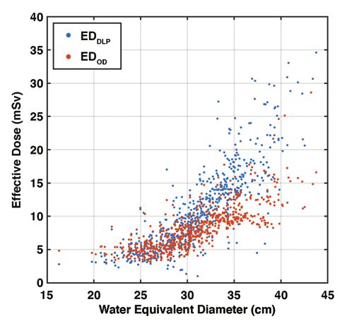
Patient-Informed Organ Dose Estimation in CT
Fig. 1—Flowchart shows clinical organ dose
Patient-CT scan estimation framework used in this study. Rectangles
indicate precomputed data; ovals represent data
generated during computation; parallelograms
represent calculation. h factors are CT dose index–
to–organ dose conversion coefficients. TCM = tube
Topogram CT images TCM profile CTDIvol current modulation, CTDIvol = volume CT dose index,
Downloaded from www.ajronline.org by DUMC Univ Med Ctr on 01/25/21 from IP address 152.3.102.254. Copyright ARRS. For personal use only; all rights reserved
WED = water equivalent diameter, DSF = dose
spread function, XCAT = extended cardiac-torso
phantom.
Detect Measure
landmarks WED
Monte Carlo— and
Anatomic size-based DSF
WED libraries
landmarks
Interpolate
size-based DSF
XCAT Monte Carlo—
phantom based
libraries h factor libraries
Patient size-
specific DFS
Interpolate
Match to a
size-based h Convolution
phantom
factors
Matched XCAT
h factors Dose ratio
phantom
Consolidate
organ geometry
and dose
Initial organ dose
Anatomy model
Exposure model
Monte Carlo—based bias and Estimate bias Accuracy calculation
uncertainty benchmarks for and
organ dose under TCM uncertainties
Final corrected
organ dose with
CI
AJR:216, March 2021 7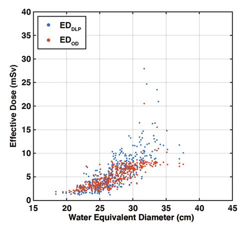
Fu et al.
Downloaded from www.ajronline.org by DUMC Univ Med Ctr on 01/25/21 from IP address 152.3.102.254. Copyright ARRS. For personal use only; all rights reserved
A
B
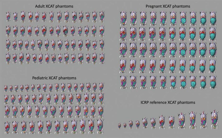
Fig. 2—Extended cardiac-torso (XCAT) phantoms used in study.
A, Chart shows frontal views of phantoms of 58 adults (age range, 18–78 years; 23 women, 35 men), 56 pediatric patients (age range, 2–18 years; 31 girls, 25 boys), five
pregnant women (gestational age range, 3–38 weeks), and 12 International Commission on Radiological Protection (ICRP) reference XCAT phantoms used in this study.
Phantom skin, head, arm, and legs were removed to enhance visualization of organs in chest-abdomen-pelvis region.
B, Graph shows body mass index and age of XCAT phantom variations within population.
8 AJR:216, March 2021Patient-Informed Organ Dose Estimation in CT
Fig. 3—Linear models of ground truth (Monte
Carlo simulation) organ dose (ODM) plotted against
estimated dose (present study) without (ODC) and
with (ODCD) bias correction for adult chest (left)
and abdomen-pelvis (right) protocols. Each dot
represents dose obtained from one phantom.
Identity line is plotted in black; ideally all points lie
Downloaded from www.ajronline.org by DUMC Univ Med Ctr on 01/25/21 from IP address 152.3.102.254. Copyright ARRS. For personal use only; all rights reserved
on this diagonal line. Dashed lines represent CIs of
linear model. Lungs (top), liver (middle), and bone
surface (bottom) were chosen to represent organ
on peripheral (inside), inside (on peripheral), or
distributed for chest (abdomen-pelvis) protocols
with respect to scan coverage.
AJR:216, March 2021 9Fu et al.
Downloaded from www.ajronline.org by DUMC Univ Med Ctr on 01/25/21 from IP address 152.3.102.254. Copyright ARRS. For personal use only; all rights reserved
Fig. 4—Maps of errors of organ dose estimation with and without bias correction, compared with ground truth (Monte Carlo) simulation averaged among phantoms
in adult, pregnant, and pediatric cohorts for each organ protocol. Error was calculated as indicated in equation at bottom of figure, where N is number of phantoms,
and ODS and ODM are organ dose estimated in this study and by Monte Carlo simulation, respectively. Asterisk represents fetus body and fetus bone tissue for pregnant
cohort. Gradient scale indicates errors of organ dose estimation (%).
10 AJR:216, March 2021Patient-Informed Organ Dose Estimation in CT
Downloaded from www.ajronline.org by DUMC Univ Med Ctr on 01/25/21 from IP address 152.3.102.254. Copyright ARRS. For personal use only; all rights reserved
A B
Fig. 5—Organ doses plotted against volume CT dose index (CTDIvol). Error bars represent 95% CI.
A, Graph shows 401 chest examinations (mean age, 61 years; range, 18–88 years) including 170 women and 231 men.
B, Graph shows 647 abdomen–pelvis examinations (mean age, 56 years; range, 18–89 years) including 354 women and 293 men.
A B
Fig. 6—Organ dose–based effective dose (EDOD) and dose-length-product–based effective dose (EDDLP) plotted against water equivalent diameter. EDDLP shows higher
values compared with EDOD for larger-sized patients and lower values for smaller-sized patients.
A, Graph shows data for 401 chest examinations (mean age, 61 years; range, 18–88 years) including 170 women and 231 men.
B, Graph shows data for 647 abdomen-pelvis examinations (mean age, 56 years; range, 18–89 years) including 354 women and 293 men.
AJR:216, March 2021 11You can also read