Sensing the world and its dangers: An evolutionary perspective in neuroimmunology - eLife
←
→
Page content transcription
If your browser does not render page correctly, please read the page content below
REVIEW ARTICLE

                                    Sensing the world and its dangers: An
                                    evolutionary perspective in
                                    neuroimmunology
                                    Aurora Kraus1, Katherine M Buckley2, Irene Salinas1*
                                    1
                                        Department of Biology, University of New Mexico, Albuquerque, United States;
                                    2
                                        Department of Biological Sciences, Auburn University, Auburn, United States

                                    Abstract Detecting danger is key to the survival and success of all species. Animal nervous and
                                    immune systems cooperate to optimize danger detection. Preceding studies have highlighted the
                                    benefits of bringing neurons into the defense game, including regulation of immune responses,
                                    wound healing, pathogen control, and survival. Here, we summarize the body of knowledge in
                                    neuroimmune communication and assert that neuronal participation in the immune response is
                                    deeply beneficial in each step of combating infection, from inception to resolution. Despite the
                                    documented tight association between the immune and nervous systems in mammals or
                                    invertebrate model organisms, interdependence of these two systems is largely unexplored across
                                    metazoans. This review brings a phylogenetic perspective of the nervous and immune systems in
                                    the context of danger detection and advocates for the use of non-model organisms to diversify the
                                    field of neuroimmunology. We identify key taxa that are ripe for investigation due to the
                                    emergence of key evolutionary innovations in their immune and nervous systems. This novel
                                    perspective will help define the primordial principles that govern neuroimmune communication
                                    across taxa.

                                    1.Introduction: animal nervous and immune systems
*For correspondence:
isalinas@unm.edu                    communicate to sense danger
                                    As multicellular organisms evolved increasing complexity, molecular mechanisms for sensing the
Competing interests: The
                                    environment arose in both the immune and nervous systems. Although the immune and nervous sys-
authors declare that no
                                    tems are traditionally studied independently, growing evidence suggests shared evolutionary
competing interests exist.
                                    requirements drove integration of both systems at the genomic, molecular, cellular, and tissue levels.
Funding: See page 20                In mammals, the immune and nervous systems deploy coordinated mechanisms for pathogen recog-
Received: 25 January 2021           nition and control. Notably, reciprocal regulation occurs at several layers: neuronal signals fine-tune
Accepted: 09 April 2021             the strength of immune responses, whereas mechanisms traditionally associated with immunity
Published: 26 April 2021            mediate neural growth, development, and function (Aurora and Olson, 2014; Cardoso et al., 2017;
                                    Chu et al., 2020; Foster et al., 2017; Gonzalez-Figueroa et al., 2021; Pinho-Ribeiro et al., 2017;
Reviewing editor: Carla V
Rothlin, Yale School of Medicine,
                                    Ramirez et al., 2020; Zigmond and Echevarria, 2019). These bidirectional, complex interactions
United States                       are the product of millennia of coevolution and lie at the heart of many physiological processes
                                    (Chesné et al., 2019; Huh and Veiga-Fernandes, 2020; Jain et al., 2020). Throughout metazoan
   Copyright Kraus et al. This
                                    evolution, the immune and nervous systems have developed into complementary processes to
article is distributed under the
                                    achieve an imperative function: sense the environment and detect danger. As a result, neuroscient-
terms of the Creative Commons
Attribution License, which          ists and immunologists continue to uncover molecules with dual functions in both physiological sys-
permits unrestricted use and        tems (Table 1 and reviewed by Kolosowska et al., 2019; Minnone et al., 2017; Ni and Gilbert,
redistribution provided that the    2017; Stevens et al., 2007; Yang et al., 2016).
original author and source are          Neuroimmune cell units (NICUs) have been described in mammals as discrete anatomical loca-
credited.                           tions where immune and neuronal cells physically interact and regulate tissue physiology and

Kraus et al. eLife 2021;10:e66706. DOI: https://doi.org/10.7554/eLife.66706                                                         1 of 33
Review Article                                                                        Immunology and Inflammation Neuroscience

                                    defense (Godinho-Silva et al., 2019). Mammalian neuroimmune interactions are mediated by solu-
                                    ble factors such as neurotransmitters, neuropeptides, and cytokines (Table 1 and reviewed in
                                    Trakhtenberg and Goldberg, 2011). Neurons innervate lymphoid organs, thereby influencing
                                    immune cell migration and development in mammals (Huang et al., 2021). In turn, specialized
                                    immune cells are present throughout nervous tissues where they influence regenerative capabilities
                                    as well as regulate pain in response to noxious stimuli (Bloom, 2014; Jung et al., 2017; Pinho-
                                    Ribeiro et al., 2017; Huang et al., 2021). Specifically, in mammals, nociceptive neurons in the lung,
                                    gut, and skin detect bacteria and release neural mediators that bias the type and magnitude of the
                                    immune response toward neuroprotection from infection (Baral et al., 2018; Chiu et al., 2013;
                                    Gabanyi et al., 2016; Matheis et al., 2020). Despite the growing appreciation for NICUs in mam-
                                    mals, NICUs are largely unexplored across metazoans, although the soluble molecules and their
                                    homologs that mediate communication within NICUs are conserved across animals (Jékely, 2013;
                                    Liongue et al., 2016; Mittal et al., 2017; Roch and Sherwood, 2014).
                                       Preliminary work in invertebrate model organisms suggests that cooperation between the ner-
                                    vous and immune systems is a central aspect of animal life that extends beyond mammals. For exam-
                                    ple, C. elegans employ sensory neurons to mediate avoidance behavior in response to pathogenic
                                    bacteria and to suppress deleterious innate immune responses (Cao and Aballay, 2016;
                                    Hoffman and Aballay, 2019). Several branches of the innate immune system are missing in C. ele-
                                    gans and identification of pattern recognition receptors (PRRs) in nematodes is still elusive. Neurons
                                    and neurotypical receptors such as G-protein-coupled receptors (GPCRs) appear to compensate for
                                    this absence and govern innate immunity in C. elegans (Irazoqui et al., 2010; Pujol et al., 2001;
                                    Wani et al., 2020; Venkatesh and Singh, 2021). Another example may be Hydra, another basal
                                    metazoan with a simple body plan where the TLR signaling pathway appears to have degenerated
                                    and neuronally secreted antimicrobial neuropeptides sense and control microorganisms
                                    (Augustin et al., 2017; Table 1). Thus, while morphologically simple animals like C. elegans and
                                    Hydra may not have discrete anatomical associations of immune effector cells and neurons such as
                                    mammalian NICUs, their neurons directly regulate tissue physiology and immunity. Of note, both C.
                                    elegans and Hydra neurons have been mapped in great anatomical detail, and therefore, neuroim-
                                    mune interactions at these sites may be straight forward to identify. In Drosophila, sensory neurons
                                    contact hemocytes (a diverse population of macrophage-like cells) in hematopoietic pockets and
                                    regulate proliferation, survival, and localization (Cattenoz et al., 2020; Makhijani et al., 2017) per-
                                    haps representing small, simple NICUs. Thus, although this field remains largely unexplored, we pro-
                                    pose that NICUs are a fundamental aspect of animal physiology and are present in specialized
                                    configurations with several degrees of complexity across all metazoans. Given that adaptive immune
                                    responses emerged relatively recently (~500 million years ago [mya]), ancient NICUs must have
                                    evolved exclusively from the interactions between innate immune cells and neurons or neuroimmune
                                    cells (Cooper and Alder, 2006). Despite these hints from non-mammalian model species, the neuro-
                                    immunology field still lacks a broader phylogenetic perspective.
                                       Given the functional similarities, frequent crosstalk, and evolutionary overlap between the nervous
                                    and immune systems, this review highlights how the tight evolution of immune cells and neurons in a
                                    pathogen-laden environment led to neuroimmune cooperation early in metazoan evolution.

                                    2.Sensing danger: what came first, immune cells or neurons?
                                    Immune and neural responses are evident even in single-celled organisms, which must constantly
                                    sense and respond to the environment. The single-celled protozoan Stentor coeruleus exhibits
                                    behaviors to environmental cues that are both predictable and can be learned by habituation
                                    (Dexter et al., 2019; Tang and Marshall, 2018; Wood, 1970). Furthermore, S. coeruleus demon-
                                    strates immune capabilities such as wound healing and curation of a microbiome that is separate
                                    from the environment (Blauch et al., 2017; Lanzoni et al., 2019). The importance of bacterial sym-
                                    bionts is highlighted by choanoflagellates, which require bacterially derived lipids to create multicel-
                                    lular colonies (Woznica et al., 2016). The origin of multicellularity allowed for the evolution of
                                    specialized cell types. As multicellular animals became more complex, the need arose to transmit
                                    information among cell types and tissues, ultimately driving evolution of the complex immune and

Kraus et al. eLife 2021;10:e66706. DOI: https://doi.org/10.7554/eLife.66706                                                          2 of 33
Review Article Immunology and Inflammation Neuroscience
Table 1. Molecules with dual roles in the immune and nervous systems.
Factors classically associated with immune functions
Protein Immune system properties Nervous system properties References
Antimicrobial peptides (AMPs) . Secreted by epithelial and . Antimicrobial in nervous Hanson et al., 2019; Lezi et al., 2018;
Su et al., 2010; Zasloff, 2002
phagocytic cells system niches
. Disrupt microbial mem- . Control chemotaxis of
branes leading to destruc- immune cells and astroglia
tion of pathogen . Mediate iron homeostasis
. Modulate nerve impulses
. Implicated in aging and
neurodegeneration
Cytokines TGF-b . Produced by all leukocytes . Produced by neurons Arnold et al., 2014; Arrieta-
. Regulates hemocyte . Controls feeding behavior Bolaños et al., 2012; Eisenstein and
Williams, 2009; Hirota et al., 2015;
proliferation . Angio-suppressive roles in Makhijani et al., 2017; Morishima et al.,
. Generally anti-inflammatory the brain 2009; Singh and Aballay, 2019;
. Inhibits B cell proliferation . Regulates neuronal devel- Yi et al., 2010; You et al., 2008;
Influences development of Zheng et al., 2006
. opment and axon
Tregs and TH17 cells outgrowth
IL-4, IL-13 . Induce TH2 antiparasitic . Regulate spatial learning Fallon et al., 2002; Gadani et al., 2012;
Kolosowska et al., 2019;
immunity, tissue repair, and neurogenesis McKenzie et al., 1998; Yang et al.,
allergic responses . Bias astrocytes and micro- 2016; Zhang et al., 2019a
glia toward M2/neuropro-
tective states
. Mediate oligodendrocyte
growth and re-myelination
TNF-a . Pro-inflammatory functions . Expressed in neurons after Borsini et al., 2015; Lambertsen et al.,
2009; Liu et al., 1994; Takei and
damage for acute Laskey, 2008; Vanderheyden et al.,
protection 2018
. Long-term presence in the
CNS is associated with
decreased proliferation
and neurogenesis
. Alters permeability of the
blood-brain barrier
. Induces changes in sleep
behavior
Complement Complement . Opsonize pathogens for . Anti-inflammatory roles Hammad et al., 2018; Nonaka, 2001;
System factors Rupprecht et al., 2007; Shinjyo et al.,
Proteins
activation of innate and during CNS infection 2009; Stevens et al., 2007
adaptive immune cells . Regulate synaptic pruning
of microglia expressing C3
receptor
. Regulate adult neurogene-
sis by causing increased
maturation and migration
of progenitors in SVZ and
dentate gyrus
Perforin-like . Pore-forming proteins . ASTNs and BRINPs are Ni and Gilbert, 2017
factors
released by cytotoxic expressed in CNS and
leukocytes associated with
. Form the membrane attack neurodevelopment
complex
Table 1 continued on next page
Kraus et al. eLife 2021;10:e66706. DOI: https://doi.org/10.7554/eLife.66706 3 of 33Review Article Immunology and Inflammation Neuroscience
Table 1 continued
Factors classically associated with immune functions
Protein Immune system properties Nervous system properties References
Pattern Toll-like . Detect extra- and intracel- . Regulate neuronal devel- Chen et al., 2019c; Donnelly et al.,
Recognition receptors 2020; Foldi et al., 2017;
Receptors (TLRs)
lular pathogen and danger opment, dendrite/axon Franzenburg et al., 2012;
associated molecular growth and synapse Lemaitre et al., 1996
patterns. formation
. Recognize neurotrophins
. Sensitize nociceptive
neurons
Nod-like . Detect intra-cellular patho- . Immunomodulate glial Gharagozloo et al., 2017
receptors
(NLRs)
gen and danger associated cells
molecular patterns. . Prevents necrosis of
neurons
Peptidoglycan . Detects peptidoglycan . Controls presynaptic Harris et al., 2015
recognition
protein LC
homeostasis
(PGRP-LC)
Formyl peptide . Expressed on macrophages . Vomeronasal sensory neu- Dietschi et al., 2017
receptors Detect pathogens and
(FPRs)
. rons receptors
induce inflammation
Histamine . Released by mast cells . Modulates neurogenic Yuan and Silberstein, 2018
. Mediates vasodilation and inflammation and nocicep-
itch tive inflammation and has
been implicated in
migraines
. Induces NGF expression
by peripheral nociceptors
Factors classically associated with neuronal functions
Transient receptor potential . Expressed by lymphocytes, . Expressed by distinct sub- Alpizar et al., 2017; López-
(TRPs) Requena et al., 2017; Parenti et al.,
dendritic cells, neutrophils, sets of sensory cells 2016
monocytes, macrophages, . Mediate neuronal depolar-
and mast cells. ization and release of
. Cause changes in intracel- CGRP
lular Ca2+, which influences
cell migration, cytokine
production, phagocytosis
and proliferation
Nerve growth factor (NGF) . Released by mast cells, B . Stimulates growth, survival, Minnone et al., 2017; Pinho-
Ribeiro et al., 2017; Takei and Laskey,
lymphocytes. and differentiation of 2008
. Increases during neurons
inflammation
. Receptor (TrkA) is
expressed throughout
immune system
. Transduced NGF signal is
anti-inflammatory
Brain-derived neurotrophic . Implicated in lymphocyte . Regulates neuron growth, Fauchais et al., 2008; Lee et al., 2012;
factor (BDNF) Linker et al., 2015; Schuhmann et al.,
development and survival survival and synapse 2005
modulation
Table 1 continued on next page
Kraus et al. eLife 2021;10:e66706. DOI: https://doi.org/10.7554/eLife.66706 4 of 33Review Article Immunology and Inflammation Neuroscience
Table 1 continued
Factors classically associated with immune functions
Protein Immune system properties Nervous system properties References
Olfactory receptors . Activate pulmonary macro- . Detect chemical odorants Heimroth et al., 2020; Li et al., 2013
phage motility and CCL2
expression
. Highly expressed in sec-
ondary lymphoid organs
Calcitonin gene related . An anti-inflammatory cyto- . Mediates pain transduc- Chung, 2017; Kerage et al., 2019;
peptide (CGRP) Pinho-Ribeiro et al., 2017; Xu et al.,
kine that promotes type 2 tion by nociceptors 2019
immunity . Regulates regeneration of
. Decreases antigen presen- peripheral neurons
tation by MHC II, and
inflammatory cytokine
expression
. Increases expression of the
anti-inflammatory cytokine
IL-10
. Potent vasodilator
. Released by T and B
lymphocytes
Substance P . Secreted by microglia, T . Neuropeptide involved in Mashaghi et al., 2016
cells, macrophages, den- nociception and neuroin-
dritic cells and eosinophils flammation as well as
. Affects cytokine expression hypotension and muscle
by binding to neurokinin contraction
receptor
Dopamine . Lymphocytes and myeloid . Neurotransmitter Kerage et al., 2019; Matt and Gaskill,
2020
cells express the dopamine
receptor
. Enhances lymphocyte che-
motaxis and maturation
. Produced by dendritic cells
DSCAMs . Acts as a pattern recogni- . Regulates axon/dendrite Goyal et al., 2019; Hattori et al., 2009;
tion receptor that mediates segregation during neuro- Ng and Kurtz, 2020
phagocytosis in arthropods nal development
NCAM/CD56 . Present on NK cells, acti- . Neuronal cell migration Van Acker et al., 2017; Vukojevic et al.,
2020
vated T cells and other and synaptic plasticity
cytotoxic cell subsets
SNARE . Exocytosis of perforins, . Exocytosis of Ramakrishnan et al., 2012; Tang, 2015
granzymes, and cytokines neurotransmitters
nervous systems present in modern-day mammals. But which appeared first in eukaryotes: immune
cells or neurons? Here we propose that a cell with antimicrobial or phagocytic capabilities, whether
it was an immune cell or a sensory neuron, predates the emergence of the first in sensu stricto neu-
ronal cell type.
We propose two hypothetical scenarios in which evolutionary pressure from microbes coerced
ancient, conserved molecules like PRRs, soluble factors (i.e., cytokines, chemokines, and neuropepti-
des), and ion channels, to coordinate communication between neurons and immune cells forming
NICUs over 700 mya (Figure 1). This is, in part, based on observations in MyD88-deficient Hydra,
demonstraiting that TLR signaling is required for pathogen defense and microbiota colonization and
Kraus et al. eLife 2021;10:e66706. DOI: https://doi.org/10.7554/eLife.66706 5 of 33Review Article Immunology and Inflammation Neuroscience Figure 1. Hypothetical pathways that lead to the emergence of NICUs in early metazoans. (A) An ancient metazoan cell under microbial pressure acquires ion channels gaining the ability to detect danger (Elkhatib et al., 2019; Senatore et al., 2016). Eventually neuropeptides that have antimicrobial functions are released in response to microbes (Augustin et al., 2017). Innate immune cells gain neuropeptide receptors that trigger cytokine release (Pinho-Ribeiro et al., 2017). Finally, neuronal cells gain the ability to detect and respond to immune signaling cytokines Figure 1 continued on next page Kraus et al. eLife 2021;10:e66706. DOI: https://doi.org/10.7554/eLife.66706 6 of 33
Review Article                                                                               Immunology and Inflammation Neuroscience

Figure 1 continued
(Chatterjea and Martinov, 2015; Chu et al., 2020). (B) An ancient metazoan cell acquires pattern recognition receptors and gains the ability to detect
microbes (Boller and Felix, 2009; Rosenstiel et al., 2009). Eventually soluble immune mediators are released in response that trigger antimicrobial
functions (Hanson et al., 2019). Neurons gain immune ligand receptors that trigger neuropeptide release (Lezi et al., 2018). Finally, immune cells gain
the ability to detect and respond to neuronally derived neuropeptides (Chu et al., 2020; Foster et al., 2017). (C) An ancient metazoan cell under
microbial pressure acquires pathogen pattern recognition receptor pathways concurrently with acquisition of ion channels that, under selective pressure
gain the ability to detect danger (Tian et al., 2019). Eventually soluble neuroimmune mediators are secreted in response to pathogens modulate
functions of neuronal and immune systems.

                                    by the discovery of voltage-gated calcium channels in early metazoans known to regulate immune
                                    responses to pathogens (Franzenburg et al., 2012; Gupta et al., 2009; Rosenstiel et al., 2009;
                                    Senatore et al., 2016). In the first scenario (Figure 1A), an ion channel gains the ability to sense
                                    microorganisms, ultimately changing the membrane potential and signaling the cell to release neuro-
                                    peptides. This phenomenon is highlighted in placozoa where Na+ channels sense changes to the
                                    microenvironment related to microbes, such as pH (Brunet and Arendt, 2016; Elkhatib et al.,
                                    2019; Moran et al., 2015). Over evolutionary time, neuropeptides acquire antimicrobial functions
                                    that affect pathogens usually encountered by the cell as revealed in Hydra studies (Augustin et al.,
                                    2017). Surrounding cells with immune functions gained expression of neuropeptide receptors, tun-
                                    ing into danger signals from ion channel expressing proto-sensory neuronal cells and releasing cyto-
                                    kines (Satake et al., 2019). In turn, the proto-sensory neuron eventually may gain expression of
                                    cytokine receptors to monitor signals from immune cells linking immune ligand receptor binding on
                                    neuronal cells’ surface to neuropeptide secretion as reported in studies into pain and itch
                                    (Chatterjea and Martinov, 2015; Cunha et al., 2008; Oetjen et al., 2017; Pinho-Ribeiro et al.,
                                    2017; Salvador et al., 2021). Together, neuropeptides and cytokines synchronize antimicrobial
                                    responses of neuronal and immune cells, forming a simple NICU.
                                        In an alternate, inverse scenario (Figure 1B), an ancestral metazoan cell expresses PRRs and links
                                    pathogen associated molecular patterns (PAMPs) to secretion of soluble immune factors such as
                                    cytokines and antimicrobial peptides (AMPs). This chain of events is well-studied in PRRs, which are
                                    present in plants as well as animals (Boller and Felix, 2009; Rosenstiel et al., 2009). We propose
                                    that in a microbe-rich environment, the proto-immune cell releases soluble mediators to defend the
                                    host against pathogens and/or sense microbiota. Interestingly, AMPs, which potently destroy bacte-
                                    ria, are lost in the genomes of some fly lineages that live in sterile environments accentuating the
                                    importance of microbial pressure in immune repertoire evolution (Choi et al., 2015; Hanson et al.,
                                    2019). Notably, AMPs have been shown to regulate sleep and memory (Barajas-Azpeleta et al.,
                                    2018; Sinner et al., 2021; Table 1). As explained above, neuronal cells may simultaneously detect
                                    danger using prototypical immune receptors to signal the secretion of neurotransmitters which sub-
                                    sequently evolve to reciprocally modulate immune cell function (Cunha et al., 2008; Oetjen et al.,
                                    2017; Pinho-Ribeiro et al., 2017).
                                        These two scenarios are likely not mutually exclusive and may have occurred concurrently in early
                                    metazoans (Figure 1C). Both PRRs and ion channels are evolutionarily ancient (Rosenstiel et al.,
                                    2009; Senatore et al., 2016). Even studies into plants show cooperation of calcium channels and
                                    PRRs to regulate innate immunity (Jogawat et al., 2020; Tian et al., 2019). Furthermore, soluble
                                    mediators that have been traditionally classified as either neuronal or immune factors are being
                                    found to serve both systems (Table 1). We may never glean conclusive evidence as to whether PRRs
                                    or ion channels evolved and impelled mutually beneficial communication between early neuronal
                                    and immune cells. We postulate on the sequence of events using known interactions from lower
                                    metazoans and resulting NICUs from mammals to identify gaps in our current understanding of neu-
                                    roimmune communication. This novel phylogenetic perspective will help define the elemental princi-
                                    ples that govern neuroimmune communication across taxa.
                                        The dual roles of ion channels in the nervous and immune systems may also reflect ancient evolu-
                                    tionary pressures (Table 1 and Figure 2). In the calcium-rich ‘primordial soup’, injury to a single-
                                    celled eukaryote’s plasma membrane would result in rapid depolarization via environmental calcium
                                    influx. Signaling via this swift change in ion concentration links cellular repair as well as endo- and
                                    exocytosis to action potentials (Brunet and Arendt, 2016). In multicellular organisms, calcium influ-
                                    xes and depolarization also occur, but result in signals that affect both local and distant cells. Thus,

Kraus et al. eLife 2021;10:e66706. DOI: https://doi.org/10.7554/eLife.66706                                                                     7 of 33
Review Article Immunology and Inflammation Neuroscience
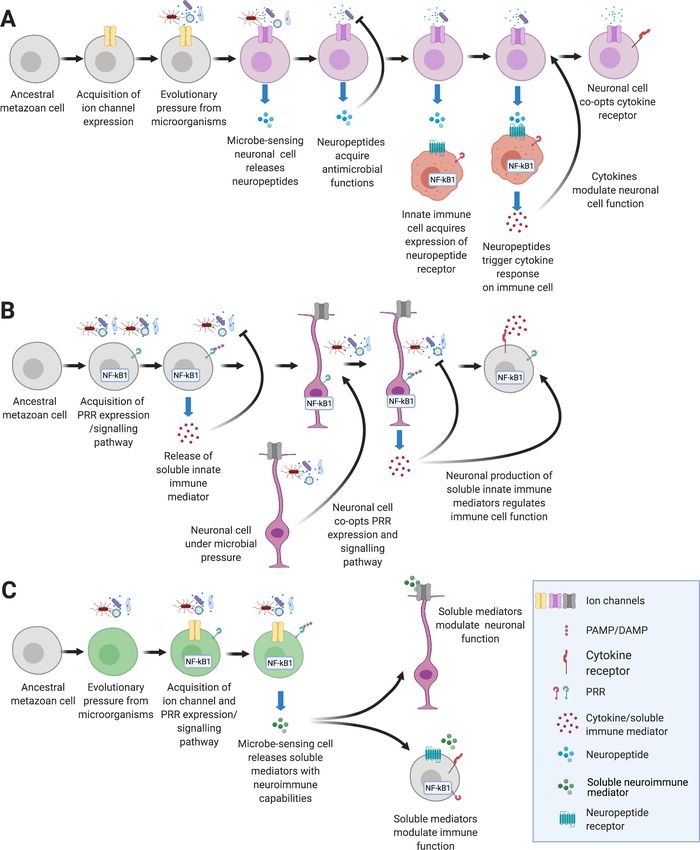
Figure 2. Emergence of the immune and nervous systems predates the emergence of Metazoa. Analysis of
genome sequences indicates that much of the cellular machinery involved in extant nervous and immune systems
was present in basal metazoan lineages. This includes ion channels, pattern recognition receptors, and
antimicrobial peptides (Franzenburg et al., 2012; Rosenstiel et al., 2009; Senatore et al., 2016). The
evolutionary origin of significant innovations is indicated by dots (immune system in orange; nervous system in
purple). Centralization of nervous systems within bilaterian phyla is indicated by small brain icons. Note CNS
structures may be very diverse among invertebrate groups. Asterisk indicates possible independent, convergent
evolution of Ctenophore neurons, a current point of debate (Moroz et al., 2014).
the role of membrane depolarization in cell defense may have predated the need to communicate
across an organism.
During infection, not only are microbes present, but the extracellular microenvironment is also
chemically altered (e.g., oxygen levels, pH, osmotic pressure) (Alam et al., 2016). Given the concur-
rence between microbes and chemical factors, it is not surprising that proteins involved in sensing
danger evolved to regulate both neural and immune processes (Elkhatib et al., 2019). One example
is the transient receptor potential (TRP) channels, which are conserved across Metazoa (Table 1 and
Figure 2). TRPs respond to a myriad of environmental cues including chemical, mechanical, and ther-
mal stimuli and are expressed in both neurons and immunocytes (López-Requena et al., 2017;
Lynagh et al., 2018; Nilius and Owsianik, 2011; Parenti et al., 2016). TRPs have been linked to
Kraus et al. eLife 2021;10:e66706. DOI: https://doi.org/10.7554/eLife.66706 8 of 33Review Article Immunology and Inflammation Neuroscience
avoidance behavior in C. elegans, in which TRPV proteins monitor oxygen levels to avoid hyperoxia
and specific pathogens (Chang et al., 2006; Singh and Aballay, 2019). Similarly, in planaria, TRPA1
directly mediates extraocular light avoidance behavior (Birkholz and Beane, 2017). Finally, TRPs
mediate inflammatory responses by controlling neuronal release of neuropeptides, such as calcitonin
gene-related peptide (CGRP), which subsequently regulates immune response and cytokine secre-
tion (Khalil et al., 2018; Marics et al., 2017; Nagashima et al., 2019; Yaraee et al., 2003;
Zhang et al., 2018; Table 1). The reciprocal relationship between neuronal and immune proteins
will be explored here in greater depth in Section 8. Furthermore, the use of immune and neuronal
building blocks by basal metazoans prompted us to question the origins and evolutionary history of
NICUs, as discussed in the next section.
3.Basal metazoans and the origins of NICUs
The field of neuroimmunology has largely been dominated by a biomedical perspective fueled by
the powerful therapeutic interventions that can stem from understanding neuroimmune communica-
tion in health and disease. However, the intricacies of neuroimmune communication can be best dis-
entangled when we investigate organisms with reduced body plan complexities and limited
numbers of cell types. NICUs have been described in detail in mammals (see review by Godinho-
Silva et al., 2019) but remain uncharacterized in most Metazoans.
The ability to respond to bacterial cues and maintain a stable microbiome is ancient within holo-
zoans, including the single-celled choanoflagellate (Salpingoeca rosetta) and porifera (Aplysina aero-
phoba) (Pita et al., 2018b; Pita et al., 2018a). Genome surveys have uncovered molecular
signatures of both vertebrate immune and nervous systems in the earliest single-celled eukaryotes,
including ion channels and post-synaptic scaffolding proteins (Csaba, 2016; Hoffmeyer and Bur-
khardt, 2016; Vig and Kinet, 2009). The presence and absence of genes encoding these proteins
can inform our evolutionary understanding of cells exhibiting immune and neural functions
(Fernández and Gabaldón, 2020). Although the relationships between the four major Metazoan lin-
eages that predate Bilateria – Porifera (sponges), Ctenophora (comb jellies), Placozoa and Cnidaria
(corals and sea anemones) – remain under debate, identifying cells with immune and nervous func-
tions in these lineages should reveal fundamental aspects of neuroimmune communication
(Nielsen, 2019).
Both poriferans and placozoans lack discrete neural structures, whereas cnidarians rely on nerve
nets consisting of neurons that resemble those found in bilaterians (Figure 2). In ctenophores, cells
with neural morphology form diffuse networks, although the absence of typical metazoan neuro-
transmitters and synaptic proteins suggests that this nervous system arose via convergent evolution
(Moroz, 2015; Moroz et al., 2014). Interestingly, ctenophores possess specialized prey capture cells
known as colloblasts, which appear to share an evolutionary progenitor with metazoan neurons
(Babonis et al., 2018).
Organisms within Porifera, the basal metazoan phylum, lack true neuronal cell types. As larvae,
these animals express photoreactive cryptochromes proposed to mediate phototactic motile behav-
ior and produce post-synaptic machinery (Rivera et al., 2012; Sakarya et al., 2007). Despite their
morphological simplicity, the poriferan immune system relies on much of the molecular machinery
that is present in the vertebrate innate immunity, including Toll-like receptors (TLRs) and nod-like
receptors (NLRs) (Rosenstiel et al., 2009). Notably, a recent transcriptomic study in the Mediterra-
nean sponge (Aplysina aerophoba) identified GPCRs among the most differentially regulated tran-
scripts in response to challenge with PAMPs (Pita et al., 2018a) opening up questions as to how
neuroimmune responses occur in the absence of bona fide neurons in this taxon.
Placozoans, primitive metazoans that emerged in the Precambrian period, consist of only six
somatic cell types and lack organs, neurons, muscle cells, immune cells, or extracellular matrix
(Laumer et al., 2018; Smith et al., 2014; Srivastava et al., 2008). Although canonical TLRs and
NLRs are absent from placozoan genomes, much of the downstream signaling pathways are present
(Kamm et al., 2019). Additionally, the Trichoplax sp. genome encodes a diverse array of scavenger
receptors (Kamm et al., 2019). Intracellular defenses in this lineage include an expansion in the
genes encoding apoptosis inducing factor (Apaf-1), suggesting that apoptotic cell death may be an
important component of immune response (Kamm et al., 2019). Despite the absence of neurons,
the placozoan genome contains many neuronal genes including SNAP25, SNARE, and 85 GPCRs
Kraus et al. eLife 2021;10:e66706. DOI: https://doi.org/10.7554/eLife.66706 9 of 33Review Article Immunology and Inflammation Neuroscience
(Srivastava et al., 2008). Interestingly, GPCRs are evolutionarily ancient, transmembrane proteins
that transduce neuropeptide signaling (Jékely, 2013; Schöneberg et al., 2007; Strotmann et al.,
2011). Placozoa have been shown to modulate ciliary movement and folding behaviors in response
to neuropeptides in their gland cells (Alzugaray et al., 2019; Varoqueaux et al., 2018). Whether
placozoans release neuropeptides in response to pathogens is unknown but this kind of experiment
may reveal unique neuroimmune pathways and perhaps unveil the most ancient NICUs in the animal
kingdom. The diversity of nervous systems in basal metazoans reflects 600 million years of ongoing
evolution, and also highlights the evolutionary flexibility of these systems (Yin et al., 2015).
Although cnidarians lack mesoderm, and therefore motile immune cells, searches for innate
immune genes reveal that while both most cnidarians have canonical a TLR signaling pathway and
complement-effector pathway, these pathways are degenerated in the model cnidarian Hydra
(Miller et al., 2007; Brennan et al., 2017). Neuroimmune cooperation has nevertheless mostly been
described in Hydra. The Hydra nerve net differentially secretes neuropeptides across the animal’s
body that exhibit specific antimicrobial properties to regulate the resident community of symbiotic
microbes (Augustin et al., 2017; Klimovich and Bosch, 2018). Using single-cell RNA-
Sequencing (sc-RNA-Seq), pacemaker neurons were recently discovered in the head region of the
hydra with conserved function and protein machinery to mammalian intestinal cells of Cajal
(Klimovich et al., 2020). Interestingly, these neurons express the PRR pathway and many AMPs
whose expression, along with pacemaker marker proteins, and contractile function are abrogated
when raised under germ-free conditions (Klimovich et al., 2020; Murillo-Rincon et al., 2017). While
the precise role of these neuropeptides in regulating the Hydra microbiome and immune system
remains unknown, it is clear that, as in mammals, the microbiome is imperative for proper neuronal
and immune function in Hydra. Neuroimmune cooperation in corals and anemones, however,
remains underexplored.
Taken together, the current body of work highlights our view that understanding neuroimmune
communication in basal metazoans is limited to a handful of studies in a small number of taxa and,
therefore, gathering new experimental evidence in basal metazoans is key to unveiling new primor-
dial mechanisms by which neurons and immune cells detect and fight danger. Genome editing, sin-
gle cell, and spatial transcriptomics can now illuminate responses to danger in non-model organisms
at unprecedented high-resolution levels. Next, we will take a deep look into the tunicates, a re-
emerging group of model organisms, and indicate exciting avenues in NICU research.
4.Unique NICUs in tunicates may govern regeneration and
colonialism
Tunicates, also known as sea squirts, are benthic marine invertebrates that emerged 550 mya (Lem-
aire, 2011). Tunicates are the closest relatives to vertebrates (Delsuc et al., 2006). There are
approximately 3000 species of tunicates including several model organisms such as Ciona intestina-
lis, Halocynthia roretzi, and Botryllus schlosseri (Holland, 2016).
Ascidians, which comprise the largest class of tunicates, have complex developmental life cycles
in which embryogenesis results in a chordate tadpole that, after hatching, metamorphoses into a
sessile form known as an oozooid that asexually buds to create a colony of adult zooids (Hol-
land, 2016; Kowarsky et al., 2021; Manni et al., 2007). The zooid has a complex body plan with
central and peripheral nervous systems, and a complex hematopoietic system with multiple immune
cell types (Braun and Stach, 2019; Burighel et al., 2005; Franchi and Ballarin, 2017; Mackie and
Burighel, 2005; Rosental et al., 2018). Detailed descriptions of the peripheral nervous system of
Botryllus reveal that each zooid of the colony possesses an individual nerve plexus that is discontinu-
ous with that of adjacent zooids (Burighel et al., 2005). Furthermore, the dense innervation of the
Botryllus gut may represent an important site for NICUs; as filter feeders, tunicate guts are regularly
exposed to the microbe-rich seawater environment (Utermann et al., 2020). Availability of sterile,
or germ-free, tunicate models offers exciting opportunities to interrogate neuroimmune interactions
in the context of microbiota recognition and regulation (Leigh et al., 2016).
Colonial tunicates have been long-studied as a model for allorecognition (De Tomaso, 2009;
Oka and Watanabe, 1967; Oka and Watanabe, 1960; Oka and Watanabe, 1957; Sabbadin, 1962;
Scofield et al., 1982). Colony fusion follows similar principles to vertebrate histocompatibility and
Kraus et al. eLife 2021;10:e66706. DOI: https://doi.org/10.7554/eLife.66706 10 of 33Review Article Immunology and Inflammation Neuroscience
only occurs when both colonies share one or more alleles of the polymorphic Botryllus Histocompati-
bility Factor (BHF) gene (De Tomaso, 2009; De Tomaso et al., 2005; Rinkevich et al., 2012;
Taketa and De Tomaso, 2015; Voskoboynik et al., 2013). When tunicate colonies with different
BHF alleles come into contact, a blood-based inflammatory reaction occurs (Taketa and De Tomaso,
2015). At the point of rejection, cytotoxic blood cells known as morula cells secrete factors that lead
to reactive oxidative damage causing cell apoptosis and cytotoxic lesions (Ballarin et al., 2002;
Ballarin et al., 1995; Menin et al., 2005; Taketa and De Tomaso, 2015). Cytotoxic natural killer
(NK) cells are important innate immune cells involved in allorecognition. A subpopulation of Botryllus
blood cells express CD94-related transmembrane receptor protein, which is a specific cell surface
marker of vertebrate NK cells (Khalturin et al., 2003). Furthermore, cytotoxicity of morula cells is
mediated by BHF self-recognition (Rosental et al., 2018). Even after fusion, chimeric colonies can
undergo rejection in a process known as allogenic resorption which involves both phagocytic and
cytotoxic pathways (Corey et al., 2016). Notably, to facilitate allorecognition reactions, cytotoxic
morula cells congregate at the tips of ampullae and exist in the tunic matrix where cilia of primary
sensory neurons also project (Mackie and Burighel, 2005; Menin et al., 2005). Although presence
of neurons or neuronal derived molecules in the point of allogenic rejection or resorption in tunicates
has not been directly observed, it is plausible that they play a role in regulation of cytotoxic events
against infection or in homeostasis (Burighel et al., 2001; De Santo and Dudley, 1969; Mackie and
Burighel, 2005; Torrence and Cloney, 1981; Zaniolo et al., 2002). Many avenues of study into
NICUs in tunicates are open for investigation.
Perhaps the best-characterized tunicate model organism is the solidary ascidian, Ciona intestina-
lis, which is able to regenerate its neural complex (NC) and syphon (Jeffery, 2015; Mingaz-
zini, 1891). Following ablation of the NC, the first step in regeneration is wound healing
(Jeffery, 2015). In the course of this process, pluripotent cells that circulate in the vasculature rap-
idly invade the wound site and, along with other progenitor cells, give rise to the regenerating brain
(Bollner, 1989; Bollner et al., 1992; Da Silva et al., 2015; Lender and Bouchard-Madrelle, 1964).
Interestingly, in crayfish, adult-born neurons are derived from hemocytes (Benton et al., 2014).
Thus, a clear connection exists between immune cells and neurons in the context of invertebrate
regeneration that deserves further investigation.
An in-depth, high-resolution developmental and cellular atlas of the immune and nervous system
of model tunicates has recently become available (Kowarsky et al., 2021), which should further facil-
itate investigation of neuroimmune interactions in tunicates. In summary, tunicates harness tremen-
dous biodiversity, unique lifestyles, and regenerative capacities that make them an ideal candidate
taxon for the discovery of novel NICUs especially as the closest extant relative to vertebrates with
central nervous systems (Holland, 2016).
5.Centralization of the nervous system in invertebrates:
crosstalk with the immune system
The evolutionary transition from diffuse to centralized nervous systems in Bilateralia is still an open
question in neuroscience. The function of the central nervous system (CNS) is to integrate peripheral
environmental inputs and orchestrate whole-body downstream behaviors. Although centralization of
the nervous system is often considered to be a vertebrate innovation, invertebrates such as echino-
derms, arthropods, nematodes, molluscs, and annelids have a CNS with various shapes and degrees
of complexity in different animal phyla (Arendt et al., 2008; Figure 2). Whether the vertebrate and
invertebrate CNS trace back to a common ancestor or evolved independently is still unknown. Con-
servation of brain patterning mechanisms suggests the mechanisms for centralization of nervous sys-
tems may be as ancient as the last common bilaterian ancestor (Denes et al., 2007;
Hartenstein and Stollewerk, 2015; Telford, 2007). Interestingly, current evidence suggests that
glial cells, also present in invertebrates, have arisen multiple times independently (Hartenstein and
Giangrande, 2018). Despite the morphological disparities between the cnidarian nerve nets, the
ventral central ganglia in arthropods and the vertebrate brain, the underlying function of nervous
systems mirrors that of immune systems: sensing the environment and responding appropriately to
maximize survival.
Kraus et al. eLife 2021;10:e66706. DOI: https://doi.org/10.7554/eLife.66706 11 of 33Review Article Immunology and Inflammation Neuroscience
Findings from protostomes suggest that connections between CNS and immune functions are
widespread among Metazoa. Drosophila neurogenesis has been studied in great detail and exhibits
many similarities to mammalian models (Buescher et al., 2002; Grens et al., 1995; Hartenstein and
Stollewerk, 2015; Heitzler et al., 1996). For example, the cell adhesion molecule dscam is involved
in axonal segregation in both Drosophila and mammals. However, this molecule has been co-opted
into a unique immune function in insect hemocytes (see Table 1 and section 8.1) (Goyal et al.,
2019; Hattori et al., 2009; Ng and Kurtz, 2020). Additionally, a Drosophila cytokine and its recep-
tor signaling through the JAK/STAT pathway are necessary for long-term odor memory (Copf et al.,
2011). To date, few studies have investigated changes in Drosophila CNS gene expression after
exposure to pathogens or PAMPs, however, the identity and homeostatic function of glial cells are
becoming elucidated (Yildirim et al., 2019). Non-model insect species have also revealed interesting
neuroimmune connections. For instance, the kissing bug produces a suite of CNS neuropeptides
that, in addition to their roles in brain development and function, may have antimicrobial activity
(Ons et al., 2009).
Annelids also have a CNS in the form of a ventral nerve cord and a prostomial brain. Evolutionary
conserved mechanisms of neuroimmune communication have been unraveled in earthworms (Oligo-
chaeta). For instance, the coelomic cytolytic factor, a PRR in annelids which is similar to mammalian
TNF, appears to interact with ion channels present on the macrophage cellular membrane, depola-
rizing the cell and triggering release of pro-inflammatory cytokines (Bilej et al., 2006). Whether this
PRR is expressed on annelid neurons in the CNS regulating immunity remains to be elucidated. The
annelid Hirudo verbena (Hirudea), or medicinal leech, has also emerged as a model for neuroim-
mune studies in invertebrates (Tasiemski and Salzet, 2010). H. verbena has a segmented, ventral
CNS that regenerates upon injury. Microbial exposure increases AMP synthesis by neurons and
microglia and results in accelerated repair of the leech CNS upon injury (Schikorski et al., 2008).
Neuronal expression of PRRs and AMPs after exposure to bacteria and the viral mimic poly(I:C) was
also shown in the leech CNS (Tasiemski and Salzet, 2017). Combined, these works underscore the
need for effective innate immune response to ensure CNS function in health and disease in inverte-
brates. Finally, neurobiology studies on the marine annelid Platynereis dumerelii (Polychaeta) have
revealed fascinating aspects of CNS evolution and neuropeptide function. For instance, the myoinhi-
bitory neuropeptide (MIP) produced by neurosecretory cells in the animal’s brain controls larval set-
tlement (Conzelmann et al., 2013). Unfortunately, the immune system of P. dumerelii has not been
studied in detail and therefore CNS immune responses remain essentially unknown. Collectively, the
current body of work offers speckled, intriguing insights as to how invertebrate CNS structures and
hemocytes cooperate to optimize danger detection and behavioral avoidance to pathogens; the
breadth of invertebrate species leaves much to be explored in the neuroimmunology field. Mapping
where NICUs are located in these taxa should reveal evolutionary conserved connections between
the CNS and the innate immune system.
Cephalochordates, commonly known as amphioxus, are basal chordates that possess a primitive
brain that is homologous to the vertebrate hindbrain but lack a telencephalon (Feinberg and Mal-
latt, 2013). Amphioxus lacks an adaptive immune system but does possess genes that support the
adaptive immune system (Feinberg and Mallatt, 2013; Holland, 2009; Sugahara et al., 2017;
Yu et al., 2005). Like invertebrates, amphioxus PRRs are expanded (Huang et al., 2008). Bulk RNA-
seq analysis of amphioxus after intraperitoneal injection with lipopolysaccharide (LPS) identified sev-
eral neuronal related genes. For instance, spondin, a gene involved in axonal guidance, was one of
the most down regulated genes, whereas nerve growth factor receptor was one of the most upregu-
lated genes 24 hr after LPS injection (Zhang et al., 2017). Furthermore, the neurogenesis-related
notch KEGG pathway was enriched 6 hr after injection and the olfactory transduction KEGG pathway
was enriched 48 hr after injection (Zhang et al., 2017). Combined, this study suggests neuronal par-
ticipation in the amphioxus innate immune response and motivates the search for NICUs in cephalo-
chordates. Such studies could elucidate unique NICUs that predate the emergence of the adaptive
immune system and vertebrate CNS structures such as the telencephalon.
Kraus et al. eLife 2021;10:e66706. DOI: https://doi.org/10.7554/eLife.66706 12 of 33Review Article Immunology and Inflammation Neuroscience
6.Centralization of the nervous system in vertebrates and the
immunological big bang: a case for investigating agnathan
neuroimmune interactions
Although centralization of the nervous system is found in protochordates, two major evolutionary
innovations occurred in the nervous and immune systems coincident with the emergence of agna-
than vertebrates (~500 mya): the emergence of a telencephalon as part of the CNS and the appear-
ance of an adaptive immune system (Guérin et al., 2009; Holland, 2015; Figure 2). While
agnathans possess a nervous system analogous to jawed vertebrates, this lineage evolved an adap-
tive immune system based on variable lymphocyte receptors (VLRs) that is functionally analogous to
the Rag1/2-mediated, immunoglobulin-based adaptive immune system (Flajnik, 2014;
Sugahara et al., 2016).
Within both vertebrate lineages, adaptive immune systems are based on lymphocyte receptors
that somatically recombine to generate nearly infinite epitope recognition. Prior to the divergence
of echinoderms and chordates, a transib-like transposon inserted into the genome of a common
ancestor to form the Rag1/2 complex (Fugmann et al., 2006; Huang et al., 2016; Zhang et al.,
2019b). Within jawed vertebrates, two lineages of mesodermally derived lymphocytes (B and T cells)
co-opted the RAG1/2 transposases to rearrange V(D)J gene segments and create complementarity
determining regions (CDRs) with clonally unique epitope specificity (Bassing et al., 2002). Notably,
one of the fundamental tenets of adaptive immune systems is self-tolerance: somatically recombined
receptors that bind host proteins are deleted during lymphocyte development. This process is medi-
ated by proteins within the MHC and has been proposed to have been co-opted from an ancient
olfactory system still used in mate choice based on peptide carrier protein phenotype
(Andreou et al., 2017; Boehm, 2006; Penn and Potts, 1998). Even after negative selection to elimi-
nate receptors that bind self-antigens, the adaptive immune system is a powerful, lethal tool in ver-
tebrates that requires tight regulation. This regulation, as described in section 9.3, is partly
mediated by the nervous system.
In parallel with the Rag1/2-based adaptive immune system of jawed vertebrates, jawless verte-
brates convergently evolved a distinct type of adaptive immunity (Flajnik, 2014). Here, VLRs exist as
gene segment clusters that are re-arranged in lymphocyte-like cells by orthologs of activation
induced cytidine deaminase (AID), known as CDA1/CDA2 (Rogozin et al., 2007). Transcriptional
profiles suggest that the two independent mechanisms of lymphocyte receptor generation arose
after the origin of three lymphocyte populations: a B cell-like progenitor, and two subsets of T-cell
progenitors. BCRs and VLRB proteins are expressed as membrane-bound receptors that are
secreted after activation. The membrane-bound VLRC and gd TCR are expressed on cells that local-
ize in skin (Flajnik, 2014; Hirano et al., 2013). Finally, T cells (both VLRA+ and VLRC+) develop in
the FoxN1+ agnathan thymoid, suggesting that VLR cells follow similar regulation mechanisms of
gnathostome adaptive immunity (Bajoghli et al., 2011). Lamprey CNS patterning during develop-
ment mirrors that of gnathostomes, but whether the agnathan CNS and meninges mount immune
responses against local or distant danger is essentially unknown (Sugahara et al., 2016). Further-
more, cross-regulation between the agnathan nervous and immune systems remains unexplored.
Recently however, CRISPR-based genome editing was used successfully in lampreys to eliminate
CDA2, the enzyme that mediates VLRB rearrangements (Morimoto et al., 2020). Consequently, this
model system is now ripe for the investigation of changes in the nervous system in the absence of
one arm of the immune system. Thus, investigation of neuroimmune interactions in the agnathan
CNS with a convergent, yet unique, adaptive immune system is an exciting avenue for this field.
7.Draining the brain: evolution of brain lymphatics
As the vertebrate CNS became larger and more compartmentalized, increased metabolic waste and
immunological demands necessitated a waste removal system to avoid deleterious effects on neuro-
nal function (Kanamori and Kipnis, 2020; Louveau et al., 2018). Lymphatic vessels present in the
meninges of the CNS of mammals drain molecules and immune cells from the subarachnoid space
into the cervical lymph nodes (Aspelund et al., 2015; Da Mesquita et al., 2018; Louveau et al.,
2015). Beyond creating an avenue for immune surveillance of antigens in the brain, meningeal
Kraus et al. eLife 2021;10:e66706. DOI: https://doi.org/10.7554/eLife.66706 13 of 33Review Article Immunology and Inflammation Neuroscience
lymphatics have been implicated in neuroinflammatory disease (Da Mesquita et al., 2018;
Louveau et al., 2015).
Although first discovered in mice, recent studies have revealed the presence of brain lymphatic
vessels in zebrafish, a cold-blooded vertebrate (Bower et al., 2017; Castranova et al., 2021;
Chen et al., 2019b; van Lessen et al., 2017). Bony fish have a similar innate and adaptive immune
system to mammals, yet a smaller CNS and lower mass-specific metabolic rates compared to those
of mammals. The presence of a meningeal lymphatic system clearing macromolecules seems to be
required in both fish and mammals for adequate function of the CNS although whether it was a
requirement for existence of a complex CNS is unclear (Louveau et al., 2015; van Lessen et al.,
2017). Agnathan vertebrates have a CNS and meninges, but the existence of lymphatics, and there-
fore a lymphatic system, associated with the CNS is unknown. Furthermore, based on current knowl-
edge, the emergence of brain lymphatics coincides with the emergence of B- and T-cell immunity in
jawed vertebrates, though whether the existence of dangerous lymphocytes obliges lymphatic evo-
lution to protect the vulnerable CNS is unknown (Figure 2). Future studies in agnathans and inverte-
brates with centralized nervous systems, such as Drosophila, may reveal unique or conserved
systems to drain the CNS in organisms where B and T cells are not present.
8.Genes and gene families that mediate both neuronal and
immune functions
Given their similar functions, it is not surprising that the immune and nervous systems rely on similar
proteins and protein domains (Table 1). Leucine-rich repeat (LRR) domains detect pathogens as part
of PRRs and VLRs and guide neuronal development (Slit proteins and LRRK). Similarly, the immuno-
globulin (Ig) domains that form the basis of jawed vertebrate adaptive immunity (i.e., BCR, TCR,
MHC) also regulate neuronal development and synapse formation (Sanes and Zipursky, 2020).
Here, we explore three examples of gene families employed by both the nervous and immune sys-
tems but many more exist, including cytokines, neurotrophins, perforins, and complement
(Table 1; Kolosowska et al., 2019; Minnone et al., 2017; Ni and Gilbert, 2017; Stevens et al.,
2007; Yang et al., 2016). Notably, many of these play crucial roles in the development of both the
nervous and immune systems.
8.1 DSCAM
The Down syndrome cell adhesion molecule (DSCAM) is a member of the Ig superfamily expressed
on the cell surface. In both mammals and arthropods, DSCAM regulates self-avoidance during neural
development (Garrett et al., 2018; Hattori et al., 2009). That is, if two dendrites from a single neu-
ron encounter one another during morphogenesis, they repel each other, preventing inappropriate
synapse formation and leading to even spatial distribution. In this context, DSCAM appears to oper-
ate in concert with two cell adhesion molecules: cadherins and protocadherins (Garrett et al.,
2018). Evolutionary analysis suggests that although the dscam gene is present throughout Bilateria,
Pancrustacea (insects and crustaceans) exhibit a distinct form of isoform diversification
(Armitage et al., 2012). While mammalian neurons only express a single dscam1 transcript, arthro-
pods have evolved a unique evolutionary strategy in which a single dscam gene is capable of pro-
ducing over 38,000 isoforms due to mutually exclusive alternative splicing (Brites and Du Pasquier,
2015). In Drosophila, the dscam gene consists of 22 exons, of which four are clusters of tandemly
arrayed sequences. All but one of these arrays are spliced out during transcript processing. Stochas-
tic expression of these hypervariable isoforms within individual neurons drives the self-avoidance
behavior.
Although mammalian dscam1 expression is restricted to neurons, dscam homologs play a unique
role in the crustacean immune system (Ng and Kurtz, 2020; Table 1). Isoform variability was origi-
nally described in transcripts isolated from Drosophila hemocytes (Watson et al., 2005). Numerous
reports have investigated transcriptional changes in the dscam isoform repertoire after pathogen
challenge in a variety of insect and crustacean species (Ng and Kurtz, 2020). Furthermore, in crabs
DSCAM acts as an opsonin; specific isoforms were shown to bind bacteria and promote phagocyto-
sis (Li et al., 2018). In contrast to the neural expression, where DSCAM hypervariability serves to
avoid self, within hemocytes, diverse DSCAM proteins function to bind to a wide array of pathogens.
Kraus et al. eLife 2021;10:e66706. DOI: https://doi.org/10.7554/eLife.66706 14 of 33Review Article Immunology and Inflammation Neuroscience
Interestingly, the expression of pathogen-specific isoforms can be upregulated in response to
immune challenge (Li et al., 2018). The hypervariable family of dscam transcripts thus highlights
how the immune and nervous systems in Pancrustacea deploy a single mechanism for generating
diversity with distinct end goals.
8.2 Toll-like receptors
TLRs are prototypic PRRs with fundamental roles in animal innate immune responses. The protein
Toll was originally described as a regulator of dorsoventral patterning in early Drosophila embryo-
genesis. However, it was observed that flies with loss-of-function mutations in the Toll signaling
pathway exhibited impaired immune responses (Lemaitre et al., 1996). Subsequently, genetic and
functional assays implicated a mammalian homolog of Toll (Toll-like receptor 4; TLR4) as a receptor
for LPS (Poltorak et al., 1998). Most vertebrate genomes contain 10–20 TLR paralogs that recognize
PAMPs, and independent expansions of TLRs occurred several times in invertebrate lineages
(Buckley and Rast, 2015). TLRs are central to innate immunity and activate adaptive responses
although they are also expressed by many cell types outside the immune system including epithelial
cells, endothelial cells, and neurons. As transmembrane receptors, TLRs localize to either the cell sur-
face, where they recognize PAMPs (e.g., LPS, flagellin) or endosomes where they largely detect
nucleic acids (Kawai and Akira, 2010). Ligand binding to the TLR ectodomain, composed of LRRs,
initiates a signaling pathway that culminates in NF-kB activation. This highly conserved pathway is
present throughout Metazoa, including poriferans (Figure 2).
Surprisingly, TLR knock-out studies in mice revealed not only impaired immune responses, but
also neural phenotypes. Of the 13 TLRs in mice, five are expressed within the CNS, where they have
roles in both neuronal development as well as neural plasticity, cognition, and behavior
(Morimoto and Nakajima, 2019). TLR2, TLR3, and TLR4 have been shown to regulate proliferation
in neural progenitor cells (NPCs) (Chen et al., 2019a; Lathia et al., 2008). TLRs 3, 7, and 8 also reg-
ulate synapse formation (Hung et al., 2018). During brain development, synapse formation is activity
dependent, leading to death of inactive axons, which creates apoptotic bodies and releases nucleic
acids that activate TLR responses (Ma et al., 2006; Zhang and Poo, 2001). It has been suggested
that these responses induce cytokine expression, which subsequently attracts phagocytic microglia
(Chen et al., 2019a; Ma et al., 2006). While nucleic acid sensing TLRs are typically restricted to
endosomes in the nervous system, these TLRs are expressed on cell surfaces, where they detect self-
antigens. This conserved detection and signaling pathway thus represents a fundamental aspect of
neuroimmune communication.
8.3 Olfactory receptor superfamily
The olfactory receptor (OR) superfamily is a diverse group of rhodopsin-type GPCRs that detect che-
mosensory cues in the environment. Mammalian genomes typically contain very large (~1,000) num-
bers of OR genes (Niimura, 2012). Rhodopsin-type GPCRs emerged 580–700 mya and are present
in Porifera and Placozoa (Churcher and Taylor, 2011). For several OR groups, orthologs can be
clearly identified in both chordates and cnidarians (Churcher and Taylor, 2011). Frequent duplica-
tion and divergence within the OR gene family early in metazoans as well as the role of rhodopsin-
type GPCRs in brain development and synapse formation suggest that ORs were essential in the
emergence of the CNS (Churcher and Taylor, 2011). Similarly, in mammals, OR expression in olfac-
tory sensory neural axons is necessary for axonal outgrowth to discrete glomeruli in the olfactory
bulb (Feinstein et al., 2004).
Within immune cells, ORs detect pathogens and pathogenic products (Li et al., 2013). Moreover,
OR expression in mucosal lymphoid tissues may contribute to tissue organization; recent transcrip-
tomic analyses reveal the enrichment of genes within the olfactory pathway in lymph nodes and
Peyer’s patches of mice suggesting co-option of ORs in mammalian lymphoid structures
(Heimroth et al., 2020; Table 1). Mammalian lymph nodes are innervated by both sympathetic and
sensory neurons, including peptidergic neurons capable of regulating gene expression in endothe-
lial, stromal, and innate immune cells (Huang et al., 2021). Given these dual roles in the nervous and
immune systems, the original OR function remains unclear. It is clear, however, that the bidirectional
neuroimmune communication in lymphatic tissues mediated by ORs is highly sophisticated and
occurs both during development and under steady-state conditions (Huang et al., 2021).
Kraus et al. eLife 2021;10:e66706. DOI: https://doi.org/10.7554/eLife.66706 15 of 33You can also read