STAN: An introduction to its use, limitations and caveats
←
→
Page content transcription
If your browser does not render page correctly, please read the page content below

STAN: An introduction to its use, limitations and caveats
    Author: Edwin Chandraharan MBBS, MS (Obs & Gyn), DFFP, DCRM, MRCOG
     Lead Consultant Labour Ward & Lead Clinical Governance in Obstetrics and
                                   Gynaecology
                        St. George’s Healthcare NHS Trust
                                 Blackshaw Road
                               London, SW17 0RE
                           E-mail: echandra@sgul.ac.uk

Introduction
Intrapartum hypoxia and subsequent metabolic acidosis is associated with short term
complications such as admission to neonatal unit, hypoxic ischaemic encephalopathy
(HIE) and neonatal death or long term implications such as cerebral palsy or learning
difficulties. The main aim of fetal monitoring is to timely identify and hence to salvage
fetuses that are at risk of intrapartum hypoxic injury, whilst avoiding unnecessary
operative intervention to fetuses that are normoxic.

Cardiotocograph (CTG) has been used for over 40 years to identify intrapartum hypoxia
and when CTG was introduced into obstetric practice it was hoped that it would help
reduce the cerebral palsy (CP) rate. Unfortunately, the incidence of cerebral palsy has
remained fairly stable over the last 40 years whereas, there has been a significant
increase in the incidence of operative delivery, since the introduction of CTG. The 4th
Confidential Enquiries into Stillbirths and Deaths in Infancy (CESDI) Report concluded
that issues with interpretation and failure to act when a CTG abnormality was detected
may have contributed to over half of all intrapartum related deaths1. It is therefore
essential to understand the pathophysiology of intrapartum fetal hypoxia to improve
outcomes2 and to explore better techniques of fetal assessment during labour.

Problems with traditional tests used for intrapartum fetal monitoring
Cardiotocograph (CTG) has been used all over the world over the last 40 years to timely
identify fetuses experiencing intrapartum hypoxic insults so that appropriate intervention
could be taken to avoid cerebral injury. However, this test has several flaws.

a. Pattern Recognition
CTG relies on pattern recognition and information management by midwives and
obstetricians. Unfortunately, not all patterns that are associated with intrapartum fetal
hypoxia are currently known. There is a vast degree of inter-observer and intra-observer
variation in pattern recognition. The 4th CESDI Report concluded that lack of knowledge
to interpret CTG traces was a major contributor to potentially avoidable intrapartum
related deaths.

b. High false positive rate and poor positive predictive value of CTG for intrapartum
hypoxia
CTG has a very good sensitivity but a very poor specificity and positive predictive value
for intrapartum hypoxic injury. Hence, the false positive rate is high. This means that
even if CTG shows all the ‘abnormal features’ such as late decelerations, complicated
baseline tachycardia and complicated variable decelerations, only 40-60% of fetuses
actually have intrapartum hypoxia3. In other words, if operative intervention is
undertaken, based on the observed CTG changes alone, 40-60% of fetuses will be born
with normal cord blood gases without any evidence of metabolic acidosis. Positive
predictive value of a pathological CTG for metabolic acidosis is approximately 30%4.
This implies that if a clinician uses the CTG alone for intrapartum fetal monitoring, it is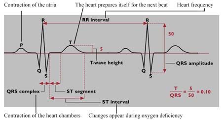
likely that unnecessary operative interventions will be increased without any discernable
benefit to perinatal outcome.

c. Requirement for additional tests of fetal wellbeing
In view of its very poor positive predictive value for metabolic acidosis and a high false
positive rate, CTG requires additional tests of fetal wellbeing such as fetal scalp blood
sampling, (FBS), fetal scalp lactate, fetal pulse oximetry and fetal electrocardiograph
(fetal ECG also called STAN or ST-analyser).

d. Problems with CTG Classification
Six years after publication of the 4th CESDI Report that highlighted substandard care
(which included lack of knowledge and failure to interpret CTG traces) in over 50% of
babies who died due to intrapartum related causes, the Royal College of Obstetricians
and Gynaecologists (RCOG) and National Institute of Health and Clinical Excellence
(NICE) produced National Guidelines on Electronic Fetal Heart Rate monitoring (EFM) in
20015. This was a very welcome and much needed step in the right direction and it
created a platform for universal classification of CTG into Normal, Suspicious and
Pathological categories. Hence, varied terminologies such as ‘good CTG’ ‘optimal CTG’,
‘sub-optimal CTG’, ‘bad CTG’, ‘non-reassuring CTG’ etc, which caused much confusion
among clinicians in the past, were avoided. However, this classification itself was riddled
with many drawbacks. First of all, it over-simplifies the complex process of labour and
assumes that there could be only three types of decelerations during labour (early due to
head compression, variable due to cord compression and late due to utero-placental
insufficiency). There has been no consideration to multiple events such as head and cord
compression occurring together during uterine contractions or the importance of fetal
reserve or its capacity to respond to hypoxia on the ultimate outcome.

Moreover, clinicians often reacted to ‘CTG Patterns’ described in the EFM Guidelines
without considering the clinical picture. Electronic Fetal Monitoring Guidelines was
subsequently revised by NICE in 2007 to include a ‘time frame’ for decelerations in order
to reduce the likelihood of clinicians reacting and thereby instituting an unnecessary
intervention to specific patterns noted in the CTG6. Hence, instead of intervening when
one or two late decelerations are noted, the current classification allows late
decelerations for 50% of contractions for up to 30 minutes to be present prior to be
considered as an abnormal feature. Although, this is a vital step in the right direction,
there has been no consideration of the type of hypoxia during labour (acute, sub acute,
gradually evolving) or emphasis of other features such as pseudo-sinusoidal patterns,
saltatory patterns, loss of cycling of fetal heart rate that may also reflect fetal
compromise, in the current NICE Classification.

Role of fetal ECG in intrapartum fetal monitoring
Fetal electrocardiograph (ECG) refers to a graphic record of the summation of electrical
activity of the myocardial cells. This in turn reflects oxygenation status of a central organ
(i.e. myocardium of the heart), which is protected until very late stages of hypoxia by a
fetus that mounts a compensatory response to lack of oxygen. Hence, indirectly, it
provides information on the oxygen status of the fetal brain, which is also a central organ
and is protected by compensatory mechanisms (re-distribution of blood from non-
essential organs such as the kidneys, skin and liver to central ‘essential’ organs such as
the heart, brain and fetal adrenal glands).
Figure 1. Fetal ECG Complex

What is STAN (ST-Analyser)?
STAN analyses changes in the fetal ECG Complex that occur secondary to myocardial
hypoxia. It is so named because it analyses the ‘ST Segment’ of the fetal ECG (Fig 1).
‘ST Segment’ and ‘T wave’ give an indication regarding the electrical changes that occur
during the repolarisation of the myocardial cells, as they prepare for the next contraction.
STAN also analyses the T/QRS Ratio (height of the T wave that reflects repolarisation of
the ventricles and QRS Complex that reflects depolarisation of the ventricles) to
determine an acute or longer lasting hypoxic insult to the myocardial cells.
                Figure 2. Scalp electrode (a), connection to skin electrode on the maternal thigh (b) and STAN
                Machine (c)

Why do we need STAN?
Cardiotocograph (CTG) has a high false positive rate of over 50%, which implies that if
CTG is used alone, it would increase interventions such as emergency caesarean
sections and operative vaginal births without any significant benefit to the perinatal
outcome. Currently available additional tests of fetal well being (FBS, fetal pulse oximetry
and fetal scalp lactate) determine evidence of hypoxia in peripheral tissue (fetal scalp)
and hence they fail to provide information on the oxygenation of central organs
(myocardium and brain). They do not help us determine the fetal responses to hypoxia,
which vary from fetus to fetus. Moreover, FBS and fetal scalp lactate do not provide
continuous information during labour and require repetition if the changes observed on
CTG, which are suggestive of suspected fetal compromise, do not improve.

ST-Analyser (STAN) aims to overcome these shortcomings by assessing fetal response
to hypoxic insults on myocardium, which is a central organ. It helps differentiate between
a fetus exposed to hypoxia that is compensating well by continuing to perfuse its central
organs from a fetus that is unable to compensate or has exhausted all the resources
available to deal with hypoxic insult. Moreover, STAN also provides continuous
information throughout labour on fetal wellbeing.
How does STAN get electrical signals from the fetal heart?
As shown in Figures 2, electrical signals from the fetal heart is captured through a fetal
scalp electrode (Fig 2a), which is connected to a skin electrode on the maternal thigh
(Fig 2b). The latter is connected to a computer that analyses the signals and displays it
on the STAN Monitor (Fig 2c).
It is important to ensure good contact between fetal scalp electrode and the fetal scalp to
obtain a good signal quality. Similarly, the skin electrode on the maternal thigh should
also have sufficient contact to ensure optimum capture of fetal ECG signals by the
computer. If there is poor contact (increased fetal hair or scalp or skin electrodes not
applied appropriately), this will be flagged up on the monitor with a suggestion to ‘check
the electrodes’.
Figure 3. Appearance on the STAN Monitor
How does STAN technology
work?
As soon as a fetus is connected to
the STAN Machine, the machine
calculates the normal baseline
T/QRS ratio for the individual fetus,
by analysing the fetal ECG
complexes that it receives through
the fetal scalp electrode. In the
presence of good signal quality, this
usually takes approximately four to
five minutes. Once this initial
calculation is completed, the
computer remembers this value as
the ‘normal baseline’ for the Figure 4. Classification of CTG using STAN Guidelines
individual fetus in question.
Subsequently, the computer
analyses every 30 fetal ECG
complexes and compares with the
original ‘baseline value’ and puts a
cross (‘x’) on the monitor. Hence, if
the fetal heart rate is 150 beats
/minute, one should expect to see
five crosses (‘x’) on the screen (Fig
3). If the computer determines that
the recent information on fetal ECG
after analysis is significantly
Figure 5. Suggested Course of Action based on ST Events noted and
different to its original calculation Classification of CTG based on STAN Guidelines
(i.e. the baseline T/QRS or ST
Segment values obtained in the
initial four minutes), this will be
flagged up as a ‘ST Event’ for
clinicians to take appropriate action.
Figure 3 shows the appearance of
fetal heart trace on the STAN Monitor which indicates the fetal heart rate (148/min), the
standard CTG trace and the crosses (‘x’) that indicate ST Analysis. Hence, the monitorprovides a continuous analysis of fetal ECG signals (i.e. an additional test of fetal wellbeing to avoid false positive rate) throughout labour. What are the ‘ST Events’ produced by STAN Technology? STAN specifically analyses T/QRS ratios and ‘ST Segment changes’ of fetal ECG complexes and produces two types of ‘ST Events’: ‘T/QRS ST Events’ and ‘Biphasic ST Events’. T/QRS ST Events These denote periods of myocardial hypoxia that results in an increase in ‘T Wave’ height. If the hypoxic insult is short lasting, it is termed ‘Episodic T/QRS Rise’ and if the hypoxia is long lasting, typically over 10 minutes duration, it is termed ‘Baseline T/QRS rise’. The underlying mechanism appears to be a ‘catecholamine surge’ from the fetal adrenal gland (emergency hormone) that occurs secondary to hypoxic stress. Catecholamines increase the heart rate and also breakdown myocardial glycogen into glucose to increase the energy substrate for the myocardium to continue to function and to continue supplying the brain and fetal adrenal gland, both of which are essential for fetal survival. This process of ‘glycogenolysis’ induced by catecholamines results in release of potassium ions along with glucose into the myocardial cell. Hence, the resultant local ‘hyperkalemia’ produces ‘tall T Waves’ and an increase in T/QRS ratio. It is vital to remember that excessive fetal movements that may result in catecholamine surges may also result in ‘Episodic T/QRS ST events’. In this case, the CTG would be otherwise normal and would show accelerations and hence, ST events should be ignored. Biphasic ST Events As mentioned before, ST Segment reflects a period of quiescence when the myocardium ‘rests’ just after a contraction (depolarisation), prior to relaxation (repolarisation). Under normal circumstances, myocardial cell membrane should not allow transfer of any ions during this ‘absolute refractory period’. Hence, the ST segment would be ‘iso-electric’ and will have a stable baseline. When there is a disturbance to myocardial pump function that may be secondary to hypoxic insults, infection, structural heart defects, myocardial dystrophies or prematurity (less contractile elements), the ST Segment of the fetal ECG segment may shift upwards or downwards leading to ‘Biphasic ST Events’. As the endocardium becomes ischaemic, sequence of repolarisation gets altered and the direction of current flow gets reversed. This results in depression of the ST segment of the fetal ECG complex with or without a negative T wave. Depending on the degree of such depression (i.e. negative ST segment due to reversal of current flow) Biphasic Grade 2 and Grade 3 ST events may be produced. These may become significant if the CTG is not normal. Breech presentation may also give rise to multiple biphasic ST events (as the heart is turned ‘upside down’ in relation to the maternal skin electrode that results in similar reversal in current flow in relation to the reference electrode), even though the fetus may not be hypoxic. STAN Machines have a ‘breech mode’ to rectify this problem and this should be activated if a decision has been made for assisted vagina breech birth and continuous electronic fetal heart rate monitoring is required. How to use STAN in clinical practice? Once a decision has been made for continuous electronic fetal heart rate monitoring, STAN Clip should be applied. As mentioned earlier, STAN technology works by determining the ‘normal baseline ECG’ for the index fetus and then compares
subsequent ECG complexes it obtains from the fetus with this calculated initial baseline.
Hence, it is important to ensure that the fetus still retains its capacity to respond to
hypoxia (i.e. can show a further change in the ECG complex, in response to hypoxia).

Therefore, analysing the initial CTG Trace prior to connecting a fetus to a STAN Machine
is very crucial. If the CTG is pre-terminal or if there is total loss of variability, the fetus
may have already exhausted all the reserves and resources available to respond to
hypoxia. Hence, it is unlikely to show any further changes in the fetal ECG Complexes
that may be determined by STAN Computer. This would mandate an immediate delivery
to salvage the fetus. In all other cases (normal CTG, CTG showing decelerations with
normal baseline heart rate and variability), the fetus is likely to have the capacity to
respond further to hypoxia (i.e. show further changes in ECG complexes) and hence,
could be connected to the STAN Machine.

Some advocate performing a fetal blood sampling (FBS) prior to connecting the fetus to
the STAN machine if all four features of the CTG are not reassuring. As one gets more
experience with the use of STAN Technology and with better understanding of
pathophysiology of fetal hypoxic changes, it is possible to connect a fetus to STAN
machine even in the presence of decelerations, if the baseline fetal heart rate and
variability have remained normal.

How to Interpret STAN Events?
Whenever a ST Event is highlighted on the STAN Monitor, it is essential to classify the
CTG (current and 30 minutes prior to the event). Classification is based on that adopted
by the International Federation of Obstetrics and Gynaecology (FIGO) and is very similar
to that recommended by National Institute of Health and Clinical Excellence (NICE), with
minor differences. Figure 4 shows current STAN Guidelines on CTG Classification.

Once the CTG is classified using the STAN Guidelines into normal, intermediary or
Abnormal, the size and magnitude of the ST Events should be noted. In the presence of
a Normal CTG or pre-terminal CTG, ST Events should be discarded and labour
continued in the former and immediate delivery carried out in the latter. Figure 5
illustrates application of STAN Guidelines based on the type and magnitude of ST
Events.

For example, if the ST Event highlighted is ‘Episodic T/QRS Rise 0.10; (type of event is
Episodic T/QRS, magnitude of the event is 0.10), and the CTG of the index fetus is
classified as ‘Intermediary’, this would not require any action. However, if the CTG has
been classified as ‘Abnormal’, Episodic T/QRS rise of 0.10 would become significant and
would therefore warrant an action (please see Figure 5).

Figure 6 illustrates an Algorithm for Interpretation of STAN Events and suggested
actions. Essentially this consists of four C’s (Check appropriateness for the use of STAN,
Classify CTG, Correlate the observed STAN Events with the CTG to determine their
significance and Cascade for timely and appropriate intervention).
Figure 6. Suggested Algorithm during use of STAN Technology

Limitations of STAN Technology

Does STAN Technology improve perinatal outcome?
Large randomised controlled trials (Plymouth7 and Swedish8) have reported significant
reductions in operative delivery for fetal distress and umbilical cord metabolic acidosis in
fetuses monitored by STAN. A recent Cochrane Review considered five trials (10,628
women) on continuous fetal monitoring alone as compared to the use of STAN9. It
reported that the use of STAN for intrapartum fetal monitoring resulted in fewer babies
with neonatal encephalopathy (four trials, risk ratio (RR) 0.37), fewer fetal scalp samples
during labour (four trials, RR 0.65) and fewer operative vaginal deliveries (five trials, RR
0.89). Recently, it has also been reported that STAN improves the inter-observer
variation among clinicians, especially on the decision to intervene for an intermediary or
abnormal CTG10.
Limitation for the use of STAN technology
STAN can be used in clinical practice only after 36 +0 weeks of gestation. This is
because preterm fetuses may have under-developed endocardial - epicardial inter-phase
that may interfere with signal conduction, leading to multiple ST Events that are usually
biphasic. STAN also cannot be used in fetuses with a structural or functional cardiac
abnormality that may interfere with generation or transmission of fetal ECG Complexes.
As it requires attachment of a fetal scalp electrode, any contra-indications to the use of
such an electrode (risks of vertical transmission or known fetal bleeding disorders) would
preclude its use. Rarely, in the presence of a large amount of fetal hair, it may not be
possible to obtain sufficient electrical signals to rely on the technology and this may be
highlighted by the STAN Machine. In this case, another additional test of fetal wellbeing
should be carried out if the CTG is pathological (i.e. using NICE Guidelines as it is not
possible to use STAN Guidelines in this case).

Use of STAN for intrapartum fetal monitoring: The caveats
Development of fetal ECG as an additional test of fetal wellbeing appears to be a
significant step in reducing the false positive rate of CTG and in improving perinatal
outcome. No test is 100% perfect and it is estimated that STAN may have a false
positive and negative rate of approximately 5%. Although the STAN technology is very
advanced, it still requires visual analysis of complex information and institution of timely
and appropriate management by attending clinicians. Hence, the system is open to
human errors. In most cases where the STAN technology did not pick up and
abnormality and the perinatal outcomes were poor, ‘human factors’ have been
identified11. These include lack of knowledge (e.g. failure to recognise a pre-terminal
CTG trace), failure to incorporate clinical picture (such as intrapartum pyrexia, fresh thick
meconium, sentinel hypoxic events during labour) and failure to follow STAN Guidelines
(including failure to take appropriate action and delays in action).

Other reported failures include the use of STAN technology in the presence of severe
group B Streptococcal infection12 thereby failure to recognise that hypoxia is not the only
pathway for brain damage and that a co-existing infection may enhance the detrimental
effect of hypoxia on the fetal brain. Following the reported three cases of adverse
outcome, the STAN Guidelines have been revised (Figure 7).

Figure 7. Revised STAN Guidelines

     •    Intervention depends on the cause of fetal compromise and the stage of labour.
          It includes qualified assessment of FHR data, alleviation of cause(s) of fetal
          distress (such as over-stimulation or maternal hypotension) and delivery
     •    During second stage with active pushing, intervention means that immediate
          operative delivery is recommended unless spontaneous delivery is anticipated
          within the next five to 10 minutes
     •    Abnormal CTG pattern for more than 60 minutes, or less if the FHR deteriorates
          rapidly, with normal ST requires qualified assessment and checking for non-
          deteriorating fetal state
     •    With a preterminal CTG pattern intervention is always indicated, irrespective of
          ST data
     •    Pause in the recording or poor signal quality with gaps in the T/QRS ratios for
          more than four minutes may result in missed ST Events: management should
          be related to the CTG pattern and clinical situation
     •    In the presence of maternal pyrexia even intermediary CTG pattern may be
          regarded as significant in combination with ST Event
Conclusion
Intrapartum fetal monitoring is aimed at identifying fetuses that are at increased risk of
hypoxic brain damage so that timely and appropriate action could be taken to improve
short term and long term outcomes. Substandard care with regard to fetal monitoring is
associated with ‘high-value’ clinical negligence claims13. They have devastating effects
on families and staff involved. Substandard care involving intrapartum fetal monitoring
increases the financial cost to the healthcare systems besides resulting in a burden to
the affected child, family and the society.
Use of STAN appears to reduce the risks of neonatal encephalopathy as well as reduce
operative interventions. Clinicians should understand the limitations as well as the
caveats, including the role of human error in the use and abuse of this technology.
Computer analysis of the fetal heart rate and ST Event signals appears to increase the
ability to predict neonatal acidaemia. Currently, a large European Multi-Centre trial is
underway assess the role of computer analysis of STAN traces in assisting clinicians to
take appropriate and timely interventions in order to improve perinatal outcome.
Acknowledgement
I would like to thank Neoventa Medical AB for their kind permission to use figures and
guidelines pertaining to the use of STAN in this article.
References
1. CESDI (1997) 4th Annual Report. London. Maternal and Child Health Research
Consortium.
2. Chandraharan E, Arulkumaran S. Prevention of birth asphyxia: responding
appropriately to cardiotocograph (CTG) traces. Best Pract Res Clin Obstet
Gynaecol. 2007 Aug;21(4):609-24.
3. Beard RW, Filshie GM, Knight CA, Roberts GM. The significance of the changes
in the continuous fetal heart rate in the first stage of labour. J Ob- stet gynaecol Br
C’wlth 1971; 78:865-81.
4. Chandraharan E, Arulkumaran S. Electronic Fetal Heart Rate Monitoring in
current and future practice. Review Article. J Obstet Gynecol India 2008; 58(2):
121-130.
5. The Royal College of Obstetricians and Gynaecologists. The Use of Electronic
Fetal Monitoring. Evidence-Based Clinical Guideline 8. RCOG Press, 2001.
6. National Institute of Clinical Excellence. Intrapartum Care: care of healthy women
and their babies during labour. NICE Clinical Guideline No.55. September, 2007.
7. Westgate J, Harris M, Curnow JSH, Greene KR Plymouth randomised trial of
cardiotocogram only versus ST waveform plus cardiotocogram for intrapartum
monitoring in 2400 cases. Am J Obstet Gynecol, 1993;169:1151-60.
8. Amer-Wåhlin I, Hellsten C, Norén H, Hagberg H, Herbst A, Kjellmer I, Lilja H,
Lindoff C, Månsson M, Mårtensson L, Olofsson P,Sundström AK, Mar_ál K
Cardiotocography only versus cardiotocography plus ST analysis of
fetalelectrocardiogram for intrapartum fetal monitoring: a Swedish ran- domised
controlled trial. Lancet 2001;358:534-38.
9. Neilson JP. Fetal electrocardiogram (ECG) for fetal monitoring during labour.
Cochrane Database of Systematic Reviews 2006, Issue 3. Art. No.: CD000116.
DOI: 10.1002/14651858.CD000116.pub2. In:The Cochrane Database ofSystematic Reviews 2010 Issue 9, Copyright © 2010 The Co chrane
Collaboration. Published by John Wiley and Sons, Ltd
10. Westerhuis ME, van Horen E, Kwee A, van der Tweel I, Visser GH, Moons KG.
Inter- and intra-observer agreement of intrapartum ST analysis of the fetal
electrocardiogram in women monitored by STAN. BJOG 2009 Mar;. 116 (4):545-
551
11. Doria V, Papageorghiou AT, Gustavsson A, Ugwumadu A, Arulkumaran S Review
of the first 1502 cases of ECG-ST waveform analysis during labour in a teaching
hospital. BJOG 2007;114:1202-1207.
12. Westerhuis ME, Kwee A, van Ginkel AA, Drogtrop AP, Gyselaers WJ, Visser GH.
Limitations of ST analysis in clinical practice: three cases of intrapartum metabolic
acidosis. BJOG. 2007 Oct;114(10):1194-201.
13. Chandraharan E, Arulkumaran S. Medico-legal problems in obstetrics. Cur- rent
Obstetrics & Gynaecology (2006); 16: 206-210.
14. Antonia Costa, Diogo Ayres-de-Campos, Fernanda Costa, Cristina Santos, Joao
Bernardes Prediction of neonatal acidemia by computer analysis of fetal heart
rate and ST event signals. Am J Obstet Gynecol 2009;201:464. e1-6.You can also read