Subtle right ventricular dysfunction in asymptomatic chronic heavy cigarette smokers: a speckle tracking case-control study
←
→
Page content transcription
If your browser does not render page correctly, please read the page content below
Janhangeer et al. The Egyptian Heart Journal
https://doi.org/10.1186/s43044-021-00151-y
                                                         (2021) 73:25
                                                                                                                                        The Egyptian Heart
                                                                                                                                                   Journal

 RESEARCH                                                                                                                                            Open Access

Subtle right ventricular dysfunction in
asymptomatic chronic heavy cigarette
smokers: a speckle tracking case-control
study
Mohammad Iqbal Janhangeer, Ghada Youssef* , Weal El Naggar and Dalia El Remisy

  Abstract
  Background: Chronic heavy cigarette smoking can affect the right ventriclular function. The standard
  echocardiography may not show early right ventricular functional changes, and a more sensitive measure is
  needed. The aim of this work was to evaluate the subtle subclinical effects of chronic heavy cigarette smoking on
  the right ventricular function. The study included 55 healthy asymptomatic chronic heavy cigarette smokers
  (smoking history of at least 5 pack-years and a daily cigarette consumption of at least 1 pack) and 35 healthy non-
  smoking control subjects. Patients underwent a full clinical assessment and a conventional as well as a 2D-speckle
  tracking transthoracic echocardiography of the right ventricle and data was compared between the 2 groups.
  Results: The mean age was 32.9 ± 7.2 years in smokers and 30.9 ± 7.9 years in non-smokers (p = 0.227). The 2
  groups showed comparable conventional right ventricular systolic and diastolic functions. Smokers showed a
  significantly lower (less negative) right ventricular global longitudinal strain (− 19.0 ± 3.2% vs. − 24.5 ± 3.5%, p <
  0.001). Patients with a higher daily cigarette consumption showed a poorer right ventricular global longitudinal
  strain (p = 0.014).
  Conclusion: Chronic heavy cigarette smoking can adversely affect the right ventricular function, a finding that can
  be easily missed by conventional echocardiography and can be better detected by the right ventricular speckle
  tracking.
  Keywords: Strain, Right ventricle, 2D echocardiography

Background                                                                              Global Adult Tobacco Survey (GATS) Egypt Country
More than one billion people, about one quarter of                                      Report 2009 [1].
adults worldwide, smoke tobacco [1]. Tobacco is cur-                                       Cigarette smoking is one of the major risk factors for
rently the greatest preventable cause of death in the                                   coronary heart disease, stroke, atherosclerosis, aortic
world, killing up to half the people who smoke it, which                                aneurysm, peripheral vascular disease, and subclinical
is more than 7 million people worldwide annually [2].                                   cerebrovascular disease [3]. Kaplan et al. reviewed the
Current data suggest that smoking is a public health                                    possible mechanisms by which cigarette smoke can dir-
problem in Egypt. Prevalence of cigarette smoking was                                   ectly affect the myocardium thereby causing smoking
46.4% among males and 0.2% among females in the                                         cardiomyopathy, and concluded that oxidative stress, in-
                                                                                        flammation, metabolic impairment, and cell death were
                                                                                        the possible factors responsible for cardiac remodeling
* Correspondence: ghadayoussef@kasralainy.edu.eg                                        after chronic cigarette exposure [4].
Cardiology Department, Kasr Al Ainy Faculty of Medicine, Cairo University,
Cairo, Egypt

                                        © The Author(s). 2021 Open Access This article is licensed under a Creative Commons Attribution 4.0 International License,
                                        which permits use, sharing, adaptation, distribution and reproduction in any medium or format, as long as you give
                                        appropriate credit to the original author(s) and the source, provide a link to the Creative Commons licence, and indicate if
                                        changes were made. The images or other third party material in this article are included in the article's Creative Commons
                                        licence, unless indicated otherwise in a credit line to the material. If material is not included in the article's Creative Commons
                                        licence and your intended use is not permitted by statutory regulation or exceeds the permitted use, you will need to obtain
                                        permission directly from the copyright holder. To view a copy of this licence, visit http://creativecommons.org/licenses/by/4.0/.
Janhangeer et al. The Egyptian Heart Journal (2021) 73:25 Page 2 of 8
The right ventricle (RV) has long been regarded as the All the standard echocardiography views were re-
forgotten side of the heart and little attention has been corded in addition to the apical RV-focused view. The
paid to its assessment [5]. Nowadays, however, there is systolic and diastolic functions of the left ventricle were
no doubt that it plays a critical role in the prognosis of assessed according to the American Society of Echocar-
different cardiovascular diseases. The function of the RV diography guidelines [16–18]. Conventional parameters
is a strong determinant of the prognosis for patients of the right ventricular systolic and diastolic functions
with congestive heart failure, ischemic heart disease, car- included tricupsid annular plane systolic excursion
diomyopathy, pulmonary arterial hypertension and con- (TAPSE), tissue Doppler imaging (TDI) tricuspid valve
genital heart defects [6–12]. Therefore, there is a great S-wave velocity, and RV Tei Index calculated as the sum
need to evaluate its function accurately. of the isovolumic contraction time (ICT) and the isovo-
There are few studies that have focused on the effects lumic relaxation time (IRT) divided by ejection time
of smoking on the right ventricular function, and they (ET) (calculated as follows: ICT + IRT/ET), and E/ and
have shown conflicting results [13–15]. A/of the lateral tricuspid annulus, right ventricular frac-
The present study aims to find out whether chronic tional area change (FAC), right atrial volume, E and A
cigarette smoking, in otherwise healthy individuals, can pulse wave Doppler velocity over the tricuspid inflow,
cause subtle dysfunction of the right ventricle by apply- diameter and inspiratory collapsibility of the inferior
ing a 2D speckle tracking echocardiography (2D-STE) as vena cava (IVC), peak tricuspid regurgitant jet velocity,
well as other conventional echocardiographic methods and estimated pulmonary artery systolic pressure (calcu-
of assessment of the right ventricular function. lated as right ventricular systolic pressure + right atrial
pressure).
Methods Video clips of the RV-focused view were recorded, en-
This is a cross-sectional, observational, case-control suring a clear definition of the RV endocardial and epi-
study which was conducted at the Cardiovascular Med- cardial borders. A minimum of three beats were
ical Department after approval from the faculty’s Ethical recorded. The depth, the sector width, and the frequency
Committee. Ninety subjects volunteered to participate in were adjusted to obtain a frame rate of at least 50 Hz.
the study of whom 55 were healthy, asymptomatic heavy The operator was blind of the smoking history of the
chronic cigarette smokers aged 45 years or less and 35 studied subjects and clips were analyzed later, using the
were healthy never-smokers control subjects. Healthy offline speckle tracking Q-lab software to determine the
current cigarette smokers should have had a smoking global longitudinal strain (GLS) of the right ventricle.
history of at least 5 pack-years and a daily cigarette con- As the available machine did not include a software
sumption of at least 1 pack (20 cigarettes) per day. Ex- for RV speckle analysis, we used the left ventricular soft-
clusion criteria included atrial fibrillation and other ware and we manually adjusted the tracked images to fit
arrhythmias, poor echocardiographic window, the pres- the RV shape. It was assumed that the aortic valve clos-
ence of > 1 segment (out of the six segments of the RV) ure time was not significantly different from the pul-
without clearly defined endocardial and epicardial bor- monary valve closure time. The software automatically
ders, other substance abuse, obesity (BMI > 30 kg/m2), calculated the aortic valve closure time from the left
chronic obstructive pulmonary disease (COPD), elevated ventricular apical three-chamber view. The region of
pulmonary artery systolic pressure (PASP) more than 35 interest (ROI) was manually selected and the software
mmHg, and greater than mild valvular lesions. After automatically traced the endocardial and epicardial bor-
obtaining an informed verbal consent from all subjects, ders of the RV. Necessary adjustments were made to en-
a detailed physical assessment was performed followed sure adequate tracking of the right ventricular free wall
by a transthoracic echocardiography. and the interventricular septum. Care was taken not to
Subjects were asked to stop smoking for at least 30 include the right atrial wall and the epicardium in the
min before undergoing echocardiography to minimize region of interest. The RV speckle tracking images in-
the effects of acute smoking on the heart. They were cluded analysis of 6 segments; the basal, mid, and apical
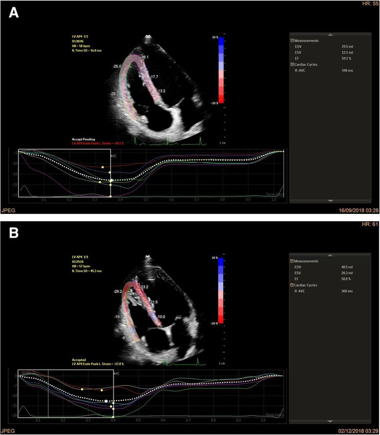
also requested not to consume any foods or drinks con- segments of the RV septal and free walls. Two examples
taining caffeine (e.g., coffee) for at least 3 h before the of the strain of the 6 segments of the RV together with
echocardiographic study. the RV GLS of both groups are shown in Fig. 1.
Echocardiography was performed using the commer- Finally, the software automatically calculated the La-
cially available machine (PHILIPS, Epic 7), equipped grangian strain of each of the 6 segments using the end-
with a 2.5-MHz phased array transducer. Analysis of the diastole as the reference point. The GLS of the RV was
Speckle images was performed using the Q-lab10 soft- also automatically calculated. Pooled data suggest that
ware available both on the echocardiography machine as global longitudinal RV free wall strain > − 20% (i.e., <
well as on a laptop. 20% in absolute value) is likely abnormal [16].Janhangeer et al. The Egyptian Heart Journal (2021) 73:25 Page 3 of 8
Fig. 1 Strain of the six segments of the right ventricle and GLS of the right ventricle of one of the non-smoking control subjects (a) and one of
the chronic cigarette smokers (b)
Statistical analysis
Categorical data was presented as numbers and percent-
ages while continuous data was presented as mean and
standard deviation (SD). Comparison of means between
groups was performed using the independent student t
Table 1 Demographic data and clinical characteristics of the 2
test or one way ANOVA test, as convenient. Correlation
groups
between continuous variables was done using Pearson
Variable Smokers Non-smokers p value
correlation test. All statistical analysis was done using an (mean ± SD) (mean ± SD)
SPSS 26 program. A p value of < 0.05 was considered Age/year 32.9 ± 7.2 30.9 ± 7.9 0.227
significant. 2
BMI (kg/m ) 24.5 ± 3.1 25.0 ± 2.5 0.481
Results BSA/m2 1.90 ± 0.16 1.89 ± 0.19 0.902
The demographic data of the 2 groups were comparable SBP/mmHg 120 ± 9 117 ± 7 0.132
(see Table 1). While chronic heavy cigarette smokers DBP/mmHg 77 ± 7 75 ± 7 0.359
had a higher pulse rate than non-smokers, the difference Pulse (beats/min) 75 ± 9 69 ± 8 0.001
in the blood pressure between the two groups was not BMI body mass index, BSA body surface area, DBP diastolic blood pressure, SBP
significant. systolic blood pressureJanhangeer et al. The Egyptian Heart Journal (2021) 73:25 Page 4 of 8
Both smokers and non-smokers had normal LV sys- duration of smoking was 10.7 ± 5.9 years, and the mean
tolic and diastolic functions. The conventional methods consumption of cigarettes was 13.5 ± 10.4 pack-years.
for the assessment of the systolic and diastolic functions RV GLS was more adversely affected among those who
of the RV were normal and comparable for both groups smoked more cigarettes per day (Fig. 2). The impair-
as shown in Table 2. Chronic heavy cigarette smokers ment of RV GLS could not be correlated to the number
were found to have a statistically significant higher tri- of years of smoking (r = 0.140, p = 0.308) or the number
cuspid valve (TV) a′ velocity and a lower TV e′/a′ ratio of pack-years (r = 0.256, p = 0.057).
as compared to non-smokers.
The strain of all RV segments was significantly lower Discussion
in smokers as compared to non-smokers, except for the Cigarette smoking is one of the modifiable cardiovascu-
free wall apical segment (Table 3). The basal RV free lar risk factors. Smoking has been linked to many car-
wall strain and basal septal wall strain were abnormally diovascular diseases. In general, smokers have a higher
lower than the cut-off limit of − 20, based on the latest risk of heart failure and poorer outcomes compared to
ASE guidelines [18]. The mean GLS of the RV was ab- never-smokers [19].
normal in chronic heavy cigarette smokers in contrast to The relatively new technique of Speckle Tracking
the mean RV free wall strain which, although signifi- Echocardiography has revealed that chronic heavy
cantly lower in smokers than in non-smokers, was still smokers who develop COPD show echocardiographic
within the currently accepted normal limits. The num- evidence of right ventricular dysfunction appearing even
ber of subjects with an abnormal RV GLS was signifi- before the occurrence of pulmonary hypertension and
cantly higher in the smokers’ group as compared to the cor pulmonale. The present study aims to find out
normal control group (80% versus 8.6%, p < 0.001). whether chronic cigarette smoking in otherwise healthy
In the smokers group, the mean number of cigarettes individuals can cause subtle subclinical dysfunction of
smoked per day was 24.2 ± 7.4 cigarettes, the mean the right ventricle by applying a 2-D speckle tracking
Table 2 Conventional echocardiographic parameters of the systolic and diastolic RV functions of both groups
Variable Smokers (mean ± SD) Non-smokers (mean ± SD) p value
RV basal diameter, cm 3.5 ± 0.4 3.4 ± 0.5 0.070
RV mid cavity diameter, cm 3.3 ± 0.4 3.2 ± 0.4 0.322
RV longitudinal diameter, cm 7.1 ± 0.6 7.0 ± 0.5 0.603
TAPSE, cm 2.3 ± 0.4 2.2 ± 0.3 0.271
RV S-wave (cm/s) 12.7 ± 2.1 13.0 ± 1.6 0.439
FAC, % 45.7 ± 6.1 48.3 ± 7.5 0.082
IVCT, ms 69.8 ± 9.3 70.6 ± 8.9 0.663
IVRT, ms 66.0 ± 14.6 62.9 ± 11.3 0.288
TEI Index 0.51 ± 0.08 0.48 ± 0.07 0.178
2
RA area, cm 15.3 ± 2.9 14.9 ± 2.3 0.422
RA volume, ml 46.7 ± 15.2 43.6 ± 10.3 0.289
2
RA volume Index, ml/m 24.6 ± 7.6 23.1 ± 5.6 0.336
TV E wave velocity (cm/s) 59.7 ± 10.7 60.7 ± 11.7 0.703
TV A wave velocity (cm/s) 39.2 ± 7.8 36.7 ± 7.6 0.142
TV E/A 1.57 ± 0.35 1.69 ± 0.34 0.107
TV e′ wave velocity (cm/s) 12.7 ± 3.3 12.7 ± 2.3 0.930
TV a′ wave velocity (cm/s) 11.8 ± 4.2 9.8 ± 2.4 0.004
TV e′/a′ ratio 1.18 ± 0.48 1.371 ± 0.38 0.052
TV E/e′ ratio 4.93 ± 1.14 4.89 ± 1.13 0.872
TV E wave DT, ms 155.4 ± 30.4 165.6 ± 40.1 0.204
IVC diameter, cm 1.9 ± 0.3 1.9 ± 0.2 0.887
EPASP, mmHg 23.2 ± 4.0 22.5 ± 3.2 0.353
DT deceleration time, EPASP estimated pulmonary artery systolic pressure, FAC fractional area change, IVC inferior vena cava, IVCT isovolumetric contraction time,
IVRT isovolumetric relaxation time, RV right ventricle, TAPSE tricuspid annular plane systolic excursion, TV tricuspid valveJanhangeer et al. The Egyptian Heart Journal (2021) 73:25 Page 5 of 8 Table 3 Right ventricular strain of the 2 groups Variable Smokers (mean ± SD) Non-smokers (mean ± SD) p value Basal RV free wall strain, % − 18.7 ± 6.2 − 27.2 ± 7.0 < 0.001 Mid RV free wall strain, % − 20.6 ± 6.6 − 24.5 ± 6.3 0.007 Apical RV free wall strain, % − 22.0 ± 6.6 − 24.8 ± 6.8 0.057 Basal septal strain, % − 13.5 ± 4.2 − 19.5 ± 5.5 < 0.001 Mid septal strain, % − 19.7 ± 4.6 − 25.8 ± 5.7 < 0.001 Apical septal strain, % − 23.7 ± 5.8 − 27.9 ± 6.1 0.001 RV GLS, % − 19.0 ± 3.2 − 24.5 ± 3.5 < 0.001 RV free wall strain, % − 20.4 ± 4.1 − 25.5 ± 3.6 < 0.001 RV right ventricle, GLS global longitudinal strain echocardiography as well as other established echocar- as possible by requiring the included subjects not to diographic parameters of right ventricular function. smoke for at least 30 min before the echocardiographic Two groups of subjects were included, the first study. group included healthy non-cardiac individuals with There was no significant difference in the blood pres- history of chronic heavy cigarette smoking (n = 55), sure (office BP) between the two groups. Indeed, no and the second group included healthy (never- chronic effects of smoking have been reported for office smokers) subjects (n = 35). BP [21]. In the study by Farsalinos et al. [20] mentioned In this study, the chronic heavy cigarette smokers had above, acute significant rise in office BP (by 8 mmHg a higher pulse rate than non-smokers. Nicotine and systolic and 4 mmHg diastolic) after smoking two ciga- PM2.5 are known to activate the sympathetic nervous rettes returned to baseline 30 min later. Considering the system. Nicotine’s effect is acute, while PM2.5’s effect is fact that chronic heavy cigarette smokers rarely abstain on chronic basis. As shown in the study performed by from cigarette for more than 30 min at a stretch while Farsalinos et al. [20], the increase in pulse after smoking awake, it would appear logical to expect that their blood two cigarettes returned to baseline 30 min later. Thus, pressure should be chronically raised. The fact that this the main contributor to this higher pulse rate is most was not the case in our study can be attributed to the use probably PM2.5 rather than nicotine. The acute effects of office BP rather than ambulator blood pressure moni- on the autonomic nervous system were minimized as far toring (ABPM), and the studied population abstained Fig. 2 Means of RV GLS according to the number of consumed cigarettes per day
Janhangeer et al. The Egyptian Heart Journal   (2021) 73:25                                                       Page 6 of 8

from smoking 30 min prior to their study. Studies using        smoking does not impair right ventricular systolic func-
ABPM, in contrast to those using office BP, have shown         tion in the acute period, but their study enrolled only 20
that both normotensive smokers and untreated hyperten-         subjects, used tissue Doppler imaging as the method to
sive smokers present higher daily BP values than their re-     calculate RV strain, and focused mainly on the acute ef-
spective non-smoking counterparts [22].                        fects of cigarette smoking.
  The estimated pulmonary artery systolic pressure was           Our study showed that the impairment in RV GLS
not elevated in the smokers as well as in the non-             correlated significantly to the number of cigarettes
smokers group (23.2 ± 4.0 mmHg and 22.5 ± 3.2 mmHg,            smoked per day, but could not be correlated to the num-
respectively, p = 0.353). Keeping in mind that the echo-       ber of years of smoking or the number of pack-years of
cardiographic assessment of the pulmonary artery sys-          smoking. However, it is well known that the association
tolic pressure has its limitations; nonetheless, there was     between smoking and cardiovascular diseases has been
no evidence that any of the subjects that participated in      shown to be nonlinear, such that even a few cigarettes a
this study had pulmonary hypertension.                         day disproportionately increases cardiovascular risk [24].
  The conventional echocardiographic methods used for          One possible explanation for our finding is that large
assessing the systolic and diastolic functions of the right    doses of the component/s of cigarette smoke that are re-
ventricle showed normal measurements for both groups.          sponsible for causing RV dysfunction are required to
Smokers had a lower FAC than non-smokers, but the dif-         cause saturation of the involved biochemical and cellular
ference was not statistically significant (p value = 0.082).   processes, thereby causing a linear dose-response rela-
Smokers had a higher TV a′ wave velocity and a lower TV        tionship on RV function.
e′/a′ ratio than non-smokers. These findings are in line
with Eroglu et al. [23] who found no difference in the         Study limitations
standard echocardiographic measurements of the right           As dedicated software for the speckle tracking of the right
ventricle between a healthy group of 40 smokers and 40         ventricle is not available; the software of the left ventricle
non-smokers. In contrast, Ilgenli and Akpınar [13] found       was adapted to the right side. In theory, this should not
that cigarette smoking led to an impaired right ventricular    have affected the study significantly as the same software
diastolic function but not right ventricular systolic func-    was used for both smoking and non-smoking subjects.
tion in the acute period. This study is different from ours,   Furthermore, the region of interest was traced and manu-
since they investigated only the acute effects of smoking      ally adjusted to fit the right ventricle and the principles of
one cigarette on ventricular function and these effects        speckle tracking remain unchanged irrespective of the site
were shown to disappear after 30 min.                          being studied. Another limitation of the study is that al-
  In our current study, both groups were compared              though all the subjects included in the study were asymp-
using the 2D-STE; healthy chronic heavy cigarette              tomatic, we could not exclude that some chronic heavy
smokers had mild impairment in the mean GLS of the             smokers had grade I COPD according to the latest gold
RV (mean value of − 19%), while non-smokers had a              criteria (GOLD grade I COPD is asymptomatic). This rea-
normal mean RV GLS (mean value of − 24.5%); this dif-          soning could be extended to include asymptomatic coron-
ference was statistically significant (p < 0.001). The RV      ary artery disease as well as other diseases. Indeed, it
segmental analysis showed a significant difference be-         would be impractical and beyond the scope of this study
tween both groups. The basal RV free wall strain (mean         to carry out the required diagnostic tests to exclude all
value of − 18.7) and basal septal strain (mean value of −      diseases. Moreover, the American College of Chest Physi-
13.5) were the adversely affected segments (according to       cians and the European Respiratory Society do not recom-
the currently accepted cut-off level) [16]. These two seg-     mend screening of asymptomatic smokers by spirometry
ments were mostly responsible for the mild impairment          [25]. Indeed, the incidence of COPD in asymptomatic
in the RV GLS in heavy chronic cigarette smokers.              cigarette smokers is very low [26].
  This result is in line with the result of the study per-
formed by Eroglu et al. [23] which showed that chronic         Conclusion
cigarette smoking affects the systolic long-axis function      The current study showed that chronic heavy cigarette
of the right ventricle in healthy young subjects by strain     smoking is associated with subtle subclinical dysfunction
imaging using Doppler myocardial velocity. In contrast         of the right ventricle which can be detected only by the
to our study, Eroglu et al. also showed impairment of          more sensitive method of speckle tracking echocardiog-
the diastolic function of the RV. In our study, despite        raphy, but not by the conventional echocardiography
the lower values of the RV diastolic function in smokers       methods. Identification of these subtle changes can
as compared to non-smokers, these values were still            facilitate aggressive smoking cessation programs to
within the currently accepted normal limits. On the con-       encourage smokers to quit smoking before the devel-
trary, Ilgenli and Akpınar [13] found that cigarette           opment of clinical diseases. In order to determine
Janhangeer et al. The Egyptian Heart Journal              (2021) 73:25                                                                                    Page 7 of 8

whether the subtle smoking-induced dysfunction of                                        org/abstract/MED/26244034, https://doi.org/10.4137/CMC.S27462, https://
the right ventricle is reversible or not, further stud-                                  europepmc.org/articles/PMC4493918, https://europepmc.org/articles/PMC44
                                                                                         93918?pdf=render.
ies are recommended.                                                               6.    Warnes CA (2009) Adult congenital heart disease importance of the right
                                                                                         ventricle. J Am Coll Cardiol 54(21):1903–1910
Abbreviations                                                                      7.    D'Alonzo GE, Barst RJ, Ayres SM, Bergofsky EH, Brundage BH, Detre KM et al
2D: Two-dimensional; 2D-STE: 2D-speckle tracking echocardiography;                       (1991) Survival in patients with primary pulmonary hypertension. Results
ABPM: Ambulator blood pressure monitoring; BMI: Body mass index;                         from a national prospective registry. Ann Intern Med 115(5):343–349
BP: Blood pressure; BSA: Body surface area; COPD: Chronic obstructive
                                                                                   8.    Forfia PR, Fisher MR, Mathai SC, Housten-Harris T, Hemnes AR, Borlaug BA
pulmonary disease; DBP: Diastolic blood pressure; EPASP: Estimated
                                                                                         et al (2006) Tricuspid annular displacement predicts survival in pulmonary
pulmonary artery systolic pressure; ET: Ejection time; FAC: Fractional area
                                                                                         hypertension. Am J Respir Crit Care Med 174(9):1034–1041
change; GATS: Global Adult Tobacco Survey; GLS: Global longitudinal strain;
                                                                                   9.    Haddad F, Doyle R, Murphy DJ, Hunt SA (2008) Right ventricular function in
IVC: Inferior vena cava; IVCT: Isovolumetric contraction time;
                                                                                         cardiovascular disease, part II: pathophysiology, clinical importance, and
IVRT: Isovolumetric relaxation time; LV: Left ventricle; PASP: Pulmonary artery
                                                                                         management of right ventricular failure. Circulation. 117(13):1717–1731
systolic pressure; ROI: Region of interest; RV: Right ventricle; SBP: Systolic
                                                                                   10.   Meyer P, Filippatos GS, Ahmed MI, Iskandrian AE, Bittner V, Perry GJ et al
blood pressure; SD: Standard deviation; TAPSE: Tricuspid annular plane
                                                                                         (2010) Effects of right ventricular ejection fraction on outcomes in chronic
systolic excursion; TDI: Tissue Doppler imaging; TV: Tricuspid valve
                                                                                         systolic heart failure. Circulation. 121(2):252–258
                                                                                   11.   de Groote P, Millaire A, Foucher-Hossein C, Nugue O, Marchandise X,
Acknowledgements
                                                                                         Ducloux G et al (1998) Right ventricular ejection fraction is an independent
Authors acknowledge all members of the Cardiology Department, Cairo
                                                                                         predictor of survival in patients with moderate heart failure. J Am Coll
University Hospitals, for their help throughout this work.
                                                                                         Cardiol 32(4):948–954
                                                                                   12.   Ghio S, Gavazzi A, Campana C, Inserra C, Klersy C, Sebastiani R et al (2001)
Authors’ contributions
                                                                                         Independent and additive prognostic value of right ventricular systolic
M.J. proposed the idea, collected the data, and wrote the first draft of the
                                                                                         function and pulmonary artery pressure in patients with chronic heart
manuscript. D.E. and G.Y. did the echocardiography and critically revised the
                                                                                         failure. J Am Coll Cardiol 37(1):183–188
manuscript. G.Y. did the statistical analysis and W.E. critically revised the
manuscript. All authors have read and approved the manuscript.                     13.   Ilgenli TF, Akpinar O (2007) Acute effects of smoking on right ventricular
                                                                                         function. A tissue Doppler imaging study on healthy subjects. Swiss Med
Funding                                                                                  Wkly 137(5-6):91–96
This study was self-funded.                                                        14.   Vitarelli A, Conde Y, Cimino E, Stellato S, D'Orazio S, D'Angeli I et al (2006)
                                                                                         Assessment of right ventricular function by strain rate imaging in chronic
Availability of data and materials                                                       obstructive pulmonary disease. Eur Respir J 27(2):268–275
The datasets used and/or analyzed during the current study are available           15.   Hilde JM, Skjørten I, Grøtta OJ, Hansteen V, Melsom MN, Hisdal J et al (2013)
from the corresponding author on reasonable request.                                     Right ventricular dysfunction and remodeling in chronic obstructive
                                                                                         pulmonary disease without pulmonary hypertension. J Am Coll Cardiol
Declarations                                                                             62(12):1103–1111
                                                                                   16.   Lang RM, Badano LP, Mor-Avi V, Afilalo J, Armstrong A, Ernande L et al
Ethics approval and consent to participate                                               (2015) Recommendations for cardiac chamber quantification by
This study was approved by the ethics committee of Cairo University Kasr                 echocardiography in adults: an update from the American Society of
Alainy, Faculty of Medicine (the number is not available). A written informed            Echocardiography and the European Association of Cardiovascular Imaging.
consent had been taken from all participants.                                            J Am Soc Echocardiogr 28(1):1–39 e14
                                                                                   17.   Rudski LG, Lai WW, Afilalo J, Hua L, Handschumacher MD, Chandrasekaran K
Consent for publication                                                                  et al (2010) Guidelines for the echocardiographic assessment of the right
Not applicable.                                                                          heart in adults: a report from the American Society of Echocardiography
                                                                                         endorsed by the European Association of Echocardiography, a registered
Competing interests                                                                      branch of the European Society of Cardiology, and the Canadian Society of
The authors declare no competing interests.                                              Echocardiography. J Am Soc Echocardiogr 23(7):685–713 quiz 86-8
                                                                                   18.   Mitchell C, Rahko PS, Blauwet LA, Canaday B, Finstuen JA, Foster MC et al
Received: 2 December 2020 Accepted: 1 March 2021                                         (2019) Guidelines for performing a comprehensive transthoracic
                                                                                         echocardiographic examination in adults: recommendations from the
                                                                                         American Society of Echocardiography. J Am Soc Echocardiogr 32(1):1–64
References                                                                         19.   Gopal DM, Kalogeropoulos AP, Georgiopoulou VV, Smith AL, Bauer DC,
1. Central Agency for Public Mobilization and Statistics (CAPMAS); Ministry of           Newman AB et al (2012) Cigarette smoking exposure and heart failure risk
    Health, Cairo, Egypt; WHO Regional Office for the Eastern Mediterranean,             in older adults: the Health, Aging, and Body Composition Study. Am Heart J
    Cairo E. Global Adult Tobacco Survey (GATS) Egypt country report. 2009               164(2):236–242
    [Available from: http://www.who.int/tobacco/surveillance/survey/gats/          20.   Farsalinos K, Tsiapras D, Kyrzopoulos S, Voudris V (2013) Acute and chronic
    egypt/en/.                                                                           effects of smoking on myocardial function in healthy heavy smokers: a
2. World Health Organization. WHO report on the global tobacco epidemic,                 study of Doppler flow, Doppler tissue velocity, and two-dimensional speckle
    2017: monitoring tobacco use and prevention policies. Geneva: World                  tracking echocardiography. Echocardiography 30(3):285–292
    Health Organization; 2017. Available from: https://www.who.int/tobacco/        21.   Primatesta P, Falaschetti E, Gupta S, Marmot MG, Poulter NR (2001)
    global_report/2017/en/.                                                              Association between smoking and blood pressure: evidence from the
3. U.S. Department of Health and Human Services. The gealth consequences                 health survey for England. Hypertension. 37(2):187–193
    of smoking—50 tears of progress a report of the Surgeon General. 2014          22.   Groppelli A, Giorgi DM, Omboni S, Parati G, Mancia G (1992) Persistent
    [Available from: https://www.hhs.gov/surgeongeneral/reports-and-publica              blood pressure increase induced by heavy smoking. J Hypertens 10(5):
    tions/tobacco/index.html.                                                            495–499
4. Kaplan A, Abidi E, Ghali R, Booz GW, Kobeissy F, Zouein FA (2017) Functional,   23.   Eroglu E, Aydin S, Yalniz F, Kalkan AK, Bayrak F, Degertekin M (2009) Chronic
    cellular, and molecular remodeling of the heart under influence of oxidative         cigarette smoking affects left and right ventricular long-axis function in
    cigarette tobacco smoke. Oxidative Med Cell Longev 2017:3759186                      healthy young subjects: a Doppler myocardial imaging study.
5. Kossaify A. Echocardiographic assessment of the right ventricle, from the             Echocardiography 26(9):1019–1025
    conventional approach to speckle tracking and three-dimensional imaging,       24.   Hackshaw A, Morris JK, Boniface S, Tang JL, Milenkovic D (2018) Low
    and insights into the "right way" to explore the forgotten chamber. Clin             cigarette consumption and risk of coronary heart disease and stroke: meta-
    Med Insights Cardiol. 2015; 9:[65-75 pp.]. Available from: http://europepmc.         analysis of 141 cohort studies in 55 study reports. BMJ. 360:j5855
Janhangeer et al. The Egyptian Heart Journal (2021) 73:25 Page 8 of 8
25. Qaseem A, Wilt TJ, Weinberger SE, Hanania NA, Criner G, van der Molen T
et al (2011) Diagnosis and management of stable chronic obstructive
pulmonary disease: a clinical practice guideline update from the American
College of Physicians, American College of Chest Physicians, American
Thoracic Society, and European Respiratory Society. Ann Intern Med 155(3):
179–191
26. Sansores RH, Velázquez-Uncal M, Pérez-Bautista O, Villalba-Caloca J, Falfán-
Valencia R, Ramírez-Venegas A (2015) Prevalence of chronic obstructive
pulmonary disease in asymptomatic smokers. Int J Chronic Obstruct
Pulmon Dis 10:2357–2363
Publisher’s Note
Springer Nature remains neutral with regard to jurisdictional claims in
published maps and institutional affiliations.You can also read