Syphilis in Maria Salviati (1499-1543), Wife of Giovanni de' Medici of the Black Bands - CDC stacks
←
→
Page content transcription
If your browser does not render page correctly, please read the page content below
HISTORICAL PERSPECTIVE

             Syphilis in Maria Salviati
          (1499–1543), Wife of Giovanni
           de’ Medici of the Black Bands
                           Antonio Fornaciari, Raffaele Gaeta, Simona Minozzi, Valentina Giuffra

                                                                            Venereal syphilis is a treponematosis caused by
 Researchers from the Division of Paleopathology of Pisa
 University (Pisa, Italy) exhumed the well-preserved skel-             the bacterium Treponema pallidum subsp. pallidum,
 eton of Maria Salviati (1499–1543), wife of Giovanni de’              the most widespread disease among the 4 trepone-
 Medici, named “Giovanni of the Black Bands,” in Flor-                 matoses. Pinta (T. carateum infection) is spread only
 ence in 2012. Many lytic lesions had affected the skull               in tropical areas of the Americas, yaws (T. pallidum
 of Maria on the frontal bone and on the parietal bones.               subsp. pertenue infection) in humid tropical and sub-
 These lesions are pathognomonic for syphilis. An an-                  tropical regions, and bejel (T. pallidum subsp. endemi-
 cient diagnosis of syphilis for Maria Salviati does not               cum infection) in arid-temperate and subtropical ru-
 emerge from the historical sources, although the symp-                ral areas. All these diseases involve the human bone,
 toms manifested in her last years of life are compatible              with the exception of pinta. Three clinical stages are
 with a colorectal localization, including severe hemor-
                                                                       typical of venereal syphilis: the primary stage is a
 rhages, caused by syphilitic infection. The case of Maria
                                                                       painless lesion (chancre) on the genitals, which heals
 Salviati can be compared with those of other famous Ital-
 ian noblewomen of the Renaissance, such as Isabella of                in 2–6 weeks; some months later, the secondary stage
 Aragon (1470–1524) and Maria of Aragon (1503–1568).                   is characterized by a widespread skin rash; and sev-
 Paleopathology made it possible to directly observe a                 eral years later, the tertiary stage involves different
 “secret illness” to which noblewomen were susceptible                 organs, including the skeleton (4).
 as a result of the sexual conduct of their husbands.                       Venereal syphilis first emerged in Europe at the
                                                                       end of the 15th century, as a result of the sexual and

S   yphilis today is a reemerging infectious disease
    that affects not only the developing countries
but also the Western world. In recent years, a new
                                                                       social behavior of the time (5). Soon after the dis-
                                                                       ease’s arrival, its sexually transmitted nature was
                                                                       recognized, becoming a mark of immoral behavior
increase has occurred in the incidence of sexually                     (6); however, the social implication of syphilis was
transmitted diseases, among which syphilis is one                      not the same for men and women, especially in the
of the most common (1). Especially in the United                       aristocracy (7). In fact, the sexual conduct of the no-
States, the rates of primary and secondary syphilis                    blemen and the possible infectious diseases that fol-
have increased since 2000–2001. A total of 27,814                      lowed were not subject to moral censorship; instead,
syphilis cases were reported in 2016 (2). During                       there was severe moral judgement for venereal dis-
2015–2016, the US syphilis rate increased by 17.6%,                    eases in noblewomen (7–9). Some paleopathological
reaching 8.7 cases/100,000 population, the highest                     cases have indicated the impact of syphilis on the
rate reported since 1993 (2). Europe experienced a                     aristocratic classes of the Renaissance (10–12). Pa-
similar trend; in 2016, a total of 29,365 confirmed                    leopathology offers a source for increasing the diag-
syphilis cases were reported in 28 countries, a rate of                nosis of infection in the past and for understanding
6.1 cases/100,000 population. The highest rates (cas-                  the social and cultural impact of infectious diseases
es/100,000 population) in Europe were observed in                      in previous populations. The models obtained can be
the United Kingdom (9.9), Malta (9.2), Iceland (9.0),                  compared with what happens today with emerging
and Germany (8.7) (3).                                                 and reemerging diseases and can serve to refine the
                                                                       systems of prevention and fight against future infec-
Author affiliation: University of Pisa, Pisa, Italy                    tion outbreaks (13). The study of the skeletal remains
DOI: https://doi.org/10.3201/eid2606.180786
                                                                       of Maria Salviati (1499–1543), wife of Giovanni de’

1274                              Emerging Infectious Diseases • www.cdc.gov/eid •Vol. 26, No. 6, June 2020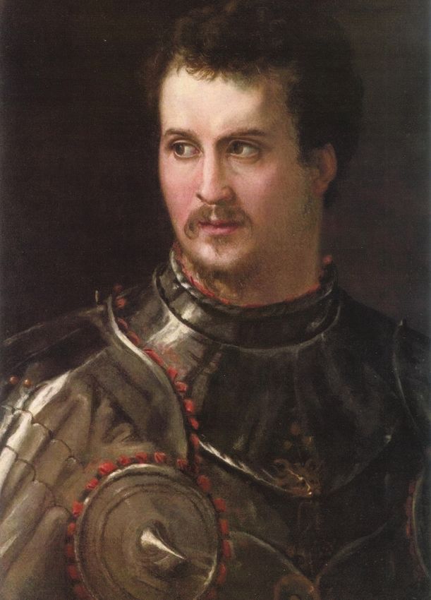
Syphilis in Maria Salviati (1499–1543

                                                                           From the late 15th to mid-16th centuries, Italy
                                                                      was a great field of war and an ideal social environ-
                                                                      ment for the spread of the disease. In the Renaissance,
                                                                      before the advent of the Catholic Counter-Reforma-
                                                                      tion, Italy experienced an increase in the volume of
                                                                      trade exchanges, new contacts between populations,
                                                                      migration from other countries, and, above all, a time
                                                                      of greater sexual liberty (5,19). The activity of pros-
                                                                      titution among the troops and the civilians in the
                                                                      towns, as well as the opportunities for extramarital
                                                                      sex created by the permanence of armies, generated a
                                                                      perfect basis for the spread of syphilis (7,9). Because
                                                                      of its sexual connotation, the disease embodied the
                                                                      concept of divine ill-punishment, which, as an arche-
                                                                      type, pervaded the popular religious sensibility of
                                                                      European populations of the early modern age (20).
                                                                      The early pandemic and violent phase of syphilis, be-
                                                                      fore the classical chronicization to three stages, had
                                                                      an impressive impact on European society (5,21).
                                                                           The first physician observing syphilis was Ales-
                                                                      sandro Benedetti, field doctor of the Italian confederate
                                                                      army fighting against the French during the battle of
                                                                      Fornovo in 1495 (22). Girolamo Fracastoro (1548) suc-
                                                                      cessfully coined the name of the disease in his famous
Figure 1. Portrait of Maria Salviati and Giulia de’ Medici depicted
by Pontormo (Jacopo Carucci) in 1537 c. Oil on panel. 34.65 ×
28.07 in. (88 × 71.3 × 1 cm). (The Walters Art Museum, Baltimore.)

Medici, which revealed lesions typical of third-stage
syphilis, has enabled us to understand the dynamics
of the infection in one of the most famous families of
the Renaissance and to examine the perception of the
illness in 16th century Europe.

Syphilis in the 16th Century
After the 1494 invasion of Italy by the troops of
Charles VIII, King of France (1470–1498), venereal
syphilis had a pandemic spread in Italy and in Eu-
rope (9). The origin of the disease remains one of the
greatest issues in the history of medicine and is still
discussed by scholars. One theory suggests the dis-
ease originated in the Americas and was introduced
by Columbus’ crew returning to Europe from the
New World in 1493. According to a second theory,
syphilis previously existed in the Old World but went
unrecognized until the late 15th century, when there
was increased prevalence and virulence of the disease
(14). In the past few years, further paleopathologic ev-
idence has indicated the presence of the disease in Eu-
rope before 1492 (15,16). However, the transmission                   Figure 2. Portrait of an Elderly Lady (Maria Salviati) depicted
at the end of the 15th century is undeniable and can                  by Agnolo Bronzino in 1542–1543 c. Oil on panel. 50 × 39.4 in.
                                                                      (127 × 100 cm). (San Francisco, The Fine Arts Museum of San
be explained in the light of the wider sociocultural                  Francisco, Gift of Mr. Samuel H. Kress, 53670. Image courtesy the
context of the period (17,18).                                        Fine Arts Museums of San Francisco).

                              Emerging Infectious Diseases • www.cdc.gov/eid • Vol. 26, No. 6, June 2020                          1275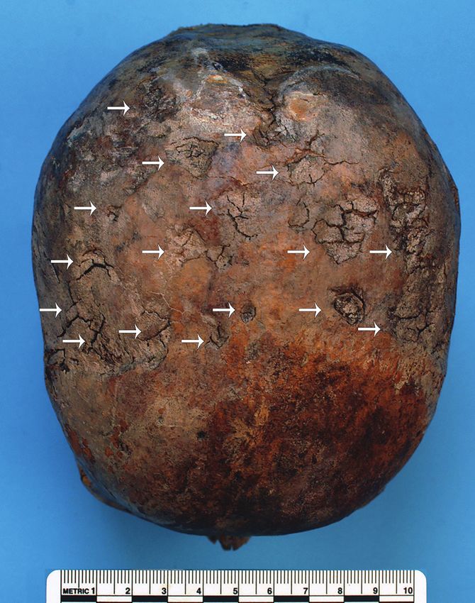
HISTORICAL PERSPECTIVE

poem “Syphilis sive morbus gallicus [Syphilis or the
French disease]” (23). Contemporary physicians de-
scribed the manifestation of the disease, consisting in
the appearance of an ulceration on the penis, followed
by pustules and sores all over the face and body with
joint pain and pruritus. The doctors quickly acknowl-
edged that the infection had been transmitted through
sexual intercourse (17). After the most aggressive first
phase of the “new” disease, syphilis quickly changed
from an acute and debilitating disease into a less se-
vere chronic infection, probably because the selection
of the less virulent strains represented an evolutionary
advantage for the pathogen (21).
     Syphilis was rife in all social classes and affected
many members of the aristocracy. Many noblemen
undertook military careers as captains of mercenary
troops, which typically involved extramarital affairs,
not only with regular lovers but also, and frequently,
with prostitutes (24). Famous are the cases of Cesare
Borgia (1476–1507), son of Pope Alexander VI, who
had to wear a leather mask covering half of his face,
which had been disfigured by syphilis in his later
years (25), and of Francesco II Gonzaga (1466–1519),
Marquis of Mantua, who had a form of tertiary syphi-
lis (23). Evidence that the disease was widespread                     Figure 3. Plan of the crypt of the Medici Chapel with the
in the 16th century aristocratic classes is also dem-                  position of the tomb of Maria Salviati. (Archive of the Division of
onstrated by paleopathology. The cases of Maria of                     Paleopathology. University of Pisa.)
Aragon, Marquise of Vasto (1503–1568) (10), Vespa-
siano Gonzaga, Duke of Sabbioneta (1531–1591) (11),                    from an injury and amputation of his right leg after
and Cardinal Giulio Della Rovere (1533–1578) (12)                      the Battle of Governolo, near Mantua, leaving his
are some of the most famous Renaissance figures for                    wife a widow at the age of 27. Maria never remarried.
whom syphilis was diagnosed.                                           Cosimo, the son of Maria and Giovanni, was called
                                                                       to govern Florence after the death of Duke Alessan-
Brief Biography and Nosography of Maria Salviati                       dro de’ Medici (1537), giving rise to the Grand Ducal
Maria Salviati, daughter of Lucrezia de’ Medici and                    Medici branch, which ruled Tuscany until 1737.
Jacopo Salviati and granddaughter of Lorenzo the                            Archival research by Gaetano Pieraccini was able
Magnificent, was born in Florence in 1499. Her mar-                    to reveal important information about Maria Salvia-
riage to Giovanni de’ Medici (1498–1526) took place                    ti, with particular regard to the last years of her life
in 1516. Giovanni died of gangrene and septicemia                      (24). Until 1540, she enjoyed good health, except for
(26) on November 30, 1526, complications resulting                     a brief episode of fever of unknown origin recorded

Figure 4. The skeletal remains of Maria Salviati at exhumation in 2012. (Archive of the Division of Paleopathology. University of Pisa.)

1276                          Emerging Infectious Diseases • www.cdc.gov/eid • Vol. 26, No. 6, June 2020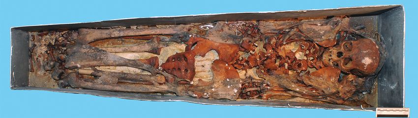
Syphilis in Maria Salviati (1499–1543

in 1517. In the last 3 years of her life, from May 1541                and abdominal colic. The letters also report “chronic
to her death on December 29, 1543, many symptoms                       weakness…, shortness of breath, frequent lipothymic
of severe illness were described in letters sent by An-                episodes, severe syncopes, cold extremities, vomit
drea Pasquali, the court physician, to Duke Cosimo,                    and agitation…” and note “the pulse was deeply re-
including abundant recurring proctorrhagias (bleed-                    duced, the frequency increased” (24). The symptoms
ing from the rectum from ½ to 3 libras of blood [i.e.,                 were certainly the expression of a severe anemia
180 g to ≈1 L]), rectal and perianal ulcers, headaches,                caused by chronic leakages of blood.
                                                                            The portraits of Maria Salviati clearly show the
                                                                       significant changes that occurred in her aspect, mark-
                                                                       ing the progress in terms both of age and of illness.
                                                                       A portrait by Jacopo Pontormo (Figure 1), painted in
                                                                       1537 and preserved at the Uffizi Museum, shows Ma-
                                                                       ria as a beautiful lady, still young; but only 6 years
                                                                       later, in a portrait by Bronzino (Figure 2), she ap-
                                                                       pears as a very old woman. Rather than to the artistic
                                                                       choices of the painter, this transformation seems to be
                                                                       strongly related to the accelerating physical decay of
                                                                       Maria, which was probably connected to the illness in
                                                                       the last years of her life.

                                                                       Historical, Archeological, and
                                                                       Taphonomic Background
                                                                       Almost all the bodies of the Medici, and also that of
                                                                       Maria Salviati, were embalmed before burial (24), but it
                                                                       was not until the mid-19th century that they were given
                                                                       a definitive grave location. Until the 19th century, the
                                                                       bodies of the Medici of the Grand Duchy dynasty were
                                                                       preserved in wooden coffins inside the 2 sacristies of the
                                                                       Basilica of San Lorenzo in Florence. During 1857–58, the
                                                                       Gran Duke of Tuscany Leopold II of Lorrain arranged
                                                                       the bodies of the Medici in the crypt of the Medici Chapel
                                                                       to give a proper and dignified burial place to the found-
                                                                       ers of the Grand Duchy of Tuscany (27). The body of
                                                                       Maria Salviati, identified in 1857 thanks to the presence
                                                                       of a copper epigraph on the coffin (28), was deposed, to-
                                                                       gether with that of her husband Giovanni “of the Black
                                                                       Bands,” in a tomb at the center of the crypt floor (Figure
                                                                       3). We have a description of this event given by Luigi
                                                                       Passerini-Rilli, director of the State Archive of Florence
                                                                       and responsible for the recognition of the bodies of the
                                                                       Grand Dukes (28): “The body, although reduced to al-
                                                                       most a skeleton in the face, was however very well pre-
                                                                       served in the other parts.... The head lay on two bricks....
                                                                       The clothing that covered it resembled that of a nun, i.e.
                                                                       a black cloth, but eaten by the moths: the leftovers of the
                                                                       wimple were still discernible, though the veil that at first
                                                                       covered the head, was worn....”
                                                                            In 1946, Gaetano Pieraccini and the anthropologist
                                                                       Giuseppe Genna conducted an exhumation (29); they
                                                                       heavily manipulated the skeletal remains of Maria
Figure 5. Present-day bones of the skeleton of Maria Salviati
                                                                       with the removal of residual soft tissues before reburial
(gray). Distribution of lesions (black). (Archive of the Division of   in a small zinc coffin (27). On this occasion, they made
Paleopathology. University of Pisa.)                                   a plaster cast of the skull, which is now in the Museum

                                Emerging Infectious Diseases • www.cdc.gov/eid • Vol. 26, No. 6, June 2020                    1277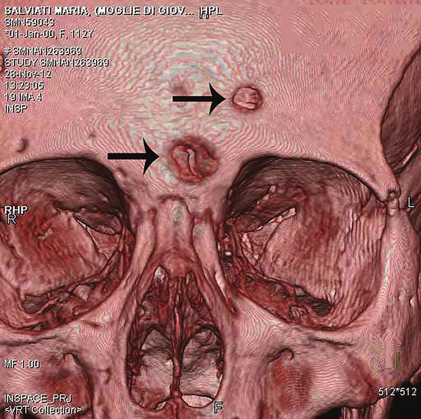
HISTORICAL PERSPECTIVE
The poor dental health of Maria is consistent with that
of the other members of the Medici family (35). We de-
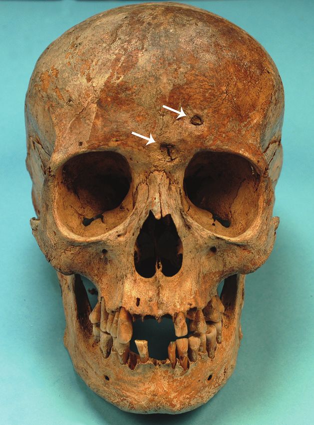
tected many lytic lesions on the skull. Two circular ec-
tocranial depressions are visible on the frontal bone, on
the glabella and above the left supraorbital ridge. They
are irregularly elliptical in shape, measuring ≈1 × 0.7 cm
and 0.5 × 0.4 cm, respectively, with a central destruc-
tive focus and a reactive compact bone formation on
the margins (Figure 6). These frontal bone lesions are
consistent with 2 destructive osteolytic inflammatory
processes, in advanced reparative phase, as clearly re-
vealed by CT examination (Figures 7, 8). Furthermore,
the cranial vault on the parietal bones shows several os-
teolytic lesions in the form of circumvallate depressed
areas with fine scar lines radiating inside the shallow
depressions (Figures 9, 10). CT examination confirms
the lytic and reparative nature of the lesions, which are
morphologically similar to internodular stellate depres-
sions. We observed no other lesions in the postcranial
bones macroscopically, by radiograph, or by CT scan.
The presence of strong bone reaction excludes meta-
static osteolytic carcinoma, multiple myeloma, tubercu-
losis, and fungal bone infections (36,37). The presence of
superficial circumvallated cavitations with radial scars
is pathognomonic of cranial syphilis (caries sicca) (38).
Discussion
Figure 6. The skull of Maria Salviati in frontal view. Cavitations The combination of crater-like lesions, such as cir-
on the frontal bone are apparent. (Archive of the Division of
cumvallate cavitations on the frontal bone (phase 4
Paleopathology. University of Pisa.)
of Anthropology of Florence. In November 2012 the
last exhumation took place, under the management
of the Division of Paleopathology of the University of
Pisa, during some architectural checks of the stability
of the floor of the Chapel. The paleopathologists found
that the remains of Maria were in excellent state of
preservation, unconnected in a small zinc coffin bear-
ing an epigraph with her name (Figure 4).
Anthropologic and Paleopathologic Study
We examined the nearly complete skeleton of Maria
Salviati macroscopically (Figure 5) and performed ra-
diographic and computed tomography (CT) scans. We
compared the skull bone recovered during exhumation
with the skull cast, and they showed the same lesions.
The anthropologic study of the skeleton revealed a fe-
male individual (30), 40–45 years of age (31–34), with
a stature of 1.56 m (34). Maria had severe periodon-
tal disease, as evidenced by the resorption of alveolar
Figure 7. Volume rendering of the skull of Maria Salviati. Two
edges and an abscess at the buccal portion of the third destructive osteolytic inflammatory processes, in advanced
right maxillary molar. Nonpenetrating caries affected reparative phase (circumvallate cavitations), are apparent.
10 teeth, of which 4 were mandibular and 6 maxillary. (Archive of the Division of Paleopathology. University of Pisa.)
1278 Emerging Infectious Diseases • www.cdc.gov/eid • Vol. 26, No. 6, June 2020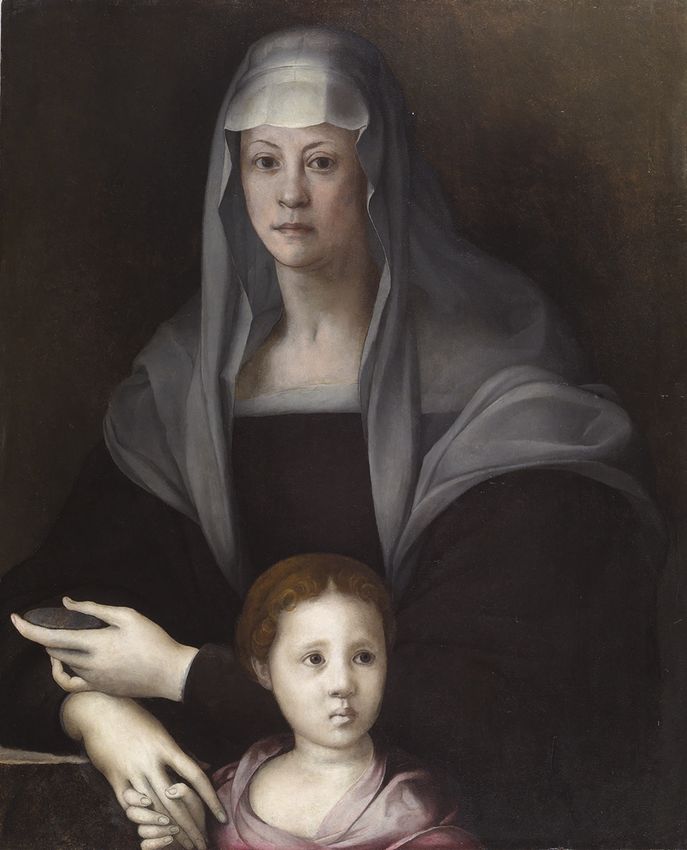
Syphilis in Maria Salviati (1499–1543

                                                                      Cosimo was laboriously trying to promote among the
                                                                      royal rank. The fact that in her last years of life Maria
                                                                      was always portrayed with a veil might indicate her in-
                                                                      tention to hide the luetic skin lesions.
                                                                           Syphilis is likely to have been more widespread
                                                                      among the noblewomen of the Renaissance than is at-
                                                                      tested by the written sources. Paleopathology has in
                                                                      some cases revealed some hidden illnesses of the Ital-
                                                                      ian noblewomen, as in the case of Isabella of Aragon,
                                                                      Duchess of Milan (1470–1524), and Maria of Aragon
                                                                      (1503–1568), Marquise of Vasto and wife of the gov-
                                                                      ernor of Milan Alfonso of Avalos (1502–1546). Isabella
                                                                      was probably affected by syphilis, despite the absence
                                                                      of bone lesions on her skeletal remains. The syphilitic
                                                                      infection was diagnosed indirectly, on the basis of the
                                                                      paleopathologic analyses performed on her teeth (42).
                                                                      On the buccal surfaces of the teeth, Isabella showed a
                                                                      strong abrasion caused by pumice powder and tooth-
                                                                      picks of cuttlebone that she used to remove the blackish
Figure 8. Computed tomography scan of frontal bone with an            patina produced by her mercurial therapy. In fact, en-
osteolytic lesion with sclerotic walls (circumvallate cavitations).   ergy-dispersive spectroscopic analysis of the dark ma-
(Archive of the Division of Paleopathology. University of Pisa.)      terial detected a massive presence of mercury, largely
                                                                      employed in the treatment of syphilis since the early
in the Caries Sicca sequence depicted in Hackett [38]),               16th century in the form of salves, fumigations, and
and circumvallate cavitations with radial scars on the                ointments (43,44). The care given to Isabella of Aragon
parietal bones (phase 5 in the Caries Sicca sequence
[38]) is pathognomonic for tertiary syphilis. Pathogra-
phy attests that Maria had abundant recurring proc-
torrhagias. This symptom strongly suggests tertiary
syphilis with cranial and possible colorectal localiza-
tion. Apart from proctorrhagias, the other symptoms,
such as frequent fever and headache, abdominal
colic, rectal and perianal ulcers, reported by the writ-
ten sources are very compatible with tertiary syphi-
lis (4,39,40) but were not attributed to syphilis by the
physicians of that time nor by the nosographic schol-
ars in recent times (24). Nevertheless, rectal syphilis is
a rare disease that usually shows proctitis with peri-
anal ulcers but lacks pathognomonic clinical symp-
toms, making diagnosis difficult. In clinical medicine,
anal syphilis could be easily misdiagnosed as cancer
or advanced stage hemorrhoids, upholding the repu-
tation of syphilis as the “great mimicker” (41).
     One hypothesis is that the contemporary physicians
might have correctly diagnosed and recognized Maria
Salviati’s disease but concealed the sexual component
of the infection. Another hypothesis is that Maria Sal-
viati, who never allowed the physicians to inspect her
genitals (24), might have hidden the symptoms of her
disease out of modesty. A further explanation, based
on political reasons, is that the mother of Duke Cosimo                Figure 9. Parietal bones of Maria Salviati, showing several
I could not appear to be affected by venereal syphilis,                radial scars typical of tertiary syphilis. (Archive of the Division of
to avoid corrupting the image of the Medici family that                Paleopathology. University of Pisa.)

                                Emerging Infectious Diseases • www.cdc.gov/eid • Vol. 26, No. 6, June 2020                               1279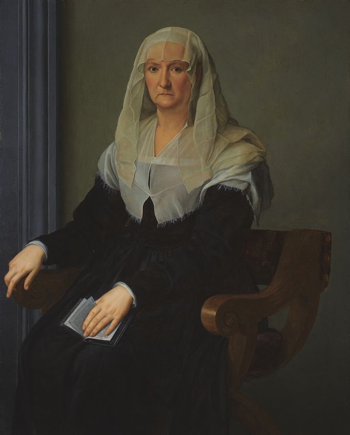
HISTORICAL PERSPECTIVE

clearly demonstrates her willingness to erase the evi-
dent traces of chronic mercury intoxication caused by
the antisyphilitic therapy. However, no references to
syphilis are reported in the written documents about
the life of Isabella. In the artificial mummy of Maria of
Aragon, the histologic, immunohistochemical, and ul-
trastructural study of a cutaneous ulcer of the left arm
led to the direct identification of Treponema pallidum
and the diagnosis of tertiary venereal syphilis (10). The
biographic sources report that Maria of Aragon peri-
odically spent time at the Agnano Baths, near Naples
(45), probably to treat a skin disease with the sulfuric
waters. However, in the written sources, there is no
mention of any possible syphilitic infection affecting
the noblewoman, who was famous at that time for her
beauty and cultural refinement.
     It is difficult to speculate on how Maria Salviati
contracted the disease, but she is likely to have been
infected by her husband Giovanni before his death in
1526, and more probably after the birth of her son Co-
simo (June 12, 1519); indeed, the historical sources do        Figure 10. Volume rendering of the skull of Maria Salviati,
not reveal any details about a possible infection of the       showing contemporary presence of lytic (superficial cavitations)
                                                               and reparative lesions (radial scars).(Archive of the Division of
child, nor do the skeletal remains of the first Grand          Paleopathology. University of Pisa.)
Duke of Tuscany show any signs of congenital syphi-
lis (46). The lifestyle of Giovanni “of the Black Bands”
(Figure 11) was characterized by intense sexual extra-         characterized by occasional relationships with pros-
marital affairs, as witnessed by many documents of             titutes. It was already a well-known fact at the time
the time preserved in the archives of Florence (24). On        that syphilis was transmitted sexually (5); therefore,
October 20, 1521, Giovanni wrote to his treasurer and          some preventive practices were undertaken by the
lieutenant Francesco Albizi to dispose of war supplies         noblewomen, wives, or lovers of the men affected
during the military campaign against the French army           by the disease. Just like Isabella d’Este, wife of Fran-
in Northern Italy: “send me that Greek whore I left in         cesco II Gonzaga, Marquise of Mantua, these women
Viterbo” (47). On September 22, 1522, during some              sometimes refused to have sexual intercourse with
military actions in the Marche region on behalf of the         their partners (7). Although the disease had no con-
Pope, Giovanni wrote again to Francesco Albizi, or-            sequences on the reputation of the men, whose sexual
dering him to kidnap “Lucrezia, courtesan of Rome”             conduct was not subject to moral censorship, the situ-
and to bring her to him by force (48). A considerable          ation was different for the women, who tried to hide
series of names of prostitutes frequented by Giovanni          their sexually transmitted infections (6).
are cited in his correspondence: Flora from Padua,
Nicolosa “the painted Jewess,” Camilla Orsini, Gi-             Conclusions
ulia, Angelica “the Venetian,” Lorenzina “the Greek            A diagnosis of syphilis for Maria Salviati does not
slave,” Baccia from Rome, Lucrezia nicknamed “Ma-              emerge from the historical sources, although the
trema non vole [Mom does not want],” and Paolina               symptoms that manifested in the last years of her
(49,50). The skeleton of Giovanni de’ Medici does not          life are compatible with a colorectal localization and
reveal any lesion of syphilis (26), probably because he        severe hemorrhages caused by syphilitic infection.
died at the age of 28 years, before the development of         In her final years, Maria had a very withdrawn life,
the tertiary stage of the disease.                             possibly to conceal the signs of the illness and cer-
     Extramarital affairs were common among aris-              tainly for the social complications caused by her re-
tocrats (7); therefore, syphilis could be considered a         current anal hemorrhages (24). However, we have
disease characterizing the pathocenosis of high so-            no reports that she was marginalized from the du-
cial classes. Noblewomen were at risk for contracting          cal court; instead, she was held in high regard as the
sexually transmitted diseases caused by the lifestyle          mother of the reigning duke. In the famous portrait
of their husbands, who led an unregulated sexual life          painted by Bronzino during 1542–1543 (Figure 2),

1280                     Emerging Infectious Diseases • www.cdc.gov/eid • Vol. 26, No. 6, June 2020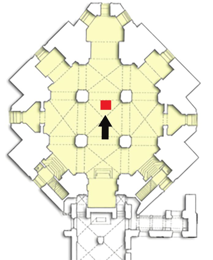
Syphilis in Maria Salviati (1499–1543

she appears veiled and in widow’s clothes, as if to                Acknowledgments
hide the signs of illness.                                         We are grateful to Gino Fornaciari for his revision of the
     The paleopathologic study of Maria Salviati re-               text, to Laura Cignoni for the linguistic revision of the
veals osteolytic and reparative lesions on the skull,              manuscript, and the anonymous consultants for their help-
which are pathognomonic for syphilis. The diagno-                  ful suggestions. We also thank Roberto Carpi for provid-
sis of syphilis for Maria Salviati enables us to directly          ing CT data.
observe the gender-based consequences of a pathol-
                                                                   The Fondazione Arpa and the Associazione Ortopedica
ogy to which the Renaissance noblewomen were sub-
                                                                   Italiana supported this research. .
jected as a consequence of the sexual conduct of their
husbands. The real nature of the disease might have
been kept secret at the time for reasons of political op-          About the Author
portunity and privacy. A disease that was not a rea-               Dr. Fornaciari is a postdoctoral fellow in History of Medi-
son of particular shame for sovereigns, princes, and               cine and Paleopathology at the Division of Paleopathol-
gentlemen, that did not affect their political role and            ogy, University of Pisa. His primary research interests are
leadership, and was therefore unnecessary to hide,                 paleopathology, archaeology, and the history of medicine
was instead jealously concealed by noblewomen as a                 of the medieval and postmedieval age.
“secret illness” that often did not seep outside the pri-
vate apartments and perhaps did not even reach the
attention of the court physicians. This attitude reveals           References
                                                                    1.   Peeling RW, Mabey D, Kamb ML, Chen XS, Radolf JD,
a disparity of perception and of mentality, symptom-                     Benzaken AS. Syphilis. Nat Rev Dis Primers. 2017;3:17073.
atic of gender discrimination that was well implanted                    https://doi.org/10.1038/nrdp.2017.73
in the heart of Renaissance society (6,20)                          2.   Centers for Disease Control and Prevention. Sexually
                                                                         transmitted disease surveillance, 2016. Atlanta: US
                                                                         Department of Health and Human Services; September 2017.
                                                                    3.   European Centre for Disease Prevention and Control.
                                                                         Syphilis. Annual epidemiological report for 2016. Stockholm:
                                                                         The Centre; 2018.
                                                                    4.   Lukehart SA. Syphilis. In: Fauci AS, Braunwald E,
                                                                         Kasper DL, Hauser SL, Longo DL, Jameson JL, et al., editors.
                                                                         Harrison’s principles of internal medicine. 17th edition. New
                                                                         York: McGraw-Hill; 2008. p. 1038–46.
                                                                    5.   Tognotti E. The rise and fall of syphilis in Renaissance
                                                                         Europe. J Med Humanit. 2009;30:99–113. https://doi.org/
                                                                         10.1007/s10912-009-9079-3
                                                                    6.   Schleiner W. Moral attitudes toward syphilis and its
                                                                         prevention in the Renaissance. Bull Hist Med. 1994;68:389–410.
                                                                    7.   Tognotti E. Prevention strategies and changes in sexual
                                                                         mores in response to the outbreak of syphilis in Europe
                                                                         in the early modern age. J Anc Dis Prev Rem. 2014;2:113.
                                                                         https://doi.org/10.4172/2329-8731.1000113
                                                                    8.   Boehrer BT. Early modern syphilis. J Hist Sex. 1990;1:
                                                                         197–214.
                                                                    9.   Foa A. The new and the old: the spread of syphilis
                                                                         (1494–1530). In: Muir E, Ruggiero G, editors. Sex and gender
                                                                         in historical perspective. Baltimore: Johns Hopkins
                                                                         University Press; 1990. p. 26–45.
                                                                   10.   Fornaciari G, Castagna M, Tognetti A, Tornaboni D, Bruno J.
                                                                         Syphilis in a Renaissance Italian mummy. Lancet. 1989;2:614.
                                                                         https://doi.org/10.1016/S0140-6736(89)90729-0
                                                                   11.   Mallegni F, Bedini E, Fornaciari G. The study of human
                                                                         remains in Vespasiano Gonzaga’s tomb 400 years later [in
                                                                         Italian]. Sabbioneta (Italy): Edizioni A Passo d’Uomo; 1991.
                                                                         p. 55–110.
                                                                   12.   Fornaciari G, Vitiello A. The tombs of Della Rovere. In:
                                                                         Vastano A, editor. Enigmas and new discoveries: the
                                                                          monastery of Santa Chiara in Urbino [in Italian]. Urbino
                                                                         (Italy): Arti Grafiche Editoriali; 2012. p. 143–66.
Figure 11. Portrait of Giovanni de’ Medici of the Black Bands,     13.   Grmek M. Diseases at the beginning of the Western
depicted by Francesco de’ Rossi circa 1546–1548. Oil on panel.           civilization [in French]. Paris: Payot; 1983.
25.6 × 18.1 in. (65 × 46 cm). (Florence, Istituti Museali della    14.   Harper KN, Zuckerman MK, Harper ML, Kingston JD,
Soprintendenza Speciale per il Polo Museale Fiorentino, Palazzo          Armelagos GJ. The origin and antiquity of syphilis

                             Emerging Infectious Diseases • www.cdc.gov/eid • Vol. 26, No. 6, June 2020                          1281
HISTORICAL PERSPECTIVE
revisited: an appraisal of Old World pre-Columbian in the human skeleton. Springfield (IL): Charles C. Thomas
evidence for treponemal infection. Am J Phys Anthropol. Publisher; 1989. p. 105–35.
2011;146(Suppl 53):99–133. https://doi.org/10.1002/ 34. Trotter M, Gleser GC. Corrigenda to “Estimation of stature
ajpa.21613 from long limb bones of American Whites and Negroes”
15. Walker D, Powers N, Connell B, Redfern R. Evidence of Am. J. Phys. Anthrop. (1952). Am J Phys Anthropol.
skeletal treponematosis from the medieval burial ground of 1977;47:355–6. https://doi.org/10.1002/ajpa.1330470216
St. Mary Spital, London, and implications for the origins of 35. Colagrande S, Villari N, Pierleoni F, Weber D, Fornaciari G,
the disease in Europe. Am J Phys Anthropol. 2015;156:90–101. Lippi D. Teeth of the Renaissance: a paleopathological and
https://doi.org/10.1002/ajpa.22630 historic-medical study of the jaws of the Medici Family.
16. Roberts CA, Millard AR, Nowell GM, Gröcke DR, Journal of Forensic Radiology and Imaging. 2013;1:193–200.
Macpherson CG, Pearson DG, et al. Isotopic tracing of the https://doi.org/10.1016/j.jofri.2013.07.004
impact of mobility on infectious disease: The origin of people 36. Waldron T. Paleopathology. Cambridge: Cambridge
with treponematosis buried in hull, England, in the late University Press; 2012.
medieval period. Am J Phys Anthropol. 2013;150:273–85. 37. Roberts CA, Buikstra JE. Bacterial Infections. In: Buikstra JE,
https://doi.org/10.1002/ajpa.22203 editor. Ortner’s identification of pathological conditions in
17. Arrizabalaga J, Henderson J, French R, French RK. The great human skeletal remains. New York: Academic Press 2019;
pox: the French disease in Renaissance Europe. New Haven p. 321–439.
(CT): Yale University Press; 1997. 38. Hackett C. Diagnostic criteria of syphilis, yaws and treponarid
18. Meyer C, Jung C, Kohl T, Poenicke A, Poppe A, Alt KW. (treponematoses) and of some other diseases in dry bones (for
Syphilis 2001—a palaeopathological reappraisal. Homo. use in osteo-archaeology). Berlin: Springer-Verlag; 1976.
2002;53:39–58. https://doi.org/10.1078/0018-442X-00037 39. Scolari EG, Giannotti B. Syphilis and other venereal diseases
19. Oriel JD. The scars of Venus. A history of venereology. [in Italian]. Turin (Italy): UTET; 1972.
London: Springer Verlag; 1984. 40. Serigado J, Lewis E, Kim G. Rectal bleeding caused
20. Siena K, editor. Sins of the flesh: responding to sexual disease by a syphilitic inflammatory mass. BMJ Case Rep.
in early modern Europe. Toronto: Centre for Reformation 2019;12:e226595. https://doi.org/10.1136/bcr-2018-226595
and Renaissance Studies; 2005. 41. Arnold CA, Roth R, Arsenescu R, Harzman A,
21. Knell RJ. Syphilis in renaissance Europe: rapid evolution of Lam-Himlin DM, Limketkai BN, et al. Sexually transmitted
an introduced sexually transmitted disease? Proc Biol Sci. infectious colitis vs inflammatory bowel disease: distinguish-
2004;271(Suppl 4):S174–6. ing features from a case-controlled study. Am J Clin Pathol.
22. Benedetti A. Diary of the Caroline War. Schullian DM, editor. 2015;144:771–81. https://doi.org/10.1309/AJCPOID4JIJ6PISC
New York: Renaissance Society of America; 1967. 42. D’Errico F, Villa G, Fornaciari G. Dental esthetics of an Ital-
23. Tognotti E. The other face of Venus. Syphilis from the early ian Renaissance noblewoman, Isabella d’Aragona. A case of
modern age to the advent of AIDS (15th–20th century) [in chronic mercury intoxication. Ossa. 1988;13:207–28.
Italian]. Milan: Franco Angeli, 2006. 43. O’Shea JG. ‘Two minutes with venus, two years with
24. Pieraccini G. The Medici of Cafaggiolo [in Italian]. Vol. 2. mercury’—mercury as an antisyphilitic chemotherapeutic
Florence (Italy): Nardini Editore, 1986. agent. J R Soc Med. 1990;83:392–5. https://doi.org/10.1177/
25. Bradford S. Ceasare Borgia: his life and times. London: 014107689008300619
Futura Publications; 1981. 44. Tilles G, Wallach D. History of the treatment of syphilis
26. Fornaciari G, Bartolozzi P, Bartolozzi C, Rossi B, Menchi I, with mercury: five centuries of uncertainty and toxicity [in
Piccioli A. A great enigma of the Italian Renaissance: French]. Rev Hist Pharm (Paris). 1996;44(suppl):347–51.
paleopathological study on the death of Giovanni dalle https://doi.org/10.3406/pharm.1996.6244
Bande Nere (1498–1526) and historical relevance of a leg 45. Fiorentino F. Studies and portraits of the Renaissance
amputation. BMC Musculoskelet Disord. 2014;15:301. [in Italian]. Bari (Italy): Giuseppe Laterza & Figli; 1911.
https://doi.org/10.1186/1471-2474-15-301 46. Fornaciari G, Vitiello A, Giusiani S, Giuffra V, Fornaciari A,
27. Lippi D. Unmourned graves. Curiosity and scientific Villari N. The Medici Project. First anthropological results of
research in history of the Medici exhumations [in Italian]. the exploration of the Medici Tombs in Florence. Med Secoli.
Florence (Italy): Firenze University Press; 2006. 2007;19:521–44.
28. Sommi Picenardi G. Exhumation and recognition of the 47. Gauthiez P. New documents about Giovanni de’ Medici
ashes of the Medici Princes in the year 1857. Minutes and named delle Bande Nere [in Italian]. Arch Stor Ital.
notes [in Italian]. Arch Stor Ital. 1888;165:333–60. 1902;227:333–60.
29. Genna G. Anthropological research on the Medici family 48. Milanesi C. Unpublished letters and testament of G. de ‘M.
[in Italian]. Atti Accademia Nazionale dei Lincei, Classe di called delle Bande Nere with others by Maria and Jacopo
Scienze Fisiche. Matematiche e Naturali. 1948;15:589–93. Salviati of princes, cardinals, captains, family members and
30. Buikstra J, Ubelaker D. Standards for data collection from soldiers gathered by cav. Filippo Moisè [in Italian]. Arch Stor
human skeletal remains. Fayetteville (AR): Arkansas Ar- Ital. 1859;9:3–29.
chaeological Survey Research Series No. 44; 1994. 49. Orlando F, Baccini G. Courtesans of the XVI century.
31. Lovejoy CO. Dental wear in the Libben population: its Letters–Curiosity–News–Anecdotes [in Italian]. Florence
functional pattern and role in the determination of adult (Italy): Il “Giornale di Erudizione” Editore; 1892.
skeletal age at death. Am J Phys Anthropol. 1985;68:47–56. 50. De Gubernatis A. Love letters of women to Giovanni dalle
https://doi.org/10.1002/ajpa.1330680105 B. N. [in Italian]. Rivista D’Italia. 1902;8.
32. Brooks ST, Suchey JM. Skeletal age determination based on
the os pubis: a comparison of the Acsadi–Nemeskeri and Address for correspondence: Antonio Fornaciari, Department of
Suchey–Brooks methods. Hum Evol. 1990;5:227–38.
Translational Research and New Technologies in Medicine and
https://doi.org/10.1007/BF02437238
33. Loth SR, Iscan MY. Morphological assessment of age in the Surgery, University of Pisa, Via Roma 57, 56126 Pisa, Italy;
adult: the thoracic region. In: Iscan MY, editor. Age markers email: antonio.fornaciari@med.unipi.it
1282 Emerging Infectious Diseases • www.cdc.gov/eid • Vol. 26, No. 6, June 2020You can also read