Syringomyelia and Chiari Syndrome Registry: advances in epidemiology, clinical phenotypes and natural history based on a North Western Italy ...
←
→
Page content transcription
If your browser does not render page correctly, please read the page content below
48
                         Ann Ist Super Sanità 2020 | Vol. 56, No. 1: 48-58
                         DOI: 10.4415/ANN_20_01_08

                         Syringomyelia and Chiari Syndrome
                         Registry: advances in epidemiology,
  articles and reviews

                         clinical phenotypes and natural history
                         based on a North Western Italy cohort
                         Palma Ciaramitaro1, Diego Garbossa2, Paola Peretta3, Gianluca Piatelli4, Luca Massimi5,
                         Laura Valentini6, Giuseppe Migliaretti7, Simone Baldovino8, Dario Roccatello8, Yllka Kodra9,
                         Domenica Taruscio9, on behalf of the Interregional Chiari and Syringomyelia Consortium*
O riginal

                         1Centro Regionale Esperto Siringomielia e Sindrome di Chiari (CRESSC), Dipartimento di Neuroscienze,
                         AOU Città della Salute e della Scienza di Torino, Turin, Italy
                         2Neurochirurgia, Università degli Studi di Torino, Turin, Italy

                         3Neurochirurgia Pediatrica, Ospedale Infantile Regina Margherita, AOU Città della Salute e della Scienza

                         di Torino, Turin, Italy
                         4Neurochirurgia, Istituto Giannina Gaslini, Genoa, Italy

                         5Neurochirurgia Pediatrica, Fondazione Ospedale Agostino Gemelli, Università Cattolica di Roma, Rome, Italy

                         6Neurochirurgia, Fondazione IRCCS Istituto Neurologico Carlo Besta, Milan, Italy

                         7Dipartimento di Salute Pubblica e Microbiologia, Unità di Statistica, Università degli Studi di Torino, Turin,

                         Italy
                         8CMID, Centro di Coordinamento Rete Interregionale per le Malattie Rare del Piemonte e della Valle d’Aosta

                         – San Giovanni Bosco Hospital and Dipartimento di Scienze Cliniche e Biologiche – Università degli Studi
                         di Torino, Turin, Italy
                         9Centro Nazionale Malattie Rare, Istituto Superiore di Sanità, Rome, Italy

                         *the members of the Interregional Chiari and Syringomyelia Consortium are listed before the references

                         Abstract                                                                                                        Key words
                         Background. Syringomyelia and Chiari Syndrome are classified as rare diseases, but                              • epidemiology
                         current known occurrence in Europe is missing. The increased ability to diagnose these                          • rare disease
                         pathologies by magnetic resonance imaging and its widespread availability has led to an                         • Syringomyelia
                         increase of reported cases, often asymptomatic, with the need to standardize definitions,                       • Chiari Malformation
                         diagnostic criteria and treatments.                                                                             • recommendations
                         Aims. We present shared Interregional Recommendations developed with the primary
                         aim to estimate Syringomyelia and Chiari Syndrome prevalence and incidence in North
                         Western Italy, with special reference to symptomatic forms.
                         Methods. An agreement for the standardization of definitions, classifications, diagnostic
                         criteria and surgical Recommendations was reached by the multidisciplinary Interregion-
                         al Piemonte and Valle d’Aosta Chiari-Syringomyelia Consortium (Delphi method); next,
                         in 2011 a census for Syringomyelia and Chiari Malformation was performed through the
                         Interregional Piemonte and Valle d’Aosta Rare Disease Registry, integrated by a dedi-
                         cated form in order to estimate prevalence and incidence.
                         Results. 436 patients, 292 females, met shared interregional diagnostic criteria. Syringo-
                         myelia prevalence was estimated in 4.84:100 000; Chiari Malformation prevalence was
                         7.74:100 000; incidence was 0.82:100 000 and 3.08:100 000 respectively. Demographics,
                         neuroradiological parameters and aetiology were reported (in symptomatic and asymp-
                         tomatic forms). Finally, symptoms and signs, familiar and natural history were analyzed.
                         Conclusions. First Italian epidemiological data (prevalence, incidence) on Chiari and
                         syringomyelia was collected, according to shared diagnostic Recommendations. Future
                         perspectives include the adoption of these Recommendations at national level to stan-
                         dardize the access to diagnosis and care process and promote multicenter clinical trials.

                         Address for correspondence: Palma Ciaramitaro, Azienda Ospedaliero-Universitaria Città della Salute e della Scienza di Torino, Presidio CTO,
                         Via Zuretti 29, Turin, Italy. E-mail: palma.ciaramitaro@gmail.com.
49
Chiari-Syringomyelia recommendations
INTRODUCTION over, it obliges the Hospital Units to report RD in the
Syringomyelia (Syr) is morphologically defined at Regional Registry [10], with epidemiological and legisla-
magnetic resonance imaging (MRI) as the presence of tive purposes (i.e. to regulate the access on exemption
single or multiple fluid-filled cavity (syrinx) within the path for RD). In 2005 because of the low prevalence
parenchyma of the spinal cord and/or the bulb (Syringo- estimates, the potential clinical severity in chronically
articles and reviews
bulbia) and classified as a rare disease (ORPHA3280). debilitating nature and the resulting significant expense
About 50% of Syr patients have severe neurological dam- for its treatment, Syr was inserted as RD involving the
age, chronic-progressive disability with complete loss nervous system in Piemonte and Valle d’Aosta [11].
of independence. Prognostically speaking, even more Now, with the adoption of the new National law on Es-
unfavourable is the presence of Syringobulbia (swal- sential Assistance Levels [12], Syr is recognized also as
lowing and breathing bulbar centers involved). Before RD in all Italian regions, but previously this was recog-
1968, neurologists diagnosed Syr only by typical neu- nized as RD only in Piemonte, Valle d’Aosta, Toscana
rological symptoms. Since the introduction of CT my- and Marche; for this reason Italian epidemiological data
elography and MRI techniques for spinal cord imaging, on Syr was not available in the RD National Registry.
the diagnosis has become easier, and many cases have Syr and CM are classified as rare diseases on Or-
been reported. In studies conducted before the advent phanet, the International reference portal for RD and
O riginal
of modern neuroimaging, prevalence ranged from 3.3 to orphan drugs, but current prevalence data is missing.
8.5/100 000 [1-3]; after the advent of MRI estimated Guidelines on diagnostic criteria and case definition
prevalence ranged from 1.9 to 8.4/100 000 [4, 5]. are missing, and consequently estimation of the preva-
Chiari Malformation (CM) includes a heterogeneous lence in Piemonte and Valle d’Aosta for symptomatic
group of abnormalities characterized by the caudal cer- and asymptomatic forms are missing.
ebellum ptosis through the foramen magnum; clinical In 2008 a consortium dedicated to the study of Syr
manifestations define the Arnold-Chiari or Chiari Syn- and CM began its activity in the Piemonte and Valle
drome (CS). The malformation can cause a wide variety D’Aosta regions, as part of the Rare Diseases Network
of neurological symptoms, often vague or nonspecific, of the Italian National Health Service, with the aims to
such as headaches, ocular disturbances, otoneurologic standardize/share definitions, classifications, care and
disturbances, lower cranial nerve signs, cerebellar ataxia, diagnostic approaches.
or spasticity [6]. Onset of symptoms is usually in the The consortium, named the Interregional Chiari-
third decade of life. However, many individuals with Syringomyelia Consortium (CSC), was composed by
CM remain asymptomatic even later in life. A US study clinicians (neurologists, neurosurgeons, neuroradi-
estimated 400 000 patients affected by Chiari type 1 ologists, physiatrists, neuro-urologists, psychologists,
Malformation (CMI), the most common CM type. The speech pathologists, spinal surgeons, pain specialists),
epidemiology of CMI malformation has been scarcely experts of public health for RD, and patient association
investigated. The true population prevalence of CMI is representatives [13]. In 2010 the CSC Recommenda-
unknown and there are no studies on CMI incidence. A tions were proposed: some of these indications derived
retrospective analysis of more than 22 000 neuroimaging from the outcomes of the CSC meetings, others were
diagnoses [7] provided indirect data of CMI prevalence, from the First International Chiari Consensus Confer-
estimable in 1:1280 (0.77%). When CM is defined by ence, held in Milan in 2009 [14].
cerebellar tonsil position 5 mm or more below the fora- Diagnostic, Surgical and Rehabilitative Recommenda-
men magnum, imaging prevalence studies estimate CM tions were approved by the members of Technical-Spe-
prevalence at between 0.24 and 3.6% of the population cialized Task-force supporting the Regional Center of
[8]; the discrepancy between these estimates is a result Coordination for RD and by the following scientific so-
of the different age groups analysed, substantially higher cieties: Italian Society of Neurology, Piemonte and Valle
in children and young adults compared with older adults. d’Aosta section; Italian Society of Neurologists, Neuro-
In Italy and in the European Community the CMI is surgeons, Neuroradiologists, Piemonte-Valle d’Aosta-
classified as a rare disease (ORPHA268882), but cur- Liguria section; Italian Society of Physical Medicine
rent known occurrence data in Europe is missing. and Rehabilitation, Piemonte section [15]. The docu-
The increased ability to diagnose CM and Syr by MRI ment was published as technical-scientific integration of
and its widespread availability has led to an increase of the Regional Legislation (DGR n. 95-13748, 29 March
reported cases, often asymptomatic or minimally symp- 2010), specifying the Institution of the first Center of Ex-
tomatic, with the need to standardize definitions, diag- pertise for Chiari and Syringomyelia in Torino [16, 17].
nostic criteria and treatments. In this study, based on the Interregional Recommen-
In 2001 DM 279/2001, National Law [9] set up the dations, we present the first estimation of prevalence
Italian National Network for rare diseases, to deal with and incidence of Syr and CM in North Western Italy
the prevention, surveillance, diagnosis and treatment of (Piemonte and Valle d’Aosta), with special reference to
rare diseases (RD), including Arnold-Chiari or Chiari symptomatic forms (Symptomatic Syringomyelia: SS;
Syndrome; the same law activated the RD National Reg- Chiari Syndrome: CS).
istry located at the Istituto Superiore di Sanità, which is
expected to receive epidemiological data from Regional MATERIAL AND METHODS
Registries. A Regional Decree (2/3/2004, n. 22-11870) Interregional recommendations
established the Piemonte Regional Network for the pre- In order to estimate the epidemiological indices
vention, screening, diagnosis and therapy of RD. More- (prevalence and incidence), the diagnostic criteria used50
Palma Ciaramitaro, Diego Garbossa, Paola Peretta et al.
for case definition was based on the Interregional Diag- Statistical analysis
nostic, Surgical and Rehabilitative Recommendations. Up to 31 December 2011, the Syr and Chiari age
The Recommendations were developed following sev- standardized prevalence and the incidence rate in 2011
eral steps: a) collecting all available evidence on clini- with 95% confidence intervals (95% CI) in Piemonte
cal studies related to Syr and CM. The following da- and Valle d’Aosta Regions was estimated. The preva-
articles and reviews
tabases were queried for literature review on Syr and lence (number of alive diagnoses up to 2011 year) and
CM: Medline (PubMed interface, www.pubmed.gov)¸ the incidence (number of new reported cases in 2011
Cochrane Library (Health Library of Piedmont, www. year) were estimated using both the Interregional
bvspiemonte.it), National Guidelines Clearinghouse Piemonte and Valle d’Aosta Rare Disease Registry and
(www.guideline.gov). The literature searching strat- the CSC census data for symptomatic forms. The CSC
egy was conducted by combining: the Medical Sub- census data was also used to analyse asymptomatic
ject Heading (MeSH): “Arnold-Chiari Malformation” forms. The standardization was performed by a direct
or “Syringomyelia” or “Arnold-Chiari Malformation” method using the Italian census population at 1st Janu-
AND “Syringomyelia”; Publication type (PT): “System- ary 2011 year (respectively 4 457 335 inhabitants in
atic Reviews”, “Practice Guideline”, “Meta-Analysis”, Piemonte and 128 230 in Valle d’Aosta, 4 585 565 to-
“Randomized Controlled Trial”; b) assessing studies for tal population), according to ISTAT census data (2012)
O riginal
relevance. All studies were selected and critically evalu- [18, 19]. Confidence intervals were calculated assum-
ated. Results with low evidence level were restricted ing a Poisson distribution.
for date (last 10 years) and language (English, French, Basic statistics in terms of frequencies (absolute and
German, Spanish, Italian); these were also selected and percentage values) were calculated for: socio-demo-
critically evaluated; c) categorising the evidence. Each graphic data, age at diagnosis, age at survey, diagnostic
expert member expressed an evaluation of the articles, delay, MRI parameters (morphology/level), diagnoses,
giving a score between 0 (strongly disagree) and 10 associated conditions, neurological symptoms/signs,
(strongly agree), according to the Delphi method [18]. types of surgery.
If there was an agreement, the document was revisited All statistical analyses were performed using the Sta-
in textual form and submitted to the working group for ta statistical software (StataCorp. Statistical Software:
the final approval and subsequent drafting of the ulti- Release 7.0. College Station, TX: Stata Corporation.
mate document; for evaluation lower or equal to 7 it 2001).
was necessary to propose an alternative text version and
relative notes. If no agreement was reached, the docu- RESULTS
ment came back to the recommendation authors with Census results
comments for necessary changes and resubmitted to Using CSC form, 436 patients (292 females and 144
the experts involved. When an agreement was reached, males) met shared diagnostic criteria for Syr and/or CM
the document in the preliminary form was prepared and (Table 3). Demographics, MRI parameters (morphol-
submitted to the judgment of the entire experts group ogy/level), diagnoses, associated conditions, estimated
for final consensus. A description of the sections of the prevalence and incidence data were reported respec-
Recommendations including classifications, radiologi- tively in 347 CM patients and in 217 patients affected
cal and clinical definitions, surgical and conservative by Syr. Percentage of symptomatic Chiari (CS) and
indications are reported in Table 1 and Table 2. symptomatic Syr (SS) are represented in Figure 2. In
2011 the estimated Syr prevalence is 4.84:100 000 and
Census study incidence is 0.82:100 000; the estimated CM preva-
According to these Recommendations, in 2011 a lence is 7.74:100 000, incidence 3.08:100 000 (Table 4
Syr and Chiari census study was performed through and Table 5).
the Interregional Piemonte and Valle d’Aosta Registry
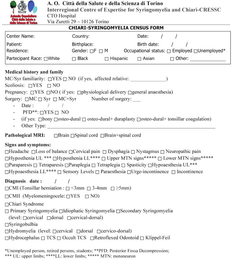
of RD, integrated by a dedicated Case Report Form, Description of clinical manifestations
reported in Figure 1; the CSC form was developed by Major neurological symptoms in CS and in SS were
the Chiari-Syringomyelia Consortium and filled out by respectively: headache (48% and 28%), cervical pain
every specialist (neurologist or neurosurgeon) involved (30% and 24%), loss of balance (30% and 18%). Major
in the diagnosis. neurological signs in CS and in SS respectively were:
We integrate the data extracted from the RD Reg- sensory disorders (48% and 70%), motor disorders
istry (minimal data set) with the CSC form in order (32% and 60%), cranial nerves (41% and 40%), auto-
to estimate Syr and Chiari prevalence and incidence; nomic bladder disorders (14% and 19%). Neuropathic
moreover, the CSC form enriched the information pro- pain, defined on the basis of a DN4 questionnaire score
viding further socio-demographics and clinical data. higher or equal than 4 [30], was 19% in CS and 32% in
The study was approved by the Local Ethic Committee SS. Familiar history was positive in 5% in CS and in 2%
(“Prospective collaborative epidemiologic, clinic and in SS (familiar forms); pregnancy was carried out by
genetic study in CM with and without Syr, hereditary vaginal delivery in 67% and 55% respectively in CS and
connective tissue disorders and tethered cord”, Proto- SS, while by caesarean delivery (general anaesthesia) in
col n. 7837, 1/2/2010, Città della Salute e della Scienza 23% and 35%. Scoliosis was reported in 29% of all CM
di Torino Hospital, Turin). All partecipants gave signed and 32% of all Syr; 43 patients (25%) presented with
informed consent at the time of inclusion in the census scoliosis, CM1 and Syr.
study. In our study females are prevalent in all groups (Table51
Chiari-Syringomyelia recommendations
Table 1
Diagnostic Recommendations for Chiari 1 Malformation and Syringomyelia (elaborated by the Interregional Chiari and Syringo-
myelia Consortium)
Chiari Malformation (CM) classification
CM is a congenital anomaly of the cerebellum associated or not with neural tube defects
articles and reviews
1. CM I: paraxial mesoderm disorder, with abnormalities of the posterior cranial fossa (mostly small) and the consequent descent of the
cerebellar tonsils
CMI-A: with Syr in MRI
CMI-B: without Syr in MRI
2. CM II: associated with myelomeningocele (prevalent in childhood), hydrocephalus, and, less frequently, hydrosyringomyelia Other types
of intracranial defects (hypoplastic tentorium cerebelli, cranial lacunae, anomalies of the Sylvius aqueduct) may exist
3. CM III: intracranial defects associated with Chiari II Malformation (very rare and severe form)
4. CM IV: cerebellar aplasia or hypoplasia, associated with aplasia of the tentorium cerebelli [21]
Chiari Malformation: “subtypes” classification
1. Classical CMI + craniosynostosis + osteopetrosis
2. CMII + Tethered Cord Syndrome (TCS)
3. CMI + inherited disorders of connective tissue-HDCT (i.e. Ehlers-Danlos syndrome)
4. Hypertension intracranial + hydrocephalus + space occupying process
O riginal
5. Hypotension intraspinal CSF + lumbo-peritoneal shunting [22]
Chiari Malformation (neuroradiological) definition
According to IHS diagnostic criteria (the second updated edition of “International Classification of Headache Disorders”, code 7.7), cerebellar
tonsillar herniation is defined by one of the following on craniocervical MRI:
• ≥ 5 mm caudal descent of the cerebellar tonsils
• ≥ 3 mm caudal descent of the cerebellar tonsils plus at least one of the following indicators of crowding of the subarachnoid space in
the area of the craniocervical junction:
• compression of the CSF spaces posterior and lateral to the cerebellum
• reduced height of the supraocciput
• increased slope of the tentorium
• kinking of the medulla oblongata [23]
Chiari Syndrome definition
CS is the clinical manifestation (symptoms and signs) of CM (radiologically defined), or “symptomatic Chiari”.
Clinical diagnostic criteria (symptoms and neurological signs) are:
1. Headache, according to IHS diagnostic criteria characterised by at least one of the following criteria:
- precipitated by cough and/or Valsalva manoeuvre
- occipital and/or sub-occipital headache
- associated with symptoms and/or signs of brainstem, cerebellar and/or cervical cord dysfunction
2. Otoneurogical symptoms and/or signs (eg, dizziness, disequilibrium, sensations of alteration in ear pressure, hypacusia or hyperacusia,
down-beat nystagmus, oscillopsia)
3. Transient visual symptoms (spark photopsias, visual blurring, diplopia or transient visual field deficits)
4. Demonstration of clinical signs relevant to cervical cord, brainstem or lower cranial nerves or of ataxia or dysmetria
Notes: for the diagnosis of Chiari Syndrome, in addition to the typical headache (criterion 1), neurological symptoms/signs (at least two of criteria
2-4), evidence of posterior fossa dysfunction, are mandatory [23]
Syringomyelia/Hydromyelia: classification and definition
1. Type I: with obstruction of the foramen magnum and dilation of the central spinal canal
A) Associated with CMI
B) Associated with other obstructive lesions of the foramen magnum
2. Type II: syringomyelia without obstruction of the foramen magnum, or idiopathic
3. Type III: syringomyelia with other diseases of the spinal cord
A) Spinal cord tumours (usually intraspinal)
B) Traumatic myelopathy
C) Spinal arachnoiditis and pachymeningitis
D) Myelomalacia due to compression of the spinal cord (tumour, spondylosis)
4. Type IV: pure hydromyelia, developmental widening of the central canal of the spinal cord, usually associated with hydrocephalus
Notes: 1) The diagnosis of Syringomyelia-Syringobulbia is attributable by neurologists or neurosurgeons in the presence of Syrinx/Syringobulbia
at MRI in addition to spinal/bulbar signs related to the syrinx level. The clinical criteria are mandatory; 2) Hydromyelia is an intramedullary,
centrally located, non-enhancing, slit-like cavitation, often localized short-segment and occurring in a non-enlarged or only slightly enlarged
spinal cord (“idiopathic localized hydromyelia”); clinically, patients present without neurological deficits but unspecific pain syndromes; they lack
electrophysiological alterations and progressive signs/symptoms specifically related to the spinal cord [24].
3), and in particular in the CM group (68%). A slight a significant social impact for the high prevalence of
prevalence of employed compared to unemployed (stu- symptomatic forms in Syr (62%), potentially severely
dent and retired person) is present in the Syr group. disabling, compared with a lower percentage (40%) of
Maybe this is due to the small sample size in the pedi- Chiari symptomatic forms (Figure 2, Tables 4, 5). Per-
atric subgroup in Syr (≤18 yrs, students = 11%) com- centage in the over 60 yrs subgroup (retired person) is
pared to CM (20%) with average age at diagnosis of similar in both groups (18% CM and 22% Syr, Table 3).
34 yrs (lower than 36 yrs of Syr group). This may have Among CM, isolated form (CMI-B) is more frequent52
Palma Ciaramitaro, Diego Garbossa, Paola Peretta et al.
Table 2
Surgical Recommendations for Chiari 1 Malformation and Syringomyelia (elaborated by the Interregional Chiari and Syringomy-
elia Consortium)
CSC Surgical Recommendations
• CM I-B symptomatic (CS isolated): children and adults with headache (typical) + auditory/cerebellar/spinal/visual signs
articles and reviews
• CM I-A (CM I + Syr): children and adults, symptomatic and asymptomatic, especially in the case of
1) holocord syringomyelia
2) evolutionary trend (clinical/MRI worsening),
3) central syringe and Vaquero Index >0.5 [25] or eccentric syringe
4) syringomyelia with syringobulbia (spinal/bulbar signes)
Notes:1 In children with CMI-A surgical indications are larger, even if asymptomatic (prognostic value of early surgery: disappearance/reduction
of syringe), while in children with CM I-B (without syringe) surgical indications are not clear in asymptomatic forms (“wait and see”, with clinical
and neuroradiological follow-up); 2) Asymptomatic and isolated Syringomyelia: in children and in adults surgical indications are not clear; if
symptomatic forms, no consensus for surgery; 3) Post-traumatic Syringomyelia: no indication for direct decompression at the time of initial injury;
a strong recommendation for surgical intervention in the presence (setting) of motor neurologic deterioration; a weak recommendation against
surgical intervention for patients developing sensory loss/pain syndrome or for asymptomatic but expanding syrinx [26, 27].
Neurosurgical strategies
O riginal
• CM I-A (with syrinx) and CMI-B symptomatic
First Line: C1 occipito-cervical decompression with dura opening and dural plastic
In children with isolated CM, surgery can be limited to the bone decompression (delamination of the atlanto-occipital ligament),
without duraplasty [28, 29]
• CM 1 and hydrocephalus
First Line: third ventriculostomy by endoscopy
Second Line: osteo-dural decompression of the posterior fossa
• Re-interventions
For patients developing neurological deterioration and expanding syringe (failure of first/second Line surgery)
Notes:1) Surgical efficacy is inversely proportional to the number of treatments; 2) Section of the filum terminale (in presence of “occult” tethered
cord) is not a procedure of choice in the treatment of Chiari Syndrome; spinal cord detethering in CMI is accepted only when a real tethered cord is
associated.
(58%), while CMI-A type (CMI and Syr) is just 36%, in approximately 60% of cases; CMI-B (isolated Chiari)
according to literature data reporting association ranges is mostly asymptomatic with just 25% of CS (Table 4).
32-74%. CM type 2 (CMII) is reported in 1%; other A high percentage of SS patients has sensory and
associated conditions, such as retroflexed odontoid, motor disturbances (respectively 70% and 60%); neu-
hydrocephalus, Klippel-Feil, Tethered Cord Syndrome, ropathic pain is relevant in Syr (32%), much more
are reported in 5% of the cohort. Syr type I (associated frequent than in CS group (19%). Percentage of auto-
to CM1) is the prevalent clinical phenotype (59%), nomic disturbances (bladder dysfunction) is similar in
while isolated Syr is at 41% (18% pure hydromyelia, both groups (19% SS vs 14% CS). Familiar forms are
14% secondary and 9% idiopathic). Males are more reported, confirming a role for genetic factors in the dis-
symptomatic than females in both symptomatic groups ease pathogenesis.
(47% in CS, 64% in SS), even if the estimations on gen-
der and on measure of tonsillar herniation don’t identify DISCUSSION
associated risk factors in CS patients. This study reports diagnostic and surgical Recom-
Hydromyelia patients (Pure-Hydromyelia included) mendations for Chiari and Syr, according to the In-
are less than a third of Syr and are mostly asymptom- ternational Consensus Conference in Milan in 2009,
atic (82%); this result on MRI morphology confirms the including a panel of experts, developed by the Inter-
trend of Hydromyelia in presenting a low risk of clinical regional Piemonte and Valle d’Aosta Chiari and Syrin-
evolution towards symptomatic forms (18%). gomyelia Consortium [13, 14, 15].
Negative prognostic factors in the Syr group, with Based on these diagnostic Recommendations, the
higher percentage (> 50%) of symptomatic patients first Italian epidemiological study for CM and Syr was
(SS), are identified (Table 5). MRI syrinx distribution: designed to estimate prevalence and incidence in symp-
cranial level (syringobulbia 100%, cervical 61%) in fo- tomatic and asymptomatic forms.
cal/single cavity; MRI syrinx extension (multilevel or Our census study involved patients diagnosed in
olocorde 81%); aetiology (Secondary 77% and Primary Piemonte and Valle d’Aosta hospitals, with a total
74%). In CS (Table 4) positive prognostic factor (poor/ population of 4 484 469 inhabitants, with a 99% Cau-
any clinical evolution) at MRI morphology (as tonsillar casian ethnic group. The prevalence estimation for Syr
herniation length ≥5 mm/3-4 mm/Figure 1.
Dedicated clinical Consortium form developed by the Interregional Chiari and Syringomyelia 53
Chiari-Syringomyelia recommendations
Consortium, including medical hystory, familial, radiological, clinical and diagnostic data
articles and reviews
O riginal
Figure 1
Dedicated clinical Consortium form developed by the Interregional Chiari and Syringomyelia Consortium, including medical hystory,
familial, radiological, clinical and diagnostic data.
The etiology of CM1 malformation is, at present, the top of the spinal column. Based on examination of
poorly understood. In some cases, CM1 may be as- skull radiographs, Aydin et al. found that the posterior
sociated with connective tissue diseases like Ehlers- fossa was smaller and shallower in patients with CM1
Danhlos [22]. In the remaining cases, a multifactorial malformation than in controls; the ratio of the poste-
inheritance is the most likely explanation of the disease. rior fossa with supratentorial volumes on MR images
Recently, a role for genetic factors in the disease patho- is smaller in CM1 patients than in controls, and those
genesis has been suggested. with smaller posterior fossa developed symptoms ear-
In CM patients, recent studies have revealed the lier and were more likely to respond to decompressive
presence of a small posterior fossa (PF) leading to a surgery. Experimentally-induced small posterior fossa
cramped cerebellum and herniation of the tonsils into was also found to lead to tonsillar herniation. So, it54
Palma Ciaramitaro, Diego Garbossa, Paola Peretta et al.
Table 3
Summary of demographic and clinical data (gender, age, occupational status, surgery) in all patients and in CM/Syr patient groups;
percentages in brackets
Patient Group
All CM a Syr a
articles and reviews
n = 436 n = 347 n = 217
Gender (%) M 144 (33) 111 (32) 78 (36)
F 292 (67) 236 (68) 139 (64)
Occupational status (%) Employed 192 (44) 132 (38) 111 (51)
Unemployedb 244 (56) 215 (62) 106 (49)
Age (%) Pediatric (≤18 yrs) 70 (16) 69 (20) 24 (11)
Adult (18-60 yrs) 283 (65) 215 (62) 145 (67)
Over 60 yrs 83 (19) 63 (18) 48 (22)
Surgery (%) Yes 113 (26) 107 (31) 67 (31)
O riginal
No 323 (74) 240 (69) 150 (69)
a CM/Syr patient group includes associated forms: 128 patients with CMI/II+Syr; b Unemployed person, retired persons, students. Abbreviations - CM: Chiari
Malformation; Syr: Syringomyelia.
Chiari Syndrome (CS) Symptomatic Syr (SS)
Asymptomatic (ACM) Asymptomatic Syr (AS)
40% 60% 62% 38%
Figure 2
Percentages of symptomatic/asymptomatic forms in Chiari and Syringomyelia: SS are prevalent (62%) whereas CS are only 40%.
has been postulated that the pathogenesis of CM1 in- proximately 1/3 of untreated patients with Syr have
volves underdevelopment of the occipital bone, perhaps minimal or no neurologic progression [33]. Progres-
due to abnormal development of the occipital somite sive motor deficits and dysesthesias tend to respond
originating from the paraxial mesoderm, resulting in more favourably to surgical intervention than sensory
overcrowding in the posterior fossa [31]. In some fami- deficits. Greater syrinx size may predict a beneficial
lies, the CM1-S phenotype is inherited as autosomal surgical response. Surgical intervention is suggested in
dominat trait. Genomewide linkage analyses of several patients with progressive motor deficits and a large syr-
families with CM1 identified candidate loci on chromo- inx. Suboccipital decompression, to alter the CSF flow
some 15q21.1-q22.3 (maximum 2-point nonparametric and pressure dynamics, is considered the most success-
exponential lod score of 3.33 at rs744318) and on chro- ful technique [34, 35]. Williams advocate concurrent
mosome 9q22.31 (maximum multipoint parametric syringe-arachnoid shunting [36-38].
lod score of 3.05 between rs1000735 and rs2895201). The strength of the study is that our estimations are
Speer et al. postulated that an underlying gene respon- based on a population based registry such as the In-
sible for CMI/Syr may have pleiotropic effects that terregional Piemonte and Valle d’Aosta Rare Diseases
influence posterior fossa volume, other skull bone ab- Registry, integrated by the specific clinical Consortium
normalities, the extent of cerebellar tonsil herniation, form. A clear and standardized criteria for clinical inclu-
and the formation of Syr [32]. At present, however, the sion was adopted.
number and the type of genes involved in CM1 with or Moreover, the dissemination of the shared recom-
without Syr are unclear. mendations has led to a greater awareness in the diag-
The indications, optimal timing, and type of surgi- nostic process, especially improving its appropriateness
cal intervention to treat Syr associated with CM1 are of symptomatic versus asymptomatic forms.
unclear; prospective, controlled trials are lacking. Ap- A limitation of the study is the geographical local55
Chiari-Syringomyelia recommendations
Table 4
Demographic, radiological and prevalence/incidence data in CS and ACM patients
Total CM (CM) Symptomatic- Asymptomatic (ACM)
n = 347 Chiari Syndrome (CS) n = 208
n = 139
articles and reviews
Age (%) 69 (20) 20 (29) 49 (71)
Pediatric (≤18 yrs) 215 (62) 87 (40) 128 (60)
Adult (18-60 yrs) 63 (18) 32 (51) 31 (49)
Over 60 yrs
Gender (%)
Male 111 (32) 52 (47) 59 (53)
Female 236 (68) 87 (37) 149 (63)
MRI Morphology (%)
Tonsillar herniation ≥5mm 323 (93) 137 (42) 186 (58)
Tonsillar herniation 3-4mm 14 (4) 12 (17) 2 (83)
O riginal
Tonsillar herniation ≤3 mm 10 (3) 0 10 (100)
Types (%)
CMI A - CMI+Syr 125 (36) 75 (60) 50 (40)
CMI B-isolated 201 (58) 50 (25) 151 (75)
CMII + Myelomeningocele a 4 (1) 4 (100) 0 (0)
Other associated conditions b 17 (5) 10 (59) 7 (41)
Prevalence c 7.74 (6.965-8.596) 3.10 (2.625-3.659) 4.64 (4.049-5.313)
[x100 000] and relative 95% Confidence Intervals
Gender
Male 5.13 (4.260-6.177) 2.40 (1.833-3.151) 2.73 (2.114-3.517)
Female 10.17(8.952-11.55) 3.75 (3.039-4.624) 6.42 (5.469-7.537)
Age
Pediatric (≤18 yrs) 9.42 (7.441-11.951) 2.73 (1.767-4.216) 6.69 (5.058-8.839)
Adult (18-60 yrs) 8.74 (7.650-9.992) 3.54 (2.868-4.363) 5.21 (4.378-6.188)
Over 60 yrs 4.87 (3.810-6.235) 2.47 (1.754-3.495) 2.40 (1.690-3.404)
2011 Incidence d 3.08 (2.605-3.635) 1.23 (0.942-1.596) 1.85 (1.493-2.294)
[x100 000] and relative 95% Confidence Intervals
Gender
Male 2.36 (1.793-3.099) 1.25 (0.858-1.816) 1.11 (0.745-1.650)
Female 3.75 (3.039-4.624) 1.21 (0.835-1.744) 2.54 (1.971-3.279)
Age
Pediatric (≤18 yrs) 4.09 (2.868-5.844) 1.09 (0.553-2.154) 3.00 (1.983-4.546)
Adult (18-60 yrs) 3.54 (2.868-4.363) 1.47 (1.057-2.027) 2.07 (1.577-2.727)
Over 60 yrs 1.62 (1.063-2.484) 0.85 (0.475-1.524) 0.77 (0.420-1.424)
aCMII + Myelomeningocele: 75% of patients present also Syr; bOther associated conditions: 41% Retroflexed Odontoid, 24% Hydrocephalus, 24% Klippel-Feil, 11%
TCS; cPrevalence cases/100 000 population who were alive in 2011 (ISTAT data); dNew reported cases /100 000 population who were alive in 2011 (ISTAT data).
Abbreviations - CM: Chiari Malformation; Syr: Syringomyelia; TCS: Tethered Cord Syndrome.
extension of the census, restricted to only a few Ital- represents a valuable tool for knowledge transfer drawn
ian regions (Piemonte and Valle d’Aosta), but with from biomedical and social and healthcare practices.
standardized access to the Syr and CM diagnosis and We propose: adoption of Consortium Recommenda-
with availability of epidemiological data, also for Syr, tions at national level to standardize the accessibility
included in the registry. More analytic and association to the diagnosis and care process; moreover, the extend
analyses will need to be performed in the future. the methodology of census study in the national con-
text to complete Italian epidemiologic data on Chiari
CONCLUSIONS and Syr. The estimated prevalence at national level
The systematization of few known facts and the dis- could have a great impact in the field of rationalization
semination of guidelines or, failing these, of recommen- of diagnostic costs and reduction of unnecessary hos-
dations, as the result of a rational consensus by experts, pitalizations/surgeries. We believe shared Interregional56
Palma Ciaramitaro, Diego Garbossa, Paola Peretta et al.
Table 5
Demographic, radiological and Prevalence/Incidence data in Symptomatic Syr (SS) and in Asymptomatic Syr (AS) patients
Total Symptomatic Syr Asymptomatic
(Syr) (SS) (AS)
n = 217 n = 135 n = 82
articles and reviews
Age (%) 24 (11) 9 (37) 15 (63)
Pediatric (≤18 yrs) 145 (67) 96 (66) 49 (34)
Adult (18-60 yrs) 48 (22) 30 (62) 18 (38)
Over 60 yrs
Gender (%)
Male 78 (36) 50 (64) 28 (36)
Female 139 (64) 85 (61) 54 (39)
MRI Morphology (%)
Syr 158 (73) 123 (78) 35 (22)
Hydro 59 (27) 12 (20) 47 (80)
O riginal
MRI Distribution (%)
Syringobulbia 2 (1) 2 (100) 0
Syr/Hydro cervical 54 (25) 33 (61) 21 (39)
Syr/Hydro thoracic 52 (24) 12 (23) 40 (77)
Syr/Hydro cervical-thoracic 109 (50) 88 (81) 21 (19)
Aetiology (%)
Type I-primary Syr 128 (59) 95 (74) 33 (26)
Type II-idiopathic Syr 20 (9) 10 (50) 10 (50)
Type III-secondary Syr 30 (14) 23 (77) 7 (23)
Type IV - pure Hydro 39 (18) 7 (18) 32 (82)
Prevalence a 4.84 ( 4.124-5.527) 3.01 (2.544-3.563) 1.83 (1.473-2.269)
[x100 000] and relative 95% Confidence Intervals
Gender
Male 3.60 (2.889-4.499) 2.31 (1.753-3.046) 1.29 (0.895-1.870)
Female 5.99 (5.073-7.071) 3.66 (2.962-4.528) 2.33 (1.783-3.036)
Age
Pediatric (≤18 yrs) 3.28 (2.201-4.873) 1.23 (0.646-2.334) 2.05 (1.240-3.378)
Adult (18-60 yrs) 5.90 (5.012-6.937) 3.90 (3.197-4.767) 2.00 (1.507-2.634)
Over 60 yrs 3.71 (2.801-4.923) 2.32 (1.626-3.313) 1.39 (0.881-2.201)
2011 Incidence b 0.82 (0.599-1.137) 0.51 (0.342-0.770) 0.31 (0.186-0.5524)
[x100 000] and relative 95% Confidence Intervals
Gender
Male 0.60 (0.351-1.028) 0.46 (0.251-0.851) 0.14 (0.047-0.408)
Female 1.03 (0.695-1.539) 0.56 (0.327-0.958) 0.47 (0.265-0.849)
Age
Pediatric (≤18 yrs) 0.82 (0.375-1.786) 0.14 (0.024-0.773) 0.68 (0.291-1.597)
Adult (18-60 yrs) 1.06 (0.722-1.549) 0.81 (0.526-1.256) 0.25 (0.112-0.532)
Over 60 yrs 0.39 (0.165-0.906) 0.16 (0.042-0.564) 0.23 (0.079-0.682)
a Prevalencecases/100 000 population who were alive in 2011 (ISTAT data); b New Reported cases /100 000 population who were alive in 2011 (ISTAT data);
Abbreviations: Syr - Syringomyelia; Hydro - Hydromyelia.
Recommendations will help the promotion of national/ fected by these conditions. In fact, the European Com-
international clinical research, i.e. multi-center pro- mission is supporting European Reference Networks
spective study to evaluate surgery efficacy in different for implementing new registries on RDs. The collabo-
clinical phenotypes (CMI with or without Syr/HDCT/ ration and the strong linkage of activities at regional
TCS). level with other initiatives at European level, such as
Finally, the design and the implementation of a spe- European Reference Networks, will provide additional
cific registry dedicated to Syr and CM will contribute to opportunities in the research and clinical aspect of Syr
better understanding the natural history of patients af- and CM.57
Chiari-Syringomyelia recommendations
Members of the Interregional Chiari and Syringomyelia Piancavallo-Neuroscience Department, Torino, Italy;
Consortium Luca Ambrogio, Neurology Division, Cuneo, Italy; Ga-
Palma Ciaramitaro, Paolo Costa, Clinical Neuro- briele Panzarasa, Neurosurgery, “Maggiore della Carità”
physiology, Neuroscience Department, AOU Città della University Hospital, Italy; Roberto Cantello, Section
Salute e della Scienza di Torino, Italy; Diego Garbossa, of Neurology, Department of Translational Medicine,
articles and reviews
Fulvio Massaro, Silvana Borgarello, Neurosurgery U, University of Eastern Piedmont, Novara, Italy; Andrea
AOU Città della Salute e della Scienza di Torino, Italy; Barbanera, Neurosurgery Division, “SS Antonio e Biagio
Consuelo Valentini, Marilena Ferraris, Neuroradiology e Cesare Arrigo” Hospital, Alessandria, Italy; Michele
Division, AOU Città della Salute e della Scienza di To- D’Agruma, Neurosurgery Division, Santa Croce e Carle
rino; Paola Peretta, Pediatric Neurosurgery, AOU Città Hospital, Cuneo, Italy; Guido Giardini, Edo Bottacchi,
della Salute e della Scienza di Torino, Italy; Salvatore Department of Neurology, Valle d’Aosta Regional Hos-
Petrozzino, Ilaria Rosso, Rehabilitation and Functional pital, Aosta, Italy.
Recovery Division, AOU Città della Salute e della Sci-
enza di Torino, Italy; Mauro Petrillo, Neuro-Urology Authors’ contributions statement
Division, AOU Città della Salute e della Scienza di To- PC: study design, focus group analysis, data inter-
rino, Italy; Stefano Aleotti, Massimo Girardo, Angela pretation, literature analysis, draft and final revision of
O riginal
Coniglio, Pasquale Cinnella, Spinal Surgery Division, the manuscript. DG, PP: data collection, focus group
AOU Città della Salute e della Scienza di Torino, Italy; analysis, final revision. GP, LM, LV: final revision of
Enrico Pira, General Medicine, AOU Città della Salute the manuscript. GM: study design and statistical analy-
e della Scienza di Torino, Italy; Salvatore Gallone, Ales- sis. SB, DR: literature analysis, data collection, final
sandro Cicolin, Innocenzo Rainero, Neurology Division, revision. YK: data interpretation, collaboration to the
Neuroscience Department, AOU Città della Salute e preparation and final revision of the manuscript. DT:
della Scienza di Torino, Italy; Massimo Spadola, Andrea data collection, literature analysis, final revision of the
Canale, Roberto Albera, Otolaryngology U., AOU Cit- manuscript. The authors read and approved the final
tà della Salute e della Scienza di Torino, Italy; Alessio manuscript.
Mattei, Carlo Albera, Pulmonology U, AOU Città della
Salute e della Scienza di Torino, Italy; Enrico Fusaro, Acknowledgmentes
Reumatology Division, AOU Città della Salute e della The authors acknowledge Adam Spielholz for his help
Scienza di Torino, Italy; Dario Roccatello, Simone Bal- with English grammar.
dovino, SCU Nefrologia e Dialisi, CMID, S.G. Bosco
Hospital, Torino, Italy; Federico Griva, Christian Carli- Conflict of interest statement
no, Neurosurgery Division, S.G. Bosco Hospital, Torino, There was no financial support nor industry affilia-
Italy; Sergio Duca, Neuroradiology Division, Koelliker tions involved in this work. None of the authors has any
Hospital, Torino, Italy; Maurizio Gionco, Headache personal or institutional financial interest in drugs, ma-
Centre, Department of Neurology, Mauriziano Hospi- terials, or devices.
tal, Torino; Dario Cocito, Federico Maria Cossa, Neu-
romotor Rehabilitation Unit, I.C.S. Maugeri, Torino, Received on 23 September 2019.
Italy; Alessandro Mauro, Neurology Division, IRCCS Accepted on 5 November 2019.
REFERENCES
1. Kurland LT. Descriptive epidemiology of selected neuro- nance imaging. J Neurosurg. 2000;92(6):920-6.
logic and myopathic disorders with a particular refrence 8. Kahn EN, Muraszko KM, Maher CO. Prevalence of Chi-
to a survey in Rochester, Minnesota. J Chronic Dis. ari I Malformation and Syringomyelia. Neurosurg Clin N
1958;8:378-415. Am. 2015;26:501-7.
2. Brewis M, Poskanzer DC, Rolland C, et al. Neurological 9. Italia. Regione Piemonte. Decreto 18 maggio 2001 n.
diseases in an English city. Acta Neurol. 1966;42(S24):1- 279 “Regolamento di istituzione della rete nazionale
89. delle malattie rare e di esenzione dalla partecipazione al
3. Gudmundsson KR. The prevalence of some neurological costo delle relative prestazioni sanitarie, ai sensi dell’art.
diseases in Iceland. Acta Neurol Scand. 1968;44:57-69. 5, comma 1, lettera b), del decreto legislativo 29 aprile
4. Brickell KL, Anderson NE, Charleston AJ, et al. Ethnic 1998, n. 124”. Integrazione disposizioni. Bollettino Uf-
differences in syringomyelia in New Zealand. J Neurol ficiale Regione Piemonte n. 20, 19 maggio 2005. Avail-
Neurosurg Psychiat. 2006;77:989-91. able from: www.regione.piemonte.it/governo/bollettino/
5. Sakushima K, Tsuboi S, Yabe I, et al. Nationwide survey abbonati/2005/20/siste/00000144.htm.
on the epidemiology of syringomyelia in Japan. J Neurol 10. Italia. Regione Piemonte. Deliberazione della Giunta
Sci. 2012;313:147-52. Regionale 2 marzo 2004 n. 22-11870. “Istituzione della
6. Milhorat TH, Chou MW, Trinidad EM et al. Chiari rete regionale per la prevenzione, sorveglianza, diagnosi
I Malformation redefined: clinical and radiographic e terapia delle malattie rare e dell’ASL 4 di Torino come
findings for 364 symptomatic patients. Neurosurgery. Centro Regionale di Coordinamento
1999;44(5):1005-17. 11. Italia. Regione Piemonte. Deliberazione della Giunta
7. Meadows J, Kraut M, Guarnieri M, et al. Asymptomatic Regionale 12 aprile 2005 n. 38 – 15326. Istituzione del
Chiari Type I malformations identified on magnetic reso- Tavolo Tecnico-specialistico di supporto al Centro Regio-58
Palma Ciaramitaro, Diego Garbossa, Paola Peretta et al.
nale di Coordinamento e integrazione dell’elenco Nazio- nance imaging studies. Acta Neurochir. 2010;152:213-9.
nale delle malattie rare. 25. Vaquero J, Martinez R, Arias A. Syringomyela-Chiari
12. Italia. Decreto del Presidente del Consiglio dei Ministri complex: magnetic resonance imaging and clinical evalu-
12 gennaio 2017. Definizione e aggiornamento dei liv- ation of surgical treatment. J Neurosurg. 1990;73(1):64-
elli essenziali di assistenza, di cui all’articolo 1, comma 8.
7, del decreto legislativo 30 dicembre 1992, n. 502. 26. Bonfield CM, Levi AD, Arnold PM, et al. Surgical
articles and reviews
(17A02015). Gazzetta Ufficiale - Serie Generale, n. 65, management of post traumatic Syringomyelia. Spine.
18 marzo 2017. 2010;35(21S):S245-58.
13. Ciaramitaro P, Baldovino S, Roccatello D, et al. Chiari 27. Guyatt GH, Oxman AD, Kunz R, et al. Rating quality of
and Syringomyelia Consortium: a model of multidisci- evidence and strength of recommendations: Going from
plinary and sharing path for Rare Diseases. Neurol Sci. evidence to recommendantions. BMJ. 2008;336:1049-
2011;32(Suppl 3):S271-72. 52.
14. Consensus Conference on Chiari Malformation. Neurol 28. Durham SR, Fjeld-Olenec K. Comparison of posterior
Sci. 2011;32 (Suppl 3) fossa decompression with and without duraplasty for
15. Italia. Regione Piemonte. Trasmissione Raccomanda- the surgical treatment of Chiari malformation Type I in
zioni del Consorzio Siringomielia-Sindrome di Chiari. pediatric patients: a meta-analysis. J Neurosurg Pediatr.
Allegato, Circolare Assessorile Regione Piemonte, pr. n. 2008;2(1):42-9.
30678. DB2005 del 23 novembre 2011. 29. Caldarelli M, Novegno F, Vassimi L, Romani R, Tam-
O riginal
16. Italia. Regione Piemonte. Percorso di continuità assisten- burrini G, Di Rocco C. The role of limited posterior fossa
ziale dei soggetti affetti da siringomielia-siringobulbia e craniectomy in the surgical treatment of Chiari Malfor-
da sindrome di Chiari. Deliberazione della Giunta Re- mation Type I: experience with a pediatric series. J Neu-
gionale 29 marzo 2010, n. 95-13748. Bollettino Ufficiale rosurg. 2006;106(Suppl 3):187-95.
n. 17 del 29 aprile 2010. Available from: www.regione. 30. Bouhassira D, Attal N, Alchaar H, Boureau F, Brochet B,
piemonte.it/governo/bollettino/abbonati/2010/17/attach/ Bruxelle J, et al. Comparison of pain syndromes associ-
dgr_13748_830_29032010.pdf. ated with nervous or somatic lesions and development of
17. Italia. Rete Interregionale Piemonte e Valle d’Aosta a new neuropathic pain diagnostic questionnaire (DN4).
Malattie Rare. Attività consortili e Centri Esperti. Avail- Pain. 2005;114:29-36.
able from: www.malattierarepiemonte.it/attivita_consor- 31. Aydin S, Hanimoglu H, Tanriverdi T, Yentur E, Kaynar
tili.pdf. MY. Chiari type I malformations in adults: a morphomet-
18. Michael A, Erio Z. Gazing into the oracle. The Delphi ric analysis of the posterior cranial fossa. Surg Neurol.
method and its application to Social Policy and Public 2005;64:237-41.
Health. London: Kingsley Publishers; 1996. 32. Speer MC, George TM, Enterline DS, Franklin A, Wol-
19. Statistics Piedmont and Valle d’Aosta Regions – ISTAT pert CM, Milhorat TH. A genetic hypothesis for Chiari I
Census Data (2012). Available from: www.regione. malformation with or without syringomyelia. Neurosurg
piemonte.it/stat/dwd/annualReport/piemonteEsplora- Focus. 2000;8:E12.
zioniStatistiche.pdf. 33. Mariani C, Cislaghi MG, Barbieri S et al. The natural his-
20. www.regione.vda.it/statistica/statistiche_per_argomento/ tory and results of surgery in 50 cases of syringomyelia. J
demografia/default_i.asp. Neurol. 1991;238(8):433-8.
21. Victor M, Ropper HA. Adams and Victor’s Principles of 34. Small JA, Sheridan PH. Research priorities for syringo-
Neurology. McGraw Hill; 2002. myelia. A national institute of neurological disorders and
22. Milhorat TH, Bolognese PA, Nishikawa M et al. Syn- stroke: workshop and summary. Neurology. 1996;46:577-
drome of occipitoatlantoaxial hypermobility, cranial set- 82.
tling and Chiari Malformation Type I in patients with 35. Ghanem IB, Londono C, Delalande O, et al. Chiari I
hereditary disorders of connective tissue. J Neurosurg malformation associated with syringomyelia and scolio-
Spine. 2007;7:601-9. sis. Spine (Phila Pa 1976). 1997;15;22(12):1313-7.
23. International Headache Society. The International Clas- 36. Williams B. Syringomyelia. Neurosurg Clin N Am.
sification of Headache Disorders, 3rd edition. Cephalal- 1990;1(3):653-85.
gia. 2018;38(1):1-211. 37. Williams B. Post-traumatic syringomyelia, an update.
24. Roser F, Ebner FH, Sixt C, et al. Defining the line be- Paraplegia. 1990;28(5):296-313.
tween hydromyelia and syringomyelia. A differentiation is 38. Williams B. Post-traumatic syringomyelia. Br J Neuro-
possible based on electrophysiological and magnetic reso- surg. 1990;4(4):356-7.You can also read