The Clinical Case for ESWL - A Summary of Peer-Reviewed Articles October 2017 - Issue #1 - Dornier MedTech
←
→
Page content transcription
If your browser does not render page correctly, please read the page content below
The Clinical Case for ESWL

      A Summary of Peer-Reviewed Articles

October 2017 - Issue #1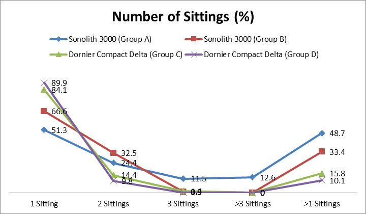
Index                                                                           Page

Outpatient basis extracorporeal shock wave lithotripsy for ureter stones:
Efficacy of the third generation lithotripter as the first line treatment          4

Extracorporeal shock wave lithotripsy for distal ureteral calculi:
Improved efficacy using a low frequency                                            7

Evolution of shockwave lithotripsy (SWL) technique:
a 25-year single centre experience of >5000 patients                               10

Anesthesia for extracorporeal shockwave lithotripsy:
Teikyo University Hospital experience using the third generation lithotripter      13

A new optical coupling control technique and application in SWL                    16

                                                                                Page 2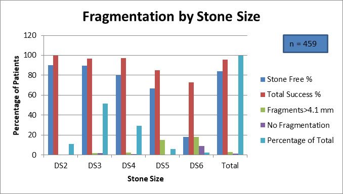
Editorial

Dear Doctor,

Extracorporeal Shock Wave Lithotripsy (ESWL) has been the cornerstone of non-in-
vasive kidney stone management for over four decades.          As the Medical Offi-
cer of Dornier MedTech, I would like to take this opportunity to introduce to you
the first issue of “The Clinical Case for ESWL”. It is a collection of summaries of
some very interesting and important peer-reviewed articles published on ESWL.

As the innovators of ESWL technology, we at Dornier MedTech continuously strive
to improve and enhance the efficacy and safety of our ESWL devices. These stud-
ies utilized Dornier Delta I and Delta II devices, and continuing this rich tradition, it
gives me immense pleasure to introduce to you the latest Dornier Delta® III lithotripter.

The Dornier Delta® III offers even more powerful imaging for improved stone visualiza-
tion, greater penetration depth to treat more stones in more patients, and greater efficien-
cy with time saving features. This semi-integrated lithotripter has everything you need to
                  best manage your patients’ stones, and perhaps our most important fea-
                     ture, Opticouple® technology—Optical Coupling Control (OCC) which
                        significantly improves stone free rates and lowers retreatment rates.

                            It is only prudent that we have a look at some of the important
                                recent evidence published on ESWL, especially with Dornier de-
                                   vices. We have made a sincere attempt to present the most
                                      relevant information in a concise and lucid manner with
                                         figures where appropriate. I am sure you will find this
                                            compendium very useful for your clinical practice.

                                                                                Happy reading!

                                                                                Yours sincerely,

                                                                            Dr. Dipen Jagatiya
                                                                                 M.B.B.S, M.P.H
                                                                                Medical Officer
                                                                             Dornier MedTech

                                                                                        Page 3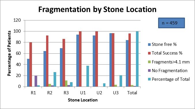
CLINICAL SUMMARY
This clinical summary reviews the following article from the International Journal of Urology.

                     International Journal of Urology (2008) 15, 210-215

                     Outpatient basis extracorporeal shock wave lithotripsy
                     for ureter stones: Efficacy of the third generation
                     lithotripter as the first line treatment
                     Akiko Murota-Kawano, Kazuhiro Ohya and Hideaki Sekine

              ESWL – A First Line Treatment for All Ureteric Stones?
Background

Extracorporeal shock wave lithotripsy (ESWL) is commonly used for the treatment of ureteral
stones. Newer Lithotripters are less invasive and are less likely to require anesthesia.

Objective

This study presented the outcomes with Dornier Compact Delta Lithotripter in treating ureteral
stones from a single center in Japan.

Methods

A total of 401 cases from December 2001 for which follow up data was available were included
in this study.

Treatment Protocol

Almost all patients (98%) received outpatient treatment. Appropriate analgesia was used and
patients were treated in the supine position. The treatment head was positioned under the ta-
ble for proximal ureteric stones and over the table for mid-distal ureteric stones. Although the
intended number of total shocks for each patient was 3000 per session, it was stopped earlier
if fluoroscopy showed sufficient fragmentation. The shocks were administered at the rate of 60
shocks per minute. The treatment was started with the lowest intensity and gradually increased
to the recommended level (≥16 mJ/shot). About 200 shots were administered at each level. Af-
ter treatment, patients were categorized as stone free, success (residual fragments ≤4 mm) or
failure (residual fragments >4 mm).

Results

•      The median age of patients was 50 years (range 3-92 years).
•      The stone free rate was 94.5% (n=379) from 401 cases.
•      Stone length (p=0.004), stone location (p=0.04) and sex (p=0.05) significantly
       predicted stone free rate in univariate analysis. However on multivariate analysis, stone
       free rate was significantly predicted by stone length only (p=0.01).
•      The stone free rate was similar for larger (≥10mm) and smaller (
Average number of sessions 1.3 (Range 1-5)
Average treatment time 49 minutes per session
Average number of shock waves 3013
Retreatment rate 15.7%
Stone free rate
1 session 81.3%
2 sessions 91.8%
3 sessions 93.5%
Table 1. Description of shock wave treament
• There was significant difference in the number of treatment sessions required between
smaller stones and larger stones on univariate analysis (1.13 vs. 1.30; p=0.006) Outcomes
after a single session
• A total of 331 patients received treatment for only one session. Only 1 patient did not
have satisfactory results. (Figure 1)
Treatment outcomes after a single session
350
318
300
250
200
150
100
50
12 1
0
Stone free Success Failure
Number of patients
Figure 1. Treatment outcomes in patients who received a single session of ESWL only
• Among stone free patients, patients with smaller stones were more likely to be stone
free after a single session as compared to larger stones. (90.1% vs. 77.5%; p=0.0008).
There was no difference on the basis of stone location.
• Overall, 22 patients did not achieve stone free status even after multiple sessions. Of
these, 18 patients were deemed success as the residual fragments were ≤4mm.
• Only one patient developed subcapsular renal hematoma (major complication)
associated with severe flank pain which resolved with conservative treatment.
Page 5Conclusion
In patients with ureteric stones, including mid-distal stones, treatment with a third generation
lithotripter is associated with a high stone free rate and small number of treatment sessions.
This non-invasive procedure can be the first line treatment of all ureteric stones.
Reference:
Murota-Kawano A et al. (2008). Outpatient basis extracorporeal shock wave lithotripsy for
ureter stones: Efficacy of the third generation lithotripter as the first line treatment. Int J Urol.
2008 Mar;15(3):210-5. doi: 10.1111/j.1442-2042.2007.01970.x.
The full article can be accessed after purchase by clicking this link:
Page 6CLINICAL SUMMARY
This clinical summary reviews the following article from the International Journal of Urology.
International Journal of Urology (2013) 20, 214-219
Extracorporeal shock wave lithotripsy for distal ureteral
calculi: Improved efficacy using a low frequency
Francisco Jose Anglada-Curado, Pablo Campos-Hernández,
Julia Carrasco-Valiente, et al.
Lower Frequency Improves Outcomes with ESWL in Distal Ureteral Calculi
Background
Literature suggests that lithotripsy with lower frequency may be associated with favorable out-
comes. Clinical trials with low frequency lithotripsy have focused on kidney and proximal ure-
teric stones.
Objective
To study the impact of two different shock wave frequencies on the fragmentation of distal
ureteral calculi.
Methods
This was a prospective study involving patients with distal ureteric calculi randomized into
two treatment groups (based on shock wave frequency), at 80 (High Frequency Group) and
60 shock waves per minute (Low Frequency Group). Only stones between the longitudinal di-
ameter of 0.5 cm and 1 cm were included to reduce the impact of size. However, patients were
divided into two groups according to stone size: stone size up to 0.7 cm and stone size between
>7 mm and ≤1 cm. Cystine, radiolucent or tenuously calcified stones were excluded.
Treatment Protocol
A Dornier Compact Delta lithotripter was used. Patients were radiologically monitored every
100 shock waves and the shock wave intensity was scaled-up according to patient tolerance. All
patients reached the maximum wave intensity of 70 mJ and majority (95%) of them achieved
it before reaching 1000 shock waves. Treatment was stopped at 3000 shock waves or if there
was evidence of stone fragmentation on monitoring. If non-fragmented calculus was seen on
x-ray at one week, a repeat session was performed at the same frequency with a maximum of
three sessions.
Primary Outcome
The total number or dose of shock waves applied.
Page 7Secondary Outcomes • The number of sessions received • Time to the elimination of the calculus • The rate of resolution • Pain perception by visual analog scale from 0 to 5 Results A total of 150 patients, 72 in the HFG group and 78 in the LFG group, recruited between Sep- tember, 2007 and September, 2009 were included in the final analysis. There was a significantly higher number of stones with size 0.7 cm or more in the HFG group as compared to the LFG group (64.3% vs. 40%; P 0.004). The investigators adjusted for this difference in their analysis. Number of shock waves and sessions The total mean number of shock waves was significantly higher in the HFG group as compared to the LFG group (5752±3121 vs. 2980±1211; p
Rate of resolution

Rate of resolution was 100% in LFG group which was significantly better than HFG group
(92.9%)(p=0.02). This difference was not significant for calculi >0.7 cm.

Pain perception and complications

Pain perception was similar between the two groups (p=0.46). Minor complications like fever
and renal colic occurred in 5 patients in the HFG group and renal colic occurred in 3 patients in
the LFG group.

Conclusion

For treatment of distal ureteric stones, lithotripsy at a rate of 60 shocks per minute is associated
with better outcomes as compared to 80 shocks per minute.

Reference

Anglada-Curado FJ et al. (2013) Extracorporeal shock wave lithotripsy for distal ureteral cal-
culi: Improved efficacy using low frequency. Int J Urol. 2013 Feb;20(2):214-9. doi: 10.1111/j.1442-
2042.2012.03133.x. Epub 2012 Sep 12

The full article can be accessed after purchase by clicking this link:

                                                                                            Page 9
CLINICAL SUMMARY
This clinical summary reviews the following article from the International Journal of Urology.
BJU International (2014) 114, 748-753
Evolution of shockwave lithotripsy (SWL) technique:
a 25-year single centre experience of >5000 patients
Jitendra Jagtap, Shashikant Mishra, Amit Bhattu, et al.
Shock Wave Lithotripsy – The Reliable Option
Introduction
Shock wave lithotripsy (SWL) is a safe and effective method of treating urolithiasis.
Objective
This study described the experience of treating more than 5,000 patients over 25 years using
shock wave lithotripsy at a single center. The authors assessed the impact of various treatment
optimizing strategies by evaluating annual SWL treatment rate, patient demographics, stone
and treatment data, re-treatment (repeat SWL), auxiliary procedures, complications and stone-
free rate (SFR) during an arbitrary four phases of development since the introduction of SWL.
Methods
The patients were divided into four groups; A (treatment period 1989-1994), B (treatment pe-
riod 1995-2000), C (treatment period 2001-2006) and D (treatment period 2007-2013) which
included 1561, 1741, 1039 and 676 patients respectively. Patients in groups A and B were treated
using Sonolith 3000 and patients in groups C and D were treated using Dornier Compact Delta
lithotripter. Optimization was done by frequent re-localization, restricting maximum number of
shocks and utilizing booster therapy in group B and Hounsfield unit estimation, power ramping
and improved coupling in group D.
Results
A total of 5,017 patients were included in the final analysis.
Stone Location and Stone Free Rate
The stone free rate was 81.3% (n=4079). The number of patients by stone location and stone
free rate by stone location are shown in the graph below (Figure 1). The stone free rate accord-
ing to stone size was 80.5% (n=99), 82.8% (n=1671), 81.0% (n=2021) and 75.4% (n=288) for sizesFigure 1. Stone location and SFR for each location Dornier Compact Delta vs. Sonolith 3000 Stone free rate was significantly better (p
Fewer patients treated with Dornier Compact Delta needed multiple sittings/repeat SWL as
compared to Sonolith 3000. (13.6% vs. 40.6%; p
CLINICAL SUMMARY

This clinical summary reviews the following article from the Hinyokika Kiyo. Acta Urologica
Japonica.

                   Acta Urologica Japonica (2007) 53: 545-549

                   Anesthesia for extracorporeal shockwave lithotripsy:
                   Teikyo University Hospital experience using the third
                   generation lithotripter
                   Koji Kurihara, Yutaka Kamiyama, Keisuke Saito, et al.

     Anesthesia & ESWL: Performance with a third generation lithotripter

Background

Newer lithotripters require a lesser degree of anesthesia or no anesthesia and they are also
more mobile as compared to the Dornier HM3 lithotripter, the world’s first commercially avail-
able lithotripter. However, they may not be as effective as the Dornier HM3 lithotripter.

Objective

To study the effectiveness of a third generation lithotripter, Dornier Compact Delta, in treating
renal and ureteral stones under anesthesia.

Methods

A total of 502 patients with symptomatic renal and ureteral stones were treated from Janu-
ary 1, 2003 to December 31, 2005. All patients were treated using the Dornier Compact Delta
lithotripter by a single surgeon who recorded all preoperative, intraoperative and postopera-
tive data to avoid inter-operator variability. Treatment success was defined as residual stone
fragments less than 4 mm on radiography. Postoperative follow up was conducted at 1 and 3
months to determine success. Stones were classified by location and size using the Japanese
Urological Association criteria.

Treatment Protocol

Patients were treated in the supine position. Seventy percent of patients were given general
anesthesia using midazolam. Remaining patients received epidural anesthesia with or without
intravenous analgesia. Shock waves were applied at a mean intensity of 5 and an average 3,471
shocks were applied. Patients with treatment failure were treated with repeat extracorporeal
shock wave lithotripsy (ESWL) or alternative procedures.

                                                                                         Page 13
Results There were 334 male and 168 female patients. About 37% of stones were located in the kidney and 63% stones were located in the ureters. A total of 61.8% (310) patients had stones
Figure 2: Fragmentation by Stone Size; Stone size: DS2, ≤4 mm; DS3, 4 mm< ≤10 mm; DS4, 10 mm< ≤20mm; DS5, 20
mm< ≤30 mm; DS6, ≥30 mm

The efficiency quotient (EQ), based on stone free rate, retreatment rate and auxiliary proce-
dures, of Dornier Compact Delta in this study was 0.65 which was similar to the EQ of 0.67 of
Dornier HM3 published previously.

Conclusion

This third generation lithotripter was found to be effective in treating renal and ureteral stones.
Treatment efficacy can be improved by performing lithotripsy under anesthesia.

Reference

Kurihara K, Kamiyama Y, Saito K, Yasuda M, Ide H, Muto S, Okada H, Horie S. (2007). Anesthesia
for extracorporeal shockwave lithotripsy: Teikyo University Hospital experience using the third
generation lithotripter. Hinyokika Kiyo. 2007;53(8):545-9

The full article can be accessed after purchase by clicking this link:

                                                                                                    Page 15
CLINICAL SUMMARY
This clinical summary reviews the following article from the journal Urolithiasis
Urolithiasis (2016) 44: 539-544
A new optical coupling control technique and
application in SWL
Jian Lin Lv
Newer advances – Optical coupling control for ESWL
Background
Modern Lithotripters use gel or oil to couple the cushion of the treatment head with the skin of
the patient. Any air which gets trapped at the coupling interface can affect the quality of stone
fragmentation by interfering with the transmission of shock waves to the patient. The therapy
heads of latest Dornier devices come fixed with a video camera and a LED light – Opticouple
imaging technology which helps to detect air bubbles and imperfect coupling.
Objective
To compare the outcomes of optical coupling control (OCC) with blind coupling during treat-
ment of renal stones with extracorporeal shock wave lithotripsy (ESWL)
Methods
A total of 336 patients with upper urinary tract stones were randomized into an optical cou-
pling control (OCC; Group A – 169 patients) and a blind coupling (Group B – 167 patients) group
between January 2014 and February 2015. A Dornier Compact Delta II UIMS was used. The
same urologist performed all the procedures. The shock waves were delivered at 70 shocks per
minute and the power was gradually increased to 100%. In the OCC group, air pockets were
removed under visual control from the video camera by gently swiping, repeatedly if needed, a
hand between the patient and the inflated water cushion. It is preferable to apply the ultrasound
gel from a large container instead of a squeeze bottle to avoid air bubbles. Stone free rates at
3 months were measured.
Results
The stone characteristics were similar between the two groups (p=ns). The overall stone free
rates at 3 months were significantly higher in Group A for both kidney stones (78.2% vs 62.8%;
p=0.027) and ureteral stones (81.7% vs 67.9%; p=0.042). The treatment results were also strat-
ified by stone location and they were significantly better in Group A for all stone locations
(pFigure 1: Stone free percentage by location
Figure 2: Retreatment percentage by stone location
Figure 3: Ancillary procedure percentage by stone location
Page 17Group A also needed significantly lesser mean number of shocks (1900±363 vs 2400±320;
p=0.013), lesser treatment time (p=0.021) and lesser energy levels (1.6±0.8 vs 2.3±1.2; p=0.036)
compared to Group B. Mean fluoroscopy time was less with Group A but not statistically signif-
icant (1.5±0.7 vs 1.8±0.8; p=0.067).
Conclusion
The new Opticouple coupling control technique is associated with significantly better out-
comes. It improved the efficiency of shock wave transmission and reduced energy loss leading
to optimization of treatment outcomes.
Reference:
Lv JL. (2016). A new optical coupling control technique and application in SWL. Urolithiasis.
2016; 44(6):539-544. https://doi.org/10.1007/s00240-016-0874-9.
The full article can be accessed after purchase by clicking this link:
Page 18DMT306-112017-REV B EN Page 19
You can also read


