The Journal of Veterinary Medical Science
←
→
Page content transcription
If your browser does not render page correctly, please read the page content below
Advance Publication

The Journal of Veterinary Medical Science

Accepted Date: 25 May 2020
J-STAGE Advance Published Date: 9 July 2020

©2020 The Japanese Society of Veterinary Science
Author manuscripts have been peer reviewed and accepted for publication but have not yet
been edited.
1                                                 Wildlife Science

 2                                                    Full paper

 3

 4   Mycological isolation from animal enclosures and environments in National Wildlife Rescue

 5   Centre and National Zoo, Malaysia

 6

 7   Running head: Mycology of captive wildlife environments

 8

 9   Sharina OMAR1),a, Fathiah Aqilah JALALUDIN1), Jacqueline Meikwei YEE1), Zubaidah

10   KAMARUDIN2), Kavitha JAYASEELAN3), Aina Nazurah Mohd KHLUBI1), Yusuf Lekko MADAKI1),

11   Hasdi HASSAN2), Mat Naim RAMLI3), Rahmat TOPANI4), Azlan CHE-AMAT1),a,*

12

     1)
13       Faculty of Veterinary Medicine, Universiti Putra Malaysia, 43400 UPM Serdang, Selangor, Malaysia.
     2)
14       National Wildlife Rescue Centre (NWRC), Department of Wildlife and National Parks Peninsular

15   Malaysia (PERHILITAN), 35600 Sungkai, Perak, Malaysia.
     3)
16       Zoo Negara (National Zoo), Hulu Kelang, 68000 Ampang, Selangor, Malaysia.
     4)
17       Ex-Situ Conservation Division, Department of Wildlife and National Parks Peninsular Malaysia

18   (PERHILITAN), KM 10 Jalan Cheras, 56100 Kuala Lumpur, Malaysia.

19

20   *Corresponding author:

21   Name: Azlan Che-Amat; Address: Faculty of Veterinary Medicine, Universiti Putra Malaysia, 43400

22   UPM Serdang, Selangor, Malaysia; Fax no.: +603 9769 1971; E-mail: c_azlan@upm.edu.my.

23

     a
24       These authors equally contributed to this work.

                                                           1
25 ABSTRACT
26
27 It is important to provide a baseline of fungal composition in the captive wildlife environment to better
28 understand their role in overall wildlife health. The objectives were to identify species of fungi existing
29 within wildlife animal enclosures and their environment at the National Wildlife Rescue Centre (NWRC)
30 and the National Zoo, Malaysia and to describe their medical and veterinary importance. Samples of air,
31 wall or floor swab, enrichment swab and soil were taken from the animal enclosures, exercise yard and
32 enrichments at NWRC and National Zoo respectively. All samples including those pre-treated samples
33 were plated onto Sabouraud’s Dextrose Agar (SDA). Numerous fungi were grown on all sampling SDA
34 plates regardless by either single or multiple growth. Samples of air in both NWRC and National Zoo
35 had the highest growth of Penicillium spp. with a prevalence of 31.2% and 83.7% respectively. Samples
36 of swab from the wall, floor and enrichments were predominantly by Candida spp. (42.6%) in NWRC
37 and Penicillium spp. (41.6%) in the National Zoo. Prevalence of multiple fungi isolated from the soil
38 samples in NWRC were 57.9% and yeast species was the most common in National Zoo with a
39 prevalence of 88.9%. Overall, 29 and 8 isolates were found in both samples from the NWRC and National
40 Zoo with a predominant species of potential zoonotic fungi have been identified in both premises. The
41 expected fungus Aspergillus spp. was not isolated in all samples in NWRC. Prevalent fungal species
42 found in this study are known to cause disease in animals and humans as primary pathogen and also as
43 opportunistic pathogens that may also cause infection. Thus, health safety precautions should be
44 considered particularly in dealing with conservation of endangered wildlife species, along with personnel
45 and public involvements.
46
47 KEY WORDS: captive, enclosure, fungal, wildlife, zoo
48
249 INTRODUCTION
50 Fungi are often overlooked as a potential cause of disease since more focus tends to fall on other
51 pathogenic agents such as bacteria or viruses. However, fungal infections are on the rise and becoming
52 an important cause of emerging diseases in wildlife worldwide [24]. In previous work by Seyedmousavi
53 et al. [26], they reported that the zoonotic fungi are naturally transmitted between animals and humans,
54 and that they can eventually cause significant public health concerns. Fungi are ubiquitous and reproduce
55 by means of spores which can be inhaled or be transmitted via direct contact with body surfaces,
56 especially the skin. Thus, fungal infections usually begin their pathogenic spread in the lungs or on the
57 skin of animals and humans [23]. These pathogenic fungi impact human health just as greatly as they do
58 with animals, leading to allergic responses, skin and mucosal infections, and even invasive diseases
59 which can be extremely fatal.
60 There are many factors that help in promoting the growth of fungus namely the level of humidity,
61 temperature, pH, presence of oxygen and absence of light [14, 27]. Fungi need water to help them to
62 obtain food, as they release enzymes to breakdown complex materials, water will dissolve these materials
63 thus aiding with absorption [16]. They grow at temperatures within the range of 5°C to 35°C with
64 optimum temperatures for growth between 25°C and 30°C [5]. Fungi in general prefer a neutral pH level
65 as more fungal species grow around the neutral pH level compared to a higher or lower pH level [27].
66 Fungi can regulate the pH in their environment by secreting acid or alkali [33]. Fungi are best grown in
67 dark places since the presence of light may induce stress for the fungal cells, thus inhibiting them from
68 growing [9]. Oxygen is a critical requirement for all eukaryotic organisms as they help in maintaining
69 the overall cellular metabolism of the fungal cells [10].
70 Captive wild animal settings or zoos are not just places for a wide range of biodiversity and
71 entertainment, but they also harbor potential emerging infectious diseases [4]. It is estimated that 75% of
72 these diseases are zoonotic and, of these, 70% originate in wildlife populations. Among these, fungal
373 pathogens are emerging at an alarming rate worldwide and pose a significant threat to all wildlife species
74 [6]. According to Sutherland et al. [29], there has been a marked increase in fatal fungal skin infections
75 in wild snakes (Snake Fungal Diseases) since 2006 which caused a decline in the population of wild
76 snakes in the eastern United States [2]. Cases of dermatophytosis in captive tigers have been reported in
77 several countries including Thailand [17] and the United States of America [30]. Cryptococcosis is
78 described in many wildlife species including wild birds [1, 19]. More recently, Aspergillus flavus has
79 been isolated from the lung lesion of a Malayan tiger in the National Wildlife Rescue Centre in Malaysia
80 (Kamarudin, Z., 2018, pers. comm.). According to Hedayati et al. [12], A. flavus is the second leading
81 cause of invasive aspergillosis after A. fumigatus and the fungus is a saprotrophic and pathogenic residing
82 in the soil.
83 It is important to mention that some fungal diseases with zoonotic potential do not receive enough
84 attention, which in turn would lead to inadequate preventive measures on a global scale. Fungi are
85 extremely ubiquitous in the environment, therefore isolating and identifying those of veterinary and
86 public health importance of protected wildlife could minimize the increase and spread of fungal diseases
87 within wildlife species [18] by implementing specific preventive measures at the enclosures. Since there
88 was lack of studies in Malaysia on this perspective, this study potentially served as an important baseline
89 reference for fungal species distribution that is present in captive animal enclosures. In addition, any
90 human and veterinary important fungi present in the captive wildlife setting can be identified earlier so
91 that safety measure can be taken to prevent infection in animals and humans. Thus, this study was carried
92 out to identify the type of fungi and to compare the presence of medical and veterinary important fungi
93 in the wildlife enclosures and the environment of selected endangered wildlife species in Malaysia.
94
95
96
497 MATERIALS AND METHODS
98 Animal and Sampling Site
99 The sampling took place at the animal enclosures and exercise yards in National Wildlife Rescue
100 Centre (NWRC), Sungkai, Perak (central south of Peninsular Malaysia), and the National Zoo, Ulu
101 Kelang, Selangor (central of Peninsular Malaysia). The study was subjected to the approval from the
102 Department of Wildlife and National Park Peninsular Malaysia (Ref. no.: JPHL&TN(IP): 100-34/1.24
103 Jld 12 (46 and 47) and National Zoo Student Research Program by the Education Department, National
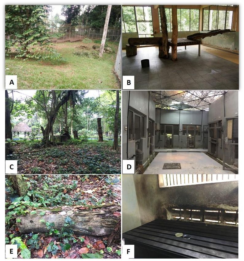
104 Zoo. Air samples, floor and enrichment swabs were taken from the captive enclosures or known as their
105 night stall while samples taken from exercise yards or exhibits were the soil and air samples. The
106 enrichment was referred to the animal enrichment provided by the two venues to the animals to enhance
107 them to explore and interacts with their environment, these include woods, balls, swing, etc. In NWRC,
108 the captive enclosures and the exercise yards of the Malayan tiger (Panthera tigris jacksoni), Malayan
109 sun bear (Helarctos malayanus) and clouded leopard (Neofelis nebulosa) were selected while Malayan
110 tiger, Malayan sun bear and Orangutan (Pongo pygmaeus) enclosures and exhibits were selected in the
111 National Zoo.
112
113 Collection and preparation of samples
114 A passive air sampling technique (gravitational sedimentation sampling) was employed by placing
115 two Sabouraud’s Dextrose Agar (SDA) plates at the animal’s resting areas in the enclosures and exercise
116 yards (Figure 1). We used the short contact time 10-15 min based on Hashimoto and Kawakami [11] and
117 our personal experience screening for the presence of fungal in our establishment (laboratory, offices,
118 staff rooms, others). The amount of time is ample to isolate multiple fungi present in the air by carrying
119 the plates around and also leaving the plates at the level of human heights and to limit the risk exposure
120 to the personnel conducting the study. Sterile cotton swabs were used to take samples from the
5121 enrichments and floor/wall surface in the animal enclosures (see Fig. 1). The swabs were then placed
122 inside a transport media before streaking onto SDA plate.
123 About 100 g soil sample from each exercise yard were collected by scraping the top soil using a clean
124 disposable spoon and kept in a clean zip-locked plastic container separately before analysed. Each soil
125 sample collected was mixed thoroughly using a Stuart scientific vortex machine and 10 g were taken and
126 diluted with 10-fold dilution method using sterilized distilled water up until the third dilution. A 10 g of
127 soil was mixed with 90 ml of sterilized distilled water for the dilution. One hundred microlitre (100 µl)
128 of the third dilution was streaked onto SDA plate, thus the detection of fungi was made from the lowest
129 dilution (10-3). The culture plate was then incubated at temperature between 25°C to 28°C in a dark
130 cabinet and was checked for fungal growth on a weekly basis until week 4. The incubation temperature
131 was established by the selected locations for sampling that had the temperature around 24°C to 32°C as
132 the areas are surrounded by thick tall trees, hence the culture temperature was selected to mimic the
133 condition present at the sampling areas.
134
135 Identification of fungus
136 The incubated plates were examined for fungal growth starting on Day 3 post inoculation (PI) and on
137 a weekly basis. The examination involved two stages: macroscopic and microscopic examination [7, 20].
138 Macroscopic examination consists of description of consistency, ridges and grooves, as well as the color
139 or pigmentation of the colony morphology observed on top and reverse side of SDA plate. Microscopic
140 examination involved the observation of fungal structures such as conidia, conidiophore, hyphae and the
141 presence of other unique characteristics of the species such as chlamydoconidia or macroconidia wet-
142 mount preparations was used to visualize the fungal structures. Briefly, a clear or colorless clean
143 cellophane tape was touched onto the surface of mycelia and placed onto a clean glass slide and stained
144 with lactophenol cotton blue (LCB). Candida spp. was identified using the available commercial
6145 identification kit API 20 C AUX (bioMérieux, Durham, NC, USA) only for isolation from the NWRC.
146 Descriptive statistics on the percentages of fungal isolation were performed for each type of sample in
147 each premise.
148
149 RESULTS
150 A total of air samples (n=57; n=25), swab samples (n=38, n=25) and soil (n=10, n=6) were collected
151 from the NWRC and National Zoo, respectively (details in Table 1). Fungi were grown on all sampling
152 SDA plates regardless by either single or multiple growth in both NWRC and National Zoo. In NWRC,
153 58.6% of fungi were isolated from the animal enclosure and 86.2% of the exercise yard with 86.2%, 36%
154 and 39.9% being isolated from the air samples, swabs and soil respectively. Multiple fungi were isolated
155 from the soil samples in NWRC with a prevalence of 57.9%. Penicillium spp. and Candida guillermondii
156 were the most prevalent in NWRC with a prevalence of 76.2% and 70.5%, respectively. Penicillium spp.
157 were mostly isolated from the air samples and enrichment swabs in the enclosure with the average
158 prevalence of 31.2% (the average % prevalence of both air samples from the enclosures and exercise
159 yards) and 30% respectively, while they represented only 3.7% of each isolate from the swabs of the
160 enclosure and soil samples. Samples of swab from the wall, floor and enrichments were predominantly
161 represented by Candida spp. (42.6%) in NWRC, especially C. guillermondii (45.3%) and C. tropicalis
162 (40%). Another 27 isolates were identified in all samples in NWRC with a prevalence range from 0.9%
163 to 49.5% (see detail in Table 2).
164 In National Zoo, fungi were isolated from all night quarters and exhibit samples with the prevalence
165 from the air, swabs of the enrichment and soil of 83.7%, 66.7% and 25% respectively. Penicillium spp.,
166 yeast species and Trichophyton spp. were predominant with a prevalence of 60.6%, 35.2% and 27.3%
167 respectively. Penicillium spp. were mostly isolated from the air samples with a prevalence of 83.7%,
168 while they were also isolated from 41.6% and 33.3% of the swabs of the enrichment and soil samples
7169 respectively. Yeast species was the most commonly isolated in soil samples with a prevalence of 88.9%,
170 while Trichophyton spp. were predominant in air samples with 39.5%. Another 5 isolates were identified
171 in samples in National Zoo with prevalence ranging from 1.2% to 11.6%, including isolation of
172 Aspergillus spp. (see detail in Table 3).
173
174 DISCUSSION
175 This study was conducted to isolate fungi species that are present in the NWRC and National Zoo
176 wildlife enclosure and environment and to identify other medical and veterinary importance fungus.
177 Because of the recent finding of A. flavus in the NWRC, this species was expected to be found,
178 surprisingly, A. flavus was not isolated in the present study. In contrast, other Aspergillus species, namely
179 A. fumigatus and A. niger, were detected at a prevalence of 1.9% and 0.9%, respectively. In the National
180 Zoo, the detection of Aspergillus spp. occurred in 5.7% of the collected samples. Fungi can easily infect
181 animals and humans as they are ubiquitous, often by inhalation and penetration through un-intact skin.
182 It is reported that previously, A. flavus had been isolated from the pulmonary lesions of an incidental
183 finding in the necropsy of a tiger in NWRC, indicating that this fungus might be present in the wildlife
184 enclosures and the environment. Aspergillus flavus produces aflatoxin, a very powerful
185 hepatocarcinogenic mycotoxin and are known to cause human and animal infection [12], being more
186 commonly found in the air compared to A. fumigatus. Aspergillus is a saprophytic mold that is closely
187 associated with agriculture and other human activities that make nutrients available to fungi.
188 Aspergillosis is not contagious, nevertheless when the human is immunocompromised, Aspergillus can
189 cause rapid developing acute infection following environmental exposure. Chronic forms of aspergillosis
190 causing respiratory tract infections in wild birds have been reported since at least back in the year 1813
191 [8]. In general, molds reported in this study are easily transmitted through inhalation of spore-containing
192 air.
8193 According to the result of this study (Table 2 and 3), the most prevalent fungus isolated from both
194 NWRC and National Zoo wildlife enclosure (floor, wall and enrichment swab) and environment (air and
195 soil) is Penicillium spp. In this study, Penicillium spp. is the most abundant fungus present in the
196 environment, it is mostly present on the air sample cultured plates from both inside animal enclosure and
197 the exercise yard of the animal. This is most probably due to the spore dispersal carried by the wind into
198 the animal enclosure from the exercise yard (outdoor). The air sample was taken by placing the plate on
199 the ground and Penicillium spp. are known to be very abundant in soil. Thus, this can explain the higher
200 number of spores present from the air sample of an exercise yard as the plates are closer to the soil. The
201 factors that might affect the prevalence of fungus species include the ease of spore dispersal as spore can
202 be widely dispersed especially by the wind intensity. In addition, animals can also be the mechanical
203 vectors that help spreading the spores. Penicillium spp. are ubiquitous soil fungi and usually regarded as
204 unimportant in terms of pathogenicity in human and animals, for most of its species. One of the
205 Penicillium spp., P. marneffei, however, is known to commonly infect immunocompromised individuals
206 [25]. It was only recognized as important when the human immunodeficiency virus (HIV) pandemic
207 occurred in Asia [32] and untreated cases are usually fatal. Infection by Penicillium spp. are rare in
208 domestic animals, however animals such as cat have been reported to get infected with Penicillium spp.
209 [28]. In addition, the organism’s natural habitat is in soil endemic to Southern China and South-East Asia
210 [3, 32]. Penicillium is globally recognized as the organism responsible for the production of the Penicillin
211 antibiotic, but little is known that one of its species, P. marneffei is an emerging pathogenic fungus
212 particularly in immunocompromised patients with HIV infection [32].
213 Conidiobolus coronatus has been isolated markedly in National Zoo while a small proportion
214 detected in NWRC. This fungus has a worldwide distribution but more often seen in tropical rainforests.
215 It is commonly found in soil and decaying leaves and possess a distinctive morphological feature for
216 identification - a short conidiophore that bears conidia that produces hair-like appendages known as
9217 villae. Since the mold is powdery, the infection may result from inhalation of the spores hence lesions
218 found in human and animals usually concentrate at the rhino-facial area. Conidiobolus coronatus is
219 known to cause entomophthoromycosis in human, that is usually characterized by nasal cavity tumor due
220 to the aggressive invasiveness of the fungus [31]. Paecilomyces spp. are also found worldwide and has
221 always been regarded as contaminants of the air, but for an immunocompromised host, it may lead to
222 fatal events [34].
223 Fusarium spp. are widely distributed in soil where they are commonly considered as a contaminant.
224 In humans, this fungus is commonly reported to cause infection in both immunocompetent and immune
225 compromised hosts [15]. It can cause superficial infection as well as invasive and disseminated infection.
226 According to Jain et al. [15], about 15 Fusarium spp. have been identified to cause infections in animals
227 and humans. Example of superficial infection by Fusarium oxysporum is onychomycosis in humans
228 while in animals, Fusarium spp. infection is uncommon. Candida guilliermondii was the second most
229 prevalent fungus identified from NWRC and markedly isolated from the swab samples from the wall,
230 floor and enrichment within the enclosure. This fungus is part of the human skin and mucosal normal
231 flora, and this fungus appeared to be the least virulent amongst Candida species [22] where the author
232 classified Candida species into 3 virulence group of decreasing pathogenicity and C. guilliermondii
233 placed on the third group with the least virulence species. The assumption of high prevalence of this
234 fungus would be the circulation and contact with personnel with the animal and the environment is higher
235 in the NWRC premises. As it is a normal inhabitant of human, the yeast becomes opportunistic pathogen
236 when immunological mechanisms are disturbed and the proliferation of the fungus is higher than normal
237 thus leading to disease formation [21]. In animal, infection with the yeast species has been reported in
238 dogs, in which the normal protective barrier was disturbed [26].
239 Wildlife populations worldwide are under increasing threat from a variety of processes, ranging from
240 climate change to habitat loss that can lead to a physiological stress response [13]. This has become a
10241 concern because captive wildlife is more prone to stress due to their unnatural environments. When
242 stressors act for a prolonged time, or when effects accumulate, it is harmful to the animal especially
243 because these environmental fungi can cause systemic mycoses and can be fatal. Zoos are places where
244 all walks of life visit all year round. Hence, it is important to ensure that proper awareness is displayed
245 at the ticket counter and provide the vulnerable visitors such as unhealthy individuals, children or elderly
246 with basic PPE (e.g. facial mask) prior to entry in addition to hand sanitizer at the exits. Zoo personnel
247 (e.g. keepers, veterinarians, curators and biologists) must also be continually reminded that all husbandry
248 practices should be based on principles which minimize stress to the wildlife.
249 Even though it is known that fungi are presented ubiquitously in the environment, most of the fungus
250 may or may not be causing disease in humans and animals. Animals may be carriers of certain agents
251 including fungi, therefore if they are translocated from one place to another, it may seed their new
252 environment with agents. Other than being presence in the environment, they need other factors such as
253 presence in extremely abundant amount to eventually cause disease. As for the fungi species found in
254 both NWRC and National Zoo animal enclosure and environment in this study, this preliminary result
255 should consider the animal and public health disease prevention especially to immunocompromised
256 animals and humans. There may not be many fungus species under a genus that may cause disease,
257 therefore it is important to identify fungus up to the species level in order to know appropriate and
258 practical action to be taken when a pathogenic fungal species is present in the environment. Molecular
259 technique is seen as the best technique to be used for fungal identification up to the species level. Besides
260 that, active air sampling technique can also be implanted for air sampling rather than passive air sampling.
261 This technique can help to capture a fungus species that is presence at a very low amount in the
262 environment. In conclusion, prevalent fungal species found in this study are known to cause disease in
263 animals and humans as primary pathogens and also as opportunistic pathogens that may also cause
264 infection, therefore health safety precautions should be emphasized by the management.
11265 FUNDING
266 This work was supported by a research project grant provided by the Faculty of Veterinary Medicine
267 Research Trust Fund, Universiti Putra Malaysia.
268
269 ACKNOWLEDGMENTS
270 The authors thank the management and staffs of the National Wildlife Rescue Centre, National Zoo and
271 Veterinary Bacteriology Laboratory, Faculty of Veterinary Medicine, Universiti Putra Malaysia and the
272 top management of PERHILITAN and Zoo Negara (Malaysian Zoological Society). The manuscript was
273 read by Luisa Helena Monteiro de Miranda, The University of Sydney, Sydney, Australia.
274
275 REFERENCES
276 1. Bolton, L.A., Lobetti, R.G., Evezard, D.N., Picard, J.A., Nesbit, J.W., Van Heerden, J. and
277 Burroughs, R.E.J. 1999. Cryptococcosis in captive Cheetah (Acinonyx jubatus): two cases: case
278 report. J. S. Afr. Vet. Assoc. 70(1): 35-39.
279 2. Burbrink, F.T., Lorch, J.M. and Lips, K.R. 2017. Host susceptibility to snake fungal disease is highly
280 dispersed across phylogenetic and functional trait space. Sci. Adv. 3(12): e1701387.
281 3. Chakrabarti, A. and Slavin, M.A. 2011. Endemic fungal infections in the Asia-Pacific region. Med.
282 Mycol. 49(4): 337-344.
283 4. Daszak, P., Cunningham, A.A. and Hyatt, A.D. 2000. Emerging infectious diseases of wildlife –
284 threats to biodiversity and human health. Science. 287: 443-449.
285 5. Dix, N.J. and Webster, J. 1995. Fungi of extreme environments. In: Fungal ecology. Springer,
286 Dordrecht. https://doi.org/10.1007/978-94-011-0693-1_12.
287 6. Fisher, M.C., Henk, D.A., Briggs, C.J., Brownstein, J.S., Madoff, L.C., McCraw, S.L. and Gurr, S.J.
288 2012. Emerging fungal threats to animal, plant and ecosystem health. Nature. 484: 186-194.
12289 7. Frey, D., Oldfield, R.J. and Bridger, R.C. (1981). A colour atlas of pathogenic fungi (2nd ed.). Wolfe
290 Medical Publications Ltd., London.
291 8. Friend, M. and Franson, J.C. 1999. Fungal diseases (Ch.3). In: Field manual of wildlife diseases.
292 General field procedures and diseases of birds. Biological Resources Division, Madison, US. 128-
293 138.
294 9. Fuller, K.K., Loros, J.J. and Dunlap, J.C. 2015. Fungal photobiology: visible light as a signal for
295 stress, space and time. Curr. Genet. 61(3): 275-288.
296 10. Grahl, N., Shepardson, K.M., Chung, D. and Cramer, R.A. 2012. Hypoxia and fungal pathogenesis:
297 to air or not to air? Eukaryot. Cell. 11(5): 560-570.
298 11. Hashimoto, K. and Kawakami, Y. 2018. Effectiveness of airborne fungi removal by using a HEPA
299 air purifier fan in houses. Biocontrol Sci. 23(4): 215-221.
300 12. Hedayati, M.T., Pasqualotto, A.C., Warn, P.A., Bowyer, P. and Denning, D.W. 2007. Aspergillus
301 flavus: human pathogen, allergen and mycotoxin producer. Microbiology. 153(6): 1677-1692.
302 13. Hing, S., Narayan, E.J., Andrew Thompson, R.C. and Godfrey, S.S. 2016. The relationship between
303 physiological stress and wildlife disease: consequences for health and conservation. Wildl. Res. 43:
304 51-60.
305 14. Hyde, K.D., Xu, J., Rapior, S., Jeewon, R., Lumyong, S., Niego, A.G.T., Abeywickrama, P.D.,
306 Aluthmuhandiram, J.V.S., Brahamanage, R.S., Brooks, S., Chaiyasen, A., Chethana, K.W.T.,
307 Chomnunti, P., Chepkirui, C. et al. (2019). The amazing potential of fungi: 50 ways we can exploit
308 fungi industrially. Fungal Divers. 97: 1-136.
309 15. Jain, P. K., Gupta, V. K., Misra, A. K., Gaur, R., Bajpai, V. and Issar, S. 2011. Current status of
310 Fusarium infection in human and animal. Asian J. Anim. Vet. Adv. 6(3): 201-227.
13311 16. Karch, C. 2008. Water and fungi: Paecilomyces species. Retrieved from
312 https://www.emlab.com/resources/education/environmental-reporter/water-and-fungi-
313 paecilomyces-species/. Assessed on 3 October 2018
314 17. Kesdangsakonwut, S., Sommanustweechai, A., Utrara, Y., Wangnitham, S. and Banlunara, W. 2006.
315 Dermatophytosis in captive Bengal tiger (Panthera tigris). Proceeding of the 2nd Symposium of the
316 Asian Zoo and Wildlife Medicine and the 1st Workshop on the Asian Zoo and Wildlife Pathology.
317 Bangkok, Thailand, November 26th-29th, 2006. p23.
318 18. Kolbert, E. 2016. What’s causing deadly outbreaks of fungal diseases in world’s wildlife. Accessed
319 https://e360.yale.edu/features/whats_causing_deadly_outbreaks_of_fungal_diseases_in_worlds_wil
320 dlife. [last date accessed on May 1, 2020].
321 19. Krockenberger, M., Stalder, K., Malik, R. and Canfield, P. 2005. Cryptococcosis in Australian
322 wildlife. Microbiol. Aust. 26(2): 69-73.
323 20. Larone, D.H. 1987. Medically important fungi: a guide to identification (2nd ed.). Elsevier, New York.
324 pp: 230.
325 21. Mueller, R.S., Bettenay, S.V. and Shipstone, M. 2002. Cutaneous candidiasis in a dog caused by
326 Candida guilliermondii. Vet. Rec. 150: 728-730.
327 22. Pasqualotto, A.C., Antunes, A.G.V. and Severo, L.C. 2006. Candida guilliermondii as the aetiology
328 of candidosis. Rev. Inst. Med. Trop. SP. 48(3): 123-127.
329 23. Revankar, S.G. 2018. Overview of fungal infections. In: online Merck Manual accessed on
330 https://www.msdmanuals.com/professional/infectious-diseases/fungi/overview-of-fungal-
331 infections. [last date accessed on May 1, 2020].
332 24. Rothenburger, J. 2017. Emerging fungal diseases threaten wildlife. The western producer. Accessed
333 from https://www.producer.com/2017/06/emerging-fungal-diseases-threaten-wildlife/. [last date
334 accessed on May 1, 2020].
14335 25. Santos, P.E., Piontelli, E., Shea, Y.R., Galluzzo, M.L., Holland, S.M., Zelazko, M.E. and
336 Rosenzweig, S.D. 2006. Penicillium piceum infection: diagnosis and successful treatment in chronic
337 granulomatous disease. Med. Mycol. 44(8): 749-753.
338 26. Seyedmousavi, S., Guillot, J., Tolooe, A., Verweij, P.E. and de Hoog, G.S. 2015. Neglected fungal
339 zoonoses: hidden threats to man and animals. Clin. Microbiol. Infect. 21(5): 416-425.
340 27. Singh, P. and Chauhan, M. 2013. Influence of environmental factors on the growth of building
341 deteriorating fungi: Aspergillus flavus and Penicillium chrysogenum. Int. J. Pharm. Sci. Rev. Res.
342 4(1): 425-429.
343 28. Soonthornsit, J., Banlunara, W., Niyomthum, W. and Pusoonthornthum, R. 2013. Penicillium
344 species-induced granuloma in a cat resulting in chronic lower urinary tract disease. J. Feline Med.
345 Surg. 15(12): 1154-1159.
346 29. Sutherland, W.J., Aveling, R., Brooks, T.M., Clout, M., Dicks, L.V., Fellman, L., Fleishman, E.,
347 Gibbons, D.W., Keim, B., Lickorish, F., Monk, K.A., Mortimer, D., Peck, L.S. et al., 2014. A horizon
348 scan of global conservation issues for 2014. Trends Ecol. Evol. 29(1): 15-22.
349 30. Sykes, J.M. and Ramsay, E.C. 2007. Attempted treatment of tigers (Panthera tigris) infected with
350 Microsporum canis. J. Zoo Wildl. Med. 38(2): 252-257.
351 31. Valle, A.C., Wanke, B., Lazéra, M.S., Monteiro, P.C. and Viegas, M.L. 2001.
352 Entomophthoramycosis by Conidiobolus coronatus. Report of a case successfully treated with the
353 combination of itraconazole and fluconazole. Rev. Inst. Med. Trop. SP. 43(4): 233-236.
354 32. Vanittanakom, N., Cooper Jr. C.R., Fisher, M.C. and Sirisanthana, T. 2006. Penicillium marneffei
355 infection and recent advances in the epidemiology and molecular biology aspects. Clin. Microbiol.
356 Rev. 19(1): 95-110.
357 33. Vylkova, S. 2017. Environmental pH modulation by pathogenic fungi as a strategy to conquer the
358 host. PLoS Pathog. 13(2):e1006149. http://doi.org/10.1371/journal.ppat.1006149.
15359 34. Walsh, T.J., Groll, A., Hiemenz, J., Fleming, R., Roilides, E. and Anaissie, E. 2004. Infections due
360 to emerging and uncommon medically important fungal pathogens. Clin. Microbiol. Infect. 10(S1):
361 48-66.
362
363
364
365
366
367
368
369
370
371
372
373
374
375
376
377
378
379
380
381
382
16383 Table 1. Distribution of samples taken from the animal environments, enclosures, enrichments and soil
384 in National Wildlife Rescue Centre (NWRC) and National Zoo (NZ)
385
Animal enclosure/night quarters Exercise yard/Display Total of
enclosure samples
Air Wall/ Enrichment Air Soil
floor swab swab
NWRC n = 37 n = 19 n = 19 n = 20 n = 10 N = 105
NZ n = 15 n = 30 n = 10 n=5 N = 60
386
387
388
389
390
391
392
393
394
395
396
397
398
399
400
401
402
403
404
405
17406 Table 2. Fungal isolates from the animal enclosure (floor, wall and enrichment swab) and environments (air and soil) in the National Wildlife
407 Rescue Centre
Isolated Prevalence of each isolated fungi from each samples
Fungal species species Prevalence Enclosure Enclosure Enclosure Exercise Exercise
(N=105) % (air) %, (wall & floor) (enrichment yard (air) yard (soil)
n=37 %, n=19 swab) %, n=19 %, n=20 %, n=10
Penicillium spp. 80 76.2 38.7 3.7 30.0 23.7 3.7
Candida guillermondii 74 70.5 5.4 52.7 37.8 4.1 0
Fusarium spp. 52 49.5 26.9 30.7 7.7 17.3 17.3
Madurella grisea 33 31.4 36.4 0 36.4 27.3 0
Paecilomyces spp. 25 23.8 16.0 32.0 16.0 24.0 12.0
Cladosporium spp. 19 18.1 42.1 0 21.1 36.8 0
Fonsecaea spp. 18 17.1 66.7 22.2 0 11.1 0
Acremonium spp. 16 15.2 6.3 0 0 62.5 31.3
Exophiala jeanselmi 15 14.3 26.7 0 0 20.0 53.3
Kloeckera spp. 14 13.3 14.3 57.1 28.6 0
Microsporum ferrugineum 10 9.5 80.0 0 0 20.0 0
Rhizopus spp. 9 8.6 0 0 0 0 100.0
Gliocladium spp. 6 5.7 0 0 0 100.0 0
Candida ciferrii 5 4.7 0 0 0 40.0 60.0
Candida tropicalis 5 4.7 0 0 80.0 20.0 0
Geotrichum klebahnii 5 4.7 40.0 0 0 0 60.0
Aspergillus fumigatus 3 2.8 33.3 0 0 66.7 0
Conidiobolus spp. 3 2.8 0 0 0 100.0 0
Exophiala werneckii 3 2.8 0 0 0 0 100.0
Microsporum gypseum 3 2.8 0 0 0 0 100.0
Trichophyton spp. 3 2.8 0 0 0 0 100.0
Aspergillus spp.* 2 1.9 0 0 0 100.0 0
Microsporum mycetomi 2 1.9 100.0 0 0 0 0
Onychocola canadensis 2 1.9 0 0 0 100.0 0
Streptomyces spp. 2 1.9 0 0 0 100.0 0
Trichosporon asahii 2 1.9 100.0 0 0 0 0
Trichoderma spp. 2 1.9 0 0 0 100.0 0
Verticillium spp. 2 1.9 0 0 0 100.0 0
Basidiobolus sp. 1 0.9 100.0 0 0 0 0
18408 *excluding Aspergillus fumigatus
409
410
411
412
413
414 Table 3. Fungal isolates from the animal enclosure (enrichment swab) and environments (air and soil) in the National Zoo
415
Animal species and Penicillium Trichophyton Conidiobolus Paecilomyces Fonsecaea Aspergillus Yeast
samples (N=60) spp. (n) % spp. (n) % coronatus spp. (n) % sp. (n) % spp. (n) % (n) %
(n) %
Orang utan
night quarters (air), n=7 (6) 85.7 (2) 28.6 (0) 0 (0) 0 (1) 14.3 (0) 0 (1) 14.3
exhibit (air), n=6 (4) 66.7 (5) 83.3 (2) 33.3 (0) 0 (0) 0 (0) 0 (2) 33.3
enrichment (swabs), n=12 (7) 58.3 (3) 25.0 (0) 0 (1) 8.3 (0) 0 (1) 8.3 (5) 41.7
exhibit (soil), n=3 (0) 0 (1) 33.3 (0) 0 (1) 33.3 (0) 0 (0) 0 (2) 66.7
Malayan tiger
night quarters (air), n=4 (4) 100 (1) 25.0 (1) 25.0 (1) 25.0 (0) 0 (2) 50.0 (0) 0
exhibit (air), n=2 (2) 100 (1) 50.0 (0) 0 (1) 50.0 (0) 0 (0) 0 (0) 0
enrichment (swabs), n=9 (4) 44.4 (1) 11.1 (0) 0 (2) 22.2 (0) 0 (3) 33.3 (4) 44.4
exhibit (soil), n=1 (1) 100 (0) 0 (0) 0 (0) 0 (0) 0 (0) 0 (1) 100
Malayan sun bear
night quarters (air), n=4 (2) 50.0 (0) 0 (0) 0 (0) 0 (0) 0 (0) 0 (0) 0
exhibit (air), n=2 (2) 100 (1) 50.0 (0) 0 (0) 0 (0) 0 (0) 0 (0) 0
enrichment (swabs), n=9 (2) 22.2 (2) 22.2 (0) 0 (0) 0 (0) 0 (4) 44.4 (2) 22.2
exhibit (soil), n=1 (0) 0 (0) 0 (0) 0 (0) 0 (0) 0 (0) 0 (1) 100
Overall % prevalence
of fungi 60.6 27.3 4.8 11.6 1.2 5.7 35.2
416
417
19418
419 Figure 1. Sample collection from the animal enclosures and their environment in National Wildlife
420 Rescue Centre (NWRC) and National Zoo. (A) Exercise yard for animal roaming during the daytime in
421 NWRC. (B) Animal enclosure or night quarters for animal resting and keeping animal at night in NWRC.
422 (C) Animal display enclosure in National Zoo. (D) Animal holding area or night quarters in National
423 Zoo. (E-F) Examples of enrichment (tree trunk) in the display enclosure and furniture (wooden platform)
424 in the night quarters.
20You can also read