The morphology of preimaginal stages and male genitalia of Cirina - Biotaxa
←
→
Page content transcription
If your browser does not render page correctly, please read the page content below
Ecologica Montenegrina 45: 12-15 (2021)
                                   This journal is available online at: www.biotaxa.org/em
                                   http://dx.doi.org/10.37828/em.2021.45.3

   The morphology of preimaginal stages and male genitalia of Cirina
         forda (Westwood, 1849) (Lepidoptera: Saturniidae)

        VITALY M. SPITSYN*, GRIGORY S. POTAPOV & ELIZAVETA A. SPITSYNA

  N. Laverov Federal Center for Integrated Arctic Research of the Ural Branch of the Russian Academy of Sciences,
                              Northern Dvina Emb. 23, 163000, Arkhangelsk, Russia
                              *Corresponding author: spitsyn.v.m.91993@yandex.ru

        Received: 10 July 2021│ Accepted by V. Pešić: 23 August 2021 │ Published online: 25 August 2021.

The preimaginal stages of Cirina forda (Westwood 1849) life cycle remain insufficiently studied, despite the
broad distribution of this species in Africa and its significance as a protein source for local communities
(Adepoju and Daboh 2013). Dried larvae of C. forda are widely marketed and commonly consumed
throughout tropical African countries such as Nigeria and Angola (Adepoju and Daboh 2013; Lautenschläger
et al. 2017). Pinhey (1956) illustrated the last instar larvae with a brief description ("caterpillar black, with
narrow yellow bands, and some sparse white hair") and gave a photo of the pupa. However, these black-and-
white photos have poor quality and it is impossible to understand the morphology of the larvae and pupae of
the species. The larvae of C. forda feed on plants from the genera Carissa (Apocynaceae) and Warburgia
(Canellaceae) (Pinhey 1956). Ande and Fasoranti (1998) used Vitellaria paradoxa (Sapotaceae) as a host
plant for larvae of this species. The latter paper presents a black-and-white photo of the early instar larvae
(most likely 3-4th instar).
         We could not find any available images of the male genitalia of this species. Here we illustrate eggs,
larvae, imago variability, and male genitalia (Fig 1-2). Furthermore, we report on feeding of the larvae on
Quercus robur (Fagaceae) under experimental conditions.
         This study was carried out in the I.A. Perfilyev Botanical Garden and based on the material from the
collection of the Russian Museum of Biodiversity Hotspots (RMBH), N. Laverov Federal Center for
Integrated Arctic Research of the Ural Branch of the Russian Academy of Sciences, Arkhangelsk, Russia.
The genitalia were dissected, mounted on temporary glass slides with 70% ethanol, and photographed using
a research stereomicroscope (AXIO Zoom.V16, Carl Zeiss, Germany). The genitalia are kept in a micro-tube
with glycerine pinned to the specimens. Images of the specimens were taken using a Canon EOS 6D camera
with a Canon MP-E 65mm f/2.8 1-5X Macro lens and a Canon EF 100mm f/2.8L Macro IS USM lens
(Canon Inc., Tokyo, Japan).
         The eggs were obtained from two females of C. forda on April 6, 2021, in Zanzibar (Tanzania). Our
larvae were kept in plastic boxes. After reaching 2 cm in size, they were transferred to cages with fine mesh.
These larvae were fed on Quercus robur. We changed leaves for fresh ones every day. The larvae developed
to the 6th instar but did not pupate.

Ecologica Montenegrina, 45, 2021, 12-15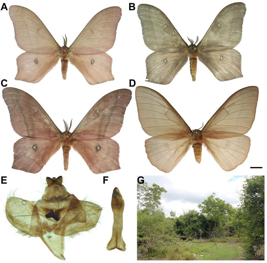
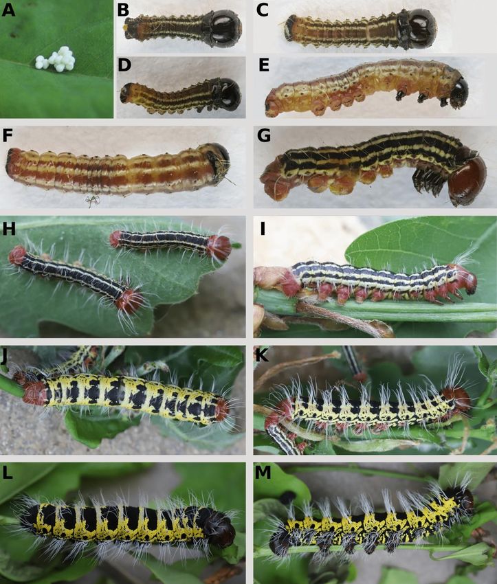
SPITSYN ET AL.

Cirina forda (Westwood 1849)

Material examined: 6♂, 2♀, TANZANIA: Zanzibar Island, Kiwengwa Forest, dry monsoon forest, 5°59'S,
39°21'E, 24.03-12.04.2021, E. Spitsyna & V. Spitsyn leg. (Fig 2).

Figure 1. Eggs and larvae of Cirina forda. (A) Eggs. (B-D) First instar larva. (E-F) Second instar larva. (G-H) Third
instar larva. (I) Fourth instar larva. (J-K) Fifth instar larva. (L-M) Sixth instar larva. (Photos: Elisaveta A. Spitsyna and
Vitaly M. Spitsyn).

Ecologica Montenegrina, 45, 2021, 12-15                                                                                   13
MORPHOLOGY OF CIRINA FORDA

         Morphology of preimaginal stages: Eggs are irregular in a shape, pale green or white (Fig 1A).
Eggs cluster together. Incubation time 8 days. L1: The length at hatching 1.5 mm. Head capsule black.
Ground colour brown, with three pale yellow stripes running along the entire body. There are 8 small warts
with short hairs on each segment (Fig 1B-D). L2: looks similar to L1. Size ca. 5 mm (Fig 1E-F). L3: Head
capsule, legs, prothoracic shield, and anal plate red-brown. Ground colour black, with three yellow-green
stripes running along the entire body. On each segment, there are 8 small warts with white hairs (Fig 1G-H).
L4: looks similar to L3 (Fig 1I). L5: Head capsule, prothoracic shield, and anal plate red-brown. Ground
colour yellow, with black pattern on each segment. On the dorsal side of each segment, there is a large black
spot with three vertices. This spot is divided into three spots on the last segments. White hairs arranged in
rows, with 4 rows on each side. There is a very large black spiracle laterally (Fig 1J-K). L6: Head capsule,
prothoracic shield, and anal plate dark brown, almost black. Ground colour yellow. On each segment, there is
a black pattern with a very large black dorsal spot. In contrast to the earlier instars, this black spot is much
larger with a less rugged contour. White hairs arranged in rows, with 4 rows on each side. There is a very
large black spiracle laterally (Fig 1L-M).

Figure 2. Cirina forda. (A-C) Male. (D) Female. (E) Male genitalia. (F) Aedeagus. (G) Habitat. (Photos: Elisaveta A.
Spitsyna and Vitaly M. Spitsyn).

                                                                                                                 14
SPITSYN ET AL.
Male genitalia: Uncus with three vertices; tegumen wide and large; saccus large, elongated, V-shaped.
Valva rounded; juxta V-shaped; anellus large, rectangular, well sclerotized (Fig 2E). Aedeagus with a wide
forked base, and an expanded apical part (Fig 2F).
Acknowledgements
This study was supported by the Young Scientists of Pomorye Region Program (Project no. 07-2021а) and
by the Russian Ministry of Science and Higher Education (Project no. АААА-А17-117033010132-2). The
authors are grateful to Dr. Elena Yu. Churakova for her valuable help in a search for food plants
References
Adepoju, O. T. & Daboh, O. O. (2013) Nutrient composition of Cirina forda (Westwood) - enriched
complementary foods. Annals of Nutrition and Metabolism, 63(1-2), 139–144.
Ande, A. T. & Fasoranti, J. O. (1998) Some aspects of the biology, foraging and defensive behaviour of the
emperor moth caterpillar, Cirina forda (Westwood). Insect Science and its Application, 18(3), 177–
181.
Lautenschläger, T., Neinhuis, C., Monizi, M., Mandombe, J. L., Förster, A., Henle, T. & Nuss, M. (2017)
Edible insects of Northern Angola. African Invertebrates, 58(2), 55–82.
Pinhey, E. C. G. (1956) The Emperor Moths of Eastern Africa. Journal of The East Africa Natural History
Society, XXIII(1), 1–62.
Ecologica Montenegrina, 45, 2021, 12-15 15You can also read