Jellyfish & Aquaculture Interactions: Last Year's Irish Experience - MARCOS-LÓPEZ M, MITCHELL S.O AND RODGER H.D
←
→
Page content transcription
If your browser does not render page correctly, please read the page content below
Jellyfish & Aquaculture
Interactions: Last Year’s
Irish Experience

     M ARCOS-LÓP EZ M, MITCHELL S.O
     AND RODGER H.D
     August 2014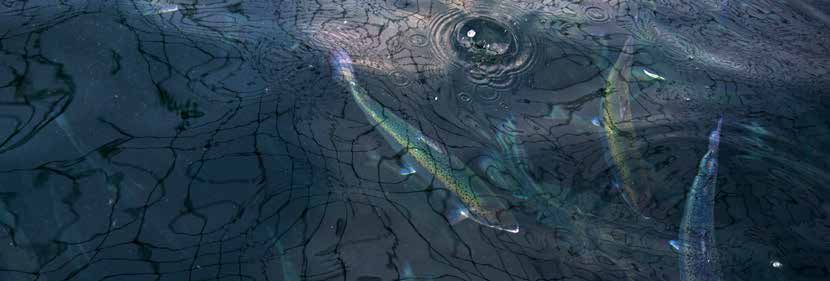
Background
Large jellyfish swarms occur naturally in our oceans but when in contact
with a fish farm they can have severe consequences. A number of mortality
events involving different jellyfish species have been reported in the
literature over the years. Small jellyfish can pass through the nets, while
bigger individuals tend to break up into pieces still capable of stinging the
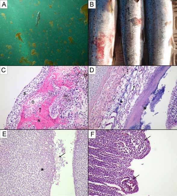
fish (Fig. 1A). Affected fish can suffer from hypoxia, mechanical damage
and toxicity via nematocysts discharge (Baxter et al. 2011). During the
summer and autumn months of 2013, large aggregations of the mauve
stinger jellyfish Pelagia noctiluca were present along the west coast of
Ireland causing significant losses to the affected Atlantic salmon farms.
Associated mortalities ranged from2 Fig. 1
Clinical Presentation & Pathology
    Affected fish showed respiratory distress, loss of appetite, lethargy and/or
    increased jumping behavior. On the most severe cases, up to 80% of the
    fish examined presented signs of jellyfish damage in the gills and skin.
    Gross skin lesions greatly varied in size and included erosion, scale loss,
    swollen and/or congested/haemorrhagic lesions and ulcers (Fig. 1B). Gill
    damage comprised necrosis, haemorrhage and/or loss of tissue. Some
    lesions showed a yellow-brownish colour suggesting bacterial infection.
    Histology samples were taken to characterize the type of pathology. Overall,
    the histopathology assessment of the skin lesions revealed a significant
    acute dermatitis with associated dermal necrosis and focal ulceration
    (Fig. 1C). The affected gill filaments showed acute haemorrhage, congestion,
    infiltration, oedema, necrosis, lamellar epithelium sloughing and/or tissue
    loss (Fig. 1E). Chronic lesions also showed lamellar epithelium hyperplasia
    and fusion, and occasional presence of giant cells within the affected
    lamellar epithelium. Bullae-like formations at the edges of the filaments
    were also observed in some samples (Fig. 1F). In some of the samples,
    large aggregations of filamentous bacteria (Tenacibaculum sp.) were
    seen colonizing the necrotic filaments (Fig. 1D). Open lesions are prone
    to secondary bacterial infections, however some zooplankton species
    including P. noctiluca have been shown to carry out and host Tenacibaculum
    maritimum (Delannoy et al. 2010). The most affected pens were treated
    with oxytetracycline (orally 8–10 days at 100mg/kg body weight), resulting
    in prevention of secondary bacterial infections due to the skin damage and
    decreased mortalities.

3
Further Insights
P. noctiluca has already been associated with mortalities in salmon
aquaculture. The most well-known episode occurred in Northern Ireland in
2007, when a large swarm of this species killed an entire Atlantic salmon
farm (~250,000 fish) (Doyle et al. 2008). Despite previous reports, the
skin and gill pathologies induced by P. noctiluca have not been previously
described. The lesions described are believed to be important and caused
by a combination of mechanical and toxic damage. We believe that the
characterization of the pathology caused by different environmental agents
(i.e. phytoplankton and zooplankton species) will improve the differential
diagnosis of gill disorders for which histopathology is a key diagnostic tool.
Unlike most terrestrial livestock farming, marine aquaculture is highly
affected by the environmental conditions. In recent years, there exists a
worldwide concern that jellyfish blooms are increasing. However, their cyclic
nature and the lack of long-term data make it difficult to draw definitive
conclusions. Increased eutrophication due to anthropogenic activities and
other human activities (e.g. over-fishing) may favour jellyfish multiplication
(Purcell et al. 2007).
4References
Baxter E.J., Sturt M.M., Ruane N.M., Doyle T.K., McAllen R., Harman L. &
Rodger H.D. (2011a) Gill damage to Atlantic salmon, Salmo salar, caused
by the common jellyfish, Aurelia aurita, under experimental challenge. PLoS
ONE, 6(4), e18529.
Delannoy C.M.J., Houghton J.D.R., Fleming N.E.C. & Ferguson H.W. (2010)
Mauve stingers (Pelagia noctiluca) as carriers of the bacterial fish pathogen
Tenacibaculum maritimum. Aquaculture 311(1–4):255–257.
Doyle T.K., De Haas H., Cotton D., Dorschel B., Cummins V., Houghton
J.D.R., Davenport J. & Hays G.C. (2008) Widespread occurrence of the
jellyfish Pelagia noctiluca in Irish coastal and shelf waters. Journal of
Plankton Research 30:963–968.
Marcos-López M., Mitchell S.O., Rodger H.D. (2014) Pathology and
mortality associated with the mauve stinger jellyfish Pelagia noctiluca in
farmed Atlantic salmon Salmo salar L. DOI: 10.1111/jfd.12267.
Purcell J.E., Uye S. & Lo W. (2007) Anthropogenic causes of jellyfish
blooms and their direct consequences for humans: a review. Marine Ecology
Progress Series 350:153–174.
5Figure Legend
Fig. 1A
Numerous P. noctiluca jellyfish inside marine Atlantic salmon pen.
Picture courtesy of Pete McDonagh.
Fig. 1B
Flank skin lesions in farmed Atlantic salmon caused by contact
with P. noctiluca.
Fig. 1C
Skin pathology caused by P. noctiluca. Note dermal necrosis (N), cell
infiltration (arrow), oedema (O), and epidermal spongiosis (S) (x20) H&E.
Fig. 1D
Severe gill pathology caused by P. noctiluca. Note lamellar epithelium
necrosis (arrow) and secondary colonization with filamentous bacteria (*).
(x20) H&E.
Fig. 1E
Jellyfish contact point in lamellar gill epithelium. Note cell infiltration
in affected epithelium (*) and remains of jellyfish tissue at the epithelial
surface (arrow) (x20) H&E.
Fig. 1F
Bullae-like lesions at the edge of proliferated affected lamellar epithelium
(arrows) (x20) H&E.
6You can also read