The pig as a model for translational research: overview of porcine animal models at Jichi Medical University
←
→
Page content transcription
If your browser does not render page correctly, please read the page content below
Kobayashi et al. Transplantation Research 2012, 1:8
http://www.transplantationresearch.com/content/1/1/8                                                                        TRANSPLANTATION
                                                                                                                            RESEARCH

 REVIEW                                                                                                                                          Open Access

The pig as a model for translational research:
overview of porcine animal models at Jichi
Medical University
Eiji Kobayashi1,2*, Shuji Hishikawa1, Takumi Teratani1 and Alan T Lefor1

  Abstract
  To improve the welfare of experimental animals, investigators seek to respect the 3R principle (Replacement,
  Reduction, and Refinement). Even when large animal studies are essential before moving to clinical trials, it is
  important to look for ways to reduce the number of experimental animals used. At the Center for the Development
  of Advanced Medical Technology, we consider ‘medical’ pigs to be ideal preclinical model systems.
  We have been using both wild-type and genetically modified pigs. We began using this approach about 10 years
  ago with a ‘total pig system’ to model human health and disease for the purposes of both medical skill education
  and the development of new devices and therapeutic strategies.
  At our Center, medical students and residents use pigs to gain experience with surgical skills and train for
  emergency procedures after appropriate simulation training. Senior clinicians have also used these models to
  advance the development of innovative tools for endo- and laparoscopic procedures. The Center focuses on
  translational research for organ transplantation and stem cell therapy. Several pig models have been established for
  liver, intestine, kidney, pancreas, and lung transplantation. Mesenchymal stromal cells have been established in
  green fluorescent protein- and red fluorescent protein-transgenic pigs and tested to trans-differentiate
  organogenesis. A program to establish induced pluripotent stem cells in the pig is ongoing at our Center.
  Here, we review our 10 years of activity in this field. Based on our experience in surgical education and research,
  experimental pigs are valuable models in translational research.
  Keywords: Experimental animals, Pig, Translational research

Introduction                                                                          limitations on using mongrel dogs for medical education
Pigs and humans have anatomical and physiological                                     and research in Japan, we decided to use a different
similarities. First, the immune system of pigs is similar                             medium-sized experimental animal as a model.
to that of humans, and second, inbred pigs such as                                      From 2001 to 2002, with the help of a grant established
Clawn minipigs have genetically defined and fixed major                               by Tochigi Prefecture to promote regional economic
histocompatibility complex, making reproducible studies                               activity, we surveyed several institutions in Japan that
of immunologic mechanisms possible [1-4]. Therefore,                                  used pigs as experimental models. Our investigation of
the pig has attracted attention as a valuable preclinical                             the numbers of pigs used in experiments by all of the
model for medical research. Although there have been                                  medical universities in Japan revealed that there was
few reports of the scientific advantages of using pigs as                             scant information available on the use of pigs. Further-
such models, there are multiple reports of the use of                                 more, the support system for using these animals as
dogs and non-human primates. Because of the ethical                                   biomedical models was poor. More than 60% of 115 uni-
                                                                                      versities surveyed had no experience in the postoperative
* Correspondence: eijikoba@jichi.ac.jp                                                care of experimental pigs, and fewer than 10 domestic or
1
 Center for Development of Advanced Medical Technology, Jichi Medical
University, 3311-1, Yakushiji, Shimotsukeshi, Tochigi 329-0498, Japan                 miniature pigs were used annually at another university.
2
 Division of Development Advanced Therapy, Center for Development of                  As a result, we started the Tochigi Pig Project to study
Advanced Medical Technology, Jichi Medical University, 3311-1, Yakushiji,             the use of pigs as biomedical models [5].
Shimotsukeshi, Tochigi 329-0498, Japan

                                        © 2012 Kobayashi et al.; licensee BioMed Central Ltd. This is an Open Access article distributed under the terms of the Creative
                                        Commons Attribution License (http://creativecommons.org/licenses/by/2.0), which permits unrestricted use, distribution, and
                                        reproduction in any medium, provided the original work is properly cited.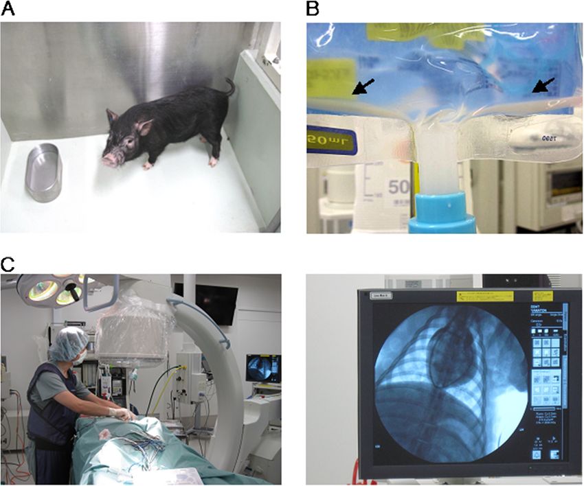
Kobayashi et al. Transplantation Research 2012, 1:8                                                                Page 2 of 9
http://www.transplantationresearch.com/content/1/1/8

Establishment of a total care system for medical pigs            welfare and public opinion in Japan, mongrel dogs are no
Over the last 10 years, we have studied several kinds of pig.    longer used for medical education and research. In 2000,
They are classified into three categories on the basis of        the use of all experimental animals in educational courses
body size. Our new pig research center, the Center for De-       at Jichi Medical University was changed from dogs to pigs
velopment of Advanced Medical Technology (CDAMTec),              (Figure 2B).
began full-scale operations in 2008 (Figure 1A). CDAMTec           At the same time, studies were initiated to investigate
has three important goals - education, surgical training, and    differences in liver metabolism between humans and pigs.
pre-clinical research - and a number of academic investiga-      A simple surgical model was established that utilizes an in-
tors and physicians have been recruited to the center            ternal shunt. First, changes in microsomal P450 isoforms
(Figure 1B). CDAMTec provides advanced-level clinical            after therapeutic liver resection were studied by using this
care and testing, including computed tomography (CT),            model [8]. Partial occlusion (portal vein and hepatic artery
magnetic resonance imaging (MRI), and an ICU                     occlusion) decreased the activities of CYP2C, CYP2E, and
(Figure 1C, Additional file 1: Movie S1). Furthermore, an        CYP3A, but not those of CYP1A and CYP2D. CYP3A,
advanced imaging system is used to combine CT and MRI            which accounts for an average of 30% of the total P450
data for surgical support (Figure 1D, Additional file 2:         content in the human liver, was most susceptible to warm
Movies S2 and Additional file 3: Movies S3). The Center is       ischemia. The metabolism of the anesthetic drug, propofol,
used for surgical training and education in high-risk tech-      was then examined [9]. Propofol is known to have intra-
niques or techniques where high skill levels are needed, or      and extrahepatic metabolic pathways, but the effect of its
before the initiation of clinical trials [6].                    continuous infusion during long-term anhepatic states had
  Jichi Medical University introduced the use of pigs as         not been determined. Hemodynamic parameters related to
part of the Bedside Learning phase of clinical surgical          the pharmacokinetics of continuously infused propofol
education at CDAMTec. The curriculum is composed of              (6 mg/kg/hr) were also investigated. Although there were
four sections: Bioethics, Orientation, Practical training,       changes in the heart rate, no significant changes in the
and Verification of surgical skills.                             concentration of hemoglobin or in hemodynamic para-
  The Advanced Trauma Operative Management course                meters were observed during the anhepatic phase when
was established in the United States in 2000 to standardize      propofol was continuously infused. Mixed venous, arterial,
the operative management of trauma patients. The course          and portal vein propofol concentrations were stable during
is given to senior surgeons and senior surgical residents,       the anhepatic phase.
by using a one-to-one instruction system. The course was           Using this model, three kinds of infusion solutions were
brought to Japan in 2008 and began at CDAMTec. We                tested, namely lactated Ringer’s solution (LR, Lactec), ace-
also offer separate training for surgical residents to im-       tated Ringer’s solution (AR, VeenF), and acetated Ringer’s
prove their skills with a dry-lab and microsurgical training     solution with 1% glucose (AR-G, Phisio140) [10]. Although
[See Additional file 4: Movie S4].                               no major difference was observed in the hemodynamic
  In addition to training in specific procedures, all users of   parameters, arterial blood gas data, or electrolyte concen-
the facility are educated regarding the welfare of ex-           trations among the three groups, a significant and progres-
perimental animals. The design of the programs con-              sive elevation of lactic acid levels was observed in the LR
ducted has reduced the overall number of experimental            group. Severe hypoglycemia was found in the LR and AR
pigs used in training [7].                                       groups, whereas the AR-G group maintained its blood glu-
                                                                 cose levels throughout the anhepatic phase.
Research using domestic pigs                                       Hepatitis E virus (HEV) is highly prevalent among do-
The animals used in these training programs are domestic         mestic pigs, and substantial attention has been given to
pigs with a mature body weight of approximately 100 kg. In       keeping our center free from this virus [11]. We investi-
the early stage of the project, we focused on the medical        gated the prevalence of IgG class antibodies to pig HEV
use of domestic pigs with help from farmers close to the         (anti-HEV) and of HEV RNA among 152 domestic pigs at
university. Domestic pigs are easily obtained and inexpen-       2 months of age and 38 miniature pigs from 4 to 10 months
sive, because they are well established as a food source.        of age; the pigs had been obtained for research purposes
They are produced in a three-breed terminal system by            from five farms in Japan. HEV RNA was detected in 38%
crossbreeding among Landrace, Large White, and Duroc             of the domestic pigs; the 22 HEV isolates recovered from
colonies (Figure 2A). Young pigs weighing approximately          the viremic pigs were 89.8% to 100% identical in the 412-
30 to 40 kg are often used for medical training and              nucleotide sequence of open reading frame 2 and segre-
research.                                                        gated into three clusters within genotype 3. In contrast, all
  At Jichi Medical University before this project was            of the miniature pigs, which came from farms different
started, medical students practiced their surgical skills on     from those where the domestic pigs were sourced, were
mongrel dogs. Because of recent changes to both animal           negative for both anti-HEV and HEV RNA. We concluded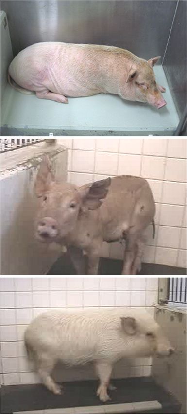
Kobayashi et al. Transplantation Research 2012, 1:8    Page 3 of 9
http://www.transplantationresearch.com/content/1/1/8

 Figure 1 (See legend on next page.)
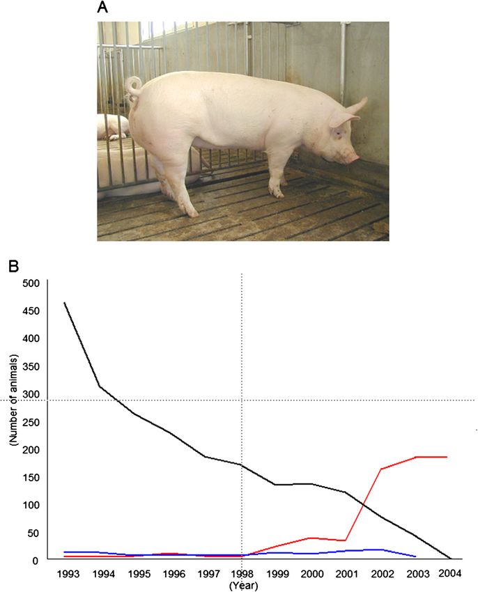
Kobayashi et al. Transplantation Research 2012, 1:8                                                                                     Page 4 of 9
http://www.transplantationresearch.com/content/1/1/8

 (See figure on previous page.)
 Figure 1 The CDAMTec research center at Jichi Medical University. A) Opening ceremony (left panel) and the outside of the pig center
 (right panel). B) Surgical training using pigs. C) CDAMTec features MRI (top panel), CT (middle panel), and an ICU (bottom panel). D) Surgical
 simulation system. Inset shows patient data. CDAMTec, Center for Development of Advanced Medical Technology; CT, computed tomography;
 MRI, magnetic resonance imaging.

that it is important to check that pigs provided for research              chronic experiments has increased; such experiments
are HEV free.                                                              accounted for 48% of experiments using pigs in 2004.
  We also began a program to share animal tissues,                         Improvement of the effective utilization of these animals
whereby researchers can reuse various organs obtained                      is required from both an economic and an ethical point
from euthanized animals [12]. In 2003, the number of                       of view. Experimental pigs undergo secondary use after
pigs used as experimental animals at our center rapidly                    being euthanized, thus reducing the total number of ex-
increased to more than 170. Moreover, the number of                        perimental animals needed for medical research. Overall

 Figure 2 Changes in use of large animals at Jichi Medical University. A) Domestic pig. B) Numbers of large animals used at our university
 for research. Pig (red), dog (black), and monkey (blue).
Kobayashi et al. Transplantation Research 2012, 1:8                                                                     Page 5 of 9
http://www.transplantationresearch.com/content/1/1/8

numbers are similarly reduced through sharing and re-
use of miniature-pig tissues and cells for research. We
recommend this system, because it improves the quality
of medical education and research and facilitates the ef-
fective use of tissues and cells through sharing and reuse
among different investigators.
  Development of an efficient system of pre-culture of
pig islets was ideal [13]. This program has continued,
resulting in a reduction in the total number of experi-
mental animals used.

Research using miniature pigs
Although miniature pigs are generally easier to handle and
more suitable for medical research than are domestic pigs,
miniature pigs are more expensive because of limited an-
nual production for experimental use in Japan. It is import-
ant to note that a mature miniature pig weighs 40 to 50 kg;
this weight is equivalent to that of an immature domestic
pig (Figure 2A).
  Platelets promote tissue repair and liver regeneration.
After observing a positive response to human thrombo-
poietin (TPO) in mature miniature pigs, we found that
platelets prevent acute liver damage after extended hepa-
tectomy in pigs [14]. Thrombocytosis was induced by the
following two methods, and an 80% hepatectomy was per-
formed. In the first method, pigs received thrombopoietin
(TPO (+) group), and were compared with a control group
(TPO (−) group). In the second method, an experimental
group underwent splenectomy (Sp (+) group) to induce re-
active thrombocytosis and was compared with a control
group (Sp (−) group). Serum concentrations of the enzyme
induced by liver damage were significantly lower in the
thrombocytotic groups than in the control groups in the
early period after hepatectomy. Histopathological examin-
ation revealed hemorrhagic necrosis with a bile plug in the
control groups, but this phenomenon was not observed in
the thrombocytotic groups. We concluded that mature
miniature pigs respond to human TPO and that an
increased platelet count prevents acute liver damage after
extended hepatectomy in this species.
  Investigators must optimize the perioperative care of ex-
                                                                Figure 3 A variety of miniture pig strains are used at Jichi
perimental animals, but little is known about the effects of    Medical University for experimental research. KCG pig (top
anesthesia and surgery on serum chemistry in KCG mini-          panel), Mexican hairless pig (middle panel), and Clawn pig
ature pigs [15] (Figure 3, top panel). Our objective in an-     (bottom panel).
other study was to examine the influence of fasting and
surgery under general anesthesia on 27 serum chemistry
parameters in KCG miniature pigs so as to improve pre-         spine was performed in two groups of animals. Those given
operative management. Crossbred KCG miniature pigs             sevoflurane anesthesia (n = 7) had significant decreases in
were used at a mean of 12.3 months of age (range, 8.6 to       serum albumin, potassium, inorganic phosphorus, gamma-
14.9 months) and 33.4 kg body weight (range, 24.0 to           glutamyltransferase peptidase, cholinesterase, and glucose
40.2 kg). Serum chemistry was evaluated at the beginning       after surgery compared with levels before surgery. Animals
and end of a 24-hour fasting period (n = 6). No significant    given isoflurane (n = 7) anesthesia had significantly decreased
differences were observed between the values tested at the     total protein, albumin, triglyceride, phospholipids, sodium,
two time points. Partial hemilaminectomy of the lumbar         potassium, calcium, alanine aminotransferase, alkaline
Kobayashi et al. Transplantation Research 2012, 1:8 Page 6 of 9 http://www.transplantationresearch.com/content/1/1/8 phosphatase, and glucose after surgery compared with decline. Increased FOXP3 expression was not observed in levels before surgery. In a separate experiment (n = 7), recipients given high-dose tacrolimus. In a miniature, such serum glucose and insulin also decreased during the post- as Clawn pig, transplantation model, FOXP3 mRNA levels operative period after isoflurane anesthesia. These results in the peripheral blood were upregulated in the early phase demonstrated that specific serum electrolytes, glucose, of rejection. Thanks to the merits of this inbred colony, and insulin were altered in KCG miniature pigs undergo- induced pluripotent stem (iPS) cells have been established ing general anesthesia. Investigators must be aware of the for development by Hanazono [20]. The established iPS cells, effects of anesthetic agents on experimental animals so as which have been confirmed to form teratomas in severe to provide optimum care and correct the interpretation of combined immunodeficiency (SCID) mice, are now being experimental data. tested in a syngeneic combination of mature Clawn pigs. Mexican hairless pigs (Figure 3, middle panel), which were developed by Japan’s National Agriculture and Food Research using genetically modified miniature pigs Research Organization, are highly suited for the evaluation As genetic modification techniques have developed, som- of topical agents, because the structure of their skin is simi- atic cloning technology has become well established in lar to that of humans [16]. To evaluate the pharmacokinet- pigs. Recently, we produced cloned miniature pigs by using ics of topical drugs, in vitro permeation studies are oocytes derived from domestic pig ovaries [21]. For the performed by using the skin of euthanized animals or production of viable somatic-cell nuclear transferred human tissues resected at surgery; however, these methods (SCNT) miniature pig embryos, the in vitro conditions for have limitations for evaluating in vivo pharmacokinetics. controlling the quality of recipient oocytes derived from Therefore, we studied the use of Mexican hairless pigs for domestic pig ovaries need to be evaluated. To obtain infor- in vivo pharmacokinetics, especially for the evaluation of mation on the optimum in vitro maturation (IVM) condi- drug concentrations in tissues. A ketoprofen patch was ap- tions for oocytes, we investigated the effect of IVM plied to the backs of Mexican hairless pigs for 24 hours; duration of recipient oocytes on the subsequent develop- this was followed by sequential collections of blood from 0 ment of SCNT miniature pig embryos. We also investi- to 36 hours. Skin, subcutaneous fat, fascia, and muscle gated maturation-promoting factor (MPF) activity in from the center of the application site were excised 12 recipient oocytes before and after SCNT, along with the hours after patch application. Ketoprofen was first detected occurrence of premature chromosome condensation in the plasma at 8 hours. The concentration increased until (PCC) and the spindle morphologies of donor nuclei fol- 24 hours and began to decrease after removal of the keto- lowing SCNT. The optimal window for the IVM period in profen patch. Ketoprofen concentrations in the tissues terms of the in vitro developmental ability of SCNT decreased with increasing tissue depth, but the amount in embryos was 36 to 40 hours after the start of IVM. Use of the deep muscles, being the lowest among the tissues recipient oocytes matured for 36 or 40 hours, but not 44 examined, was still higher than that in the plasma. Drug or 52 hours, resulted in a high level of MPF activity before concentrations are difficult to test in human tissues, and and after SCNT and increased the occurrence of PCC in the Mexican hairless pig model appears to be attractive for transferred nuclei. The proportion of abnormal spindle- in vivo pharmacokinetic studies of topically applied keto- like structures increased with prolongation of the IVM profen. This breed has also been used to develop a new period. In addition, SCNT embryos constructed from re- medical device to connect the intestinal lumen percutan- cipient cytoplasts obtained after 40 hours of maturation by eously by using a double-balloon method [17]. using fetal fibroblasts of miniature pigs were transferred to Clawn miniature pigs (Figure 3, bottom panel) commer- surrogate miniature pigs and developed to full term. These cially provided by the Japan Farm Clawn Institute are recom- results suggest that recipient oocytes matured for 36 hours mended for use in transplantation research, because their or 40 hours show effective induction of PCC with a normal SLAs (swine leukocyte antigens) have been fixed, and defin- cytoskeletal structure because of a high level of MPF activ- ite immunological reactions are observed in models of intes- ity. Furthermore, the 40 hour IVM period improves the tinal [18] and lung transplantation [19]. The outcome of in vitro development of SCNT embryos to the blastocyst highly immunogenic transplantation remains unsatisfactory, stage, resulting in the production of viable cloned mini- despite the development of potent immunosuppressants. ature pigs. There is emerging clinical evidence that, paradoxically, ex- Animal imaging sources have become indispensable in pression of forkhead box P3 (FOXP3, a specific marker for the biological sciences. Specifically, gene-encoded bio- regulatory T cells) is upregulated in graft rejection. Levels of logical probes serve as stable and high-performance tools mRNA expression of FOXP3, perforin, Fas-ligand (Fas-L), for visualizing the fate of cells in living animals. Green and interferon gamma-induced protein 10 (IL-10) were fluorescent protein (GFP)-transgenic Jinhua pigs have been quantified in the peripheral blood and reached their highest established and show normal growth and reasonable repro- value as early as postoperative day four, followed by a ductive activity [22]. In this transgenic pig model, a green
Kobayashi et al. Transplantation Research 2012, 1:8 Page 7 of 9
http://www.transplantationresearch.com/content/1/1/8
emission is observed in the body under an excitation light. dogs, monkeys, and pigs, we believe that these micromini
We use a somatic cell cloning technique to create new pigs will be suitable for use in studies of advanced medical
miniature-sized GFP-expressing Jinhua pigs; this enables treatments. We have great interest in regenerative medi-
us to characterize expression profiles in various tissues or cine and are focusing on the micromini pig model to test
organs and under various ex vivo culture conditions. its benefits and drawbacks in this field. Because of their
Strong GFP expression is observed in the skeletal muscle, moderate body size, micromini pigs are suitable for testing
pancreas, heart, and kidney. Bone-marrow-derived mesen- systemic injections of MSCs. This is not the case in
chymal stromal cells (MSCs), hepatocytes, and islet cells of rodents, which are too small, and miniature pigs, for which
the pancreas also show detectable expression, with a more time is required for stem cell expansion in vitro.
unique pattern. Moreover, the cloned pigs demonstrate MSCs are expected to have tissue-regenerative, immuno-
normal growth and fertility, and the introduced GFP gene modulatory, and anti-inflammatory effects [24].
is stably transmitted to subsequent generations. These However, a major risk of MSC therapy is pulmonary em-
GFP-expressing Jinhua pigs can be used as new cellular or bolism in the very early phases of treatment. It is important
tissue light resources for biological imaging in preclinical to note that MSCs suspended in saline or culture media eas-
research fields such as tissue engineering, experimental re- ily become sedimentary and aggregate in less than one hour
generative medicine, and transplantation. These pigs are (Figure 4B); this might be related to the risk of embolism-
also a useful source for imaging in the field of stem cell re- related complications after intravenous injection. A preclin-
search. A clinical protocol for intra-articular injections into ical study using the micromini pig clearly showed that sys-
massive meniscal defects has been tested by using synovial temic injection of MSCs (1 × 10⁷cells/kg, about 3.0 × 10⁸cells/
stem cells obtained from GFP-transgenic animals [23]. pig) immersed in normal saline without heparin (200 mL, in-
fusion time 40 minutes) caused the pulmonary arterial pres-
Research using micromini pigs sure to increase by more than 30 mm Hg, as determined by
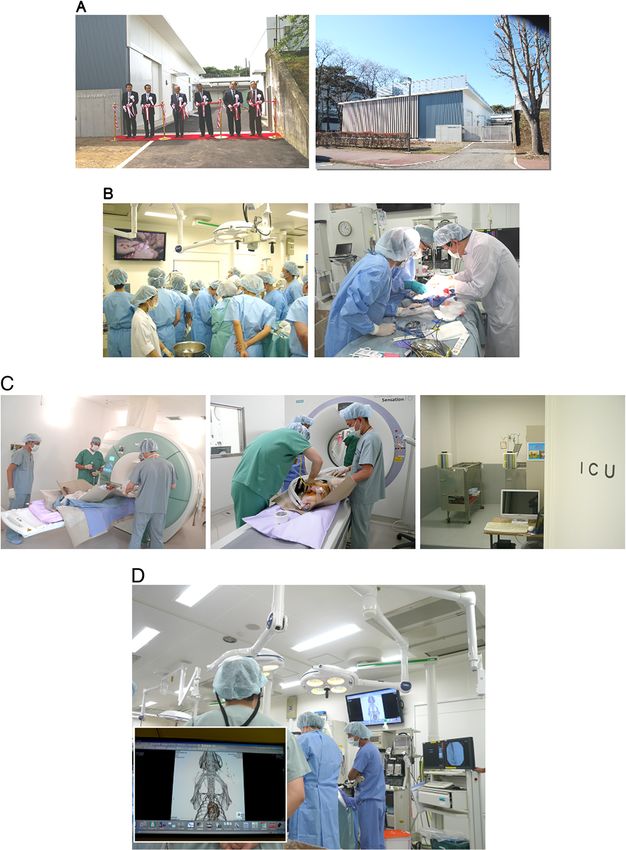
A very small pig, termed a ‘micromini pig’ is likley to prove monitoring with a pulmonary artery catheter (Figure 4C).
suitable for preclinical safety evaluations of new agents However, by using a solution that we developed in a liver is-
(Figure 4A). Although several years might be needed to chemia–reperfusion model in pigs, we found that allogenic
perform comparative studies of drug toxicology among MSC transplantation had a therapeutic effect (manuscript in
Figure 4 Cell transplantation models in the pig. A) Micromini pig. B) Cell mixture solution in an infusion pack after one hour. Arrow shows
precipitation of cells. C) Placement of a pulmonary artery catheter by using a C-arm (left panel) and monitor (right panel).Kobayashi et al. Transplantation Research 2012, 1:8 Page 8 of 9
http://www.transplantationresearch.com/content/1/1/8
preparation). Additionally, for the purpose of these proce- Ministry of Education, Culture, Sport, Science, and Technology of Japan; the
dures, the pig’s anatomy is suffiicently similar to that of CEO program of MEDEX; and Otsuka Pharmaceutical Factory Inc. (Naruto,
Japan).
humans for X-ray to be useful in monitoring blood flow to
the liver [See Additional file 5: Movie S5]. Thus, the micro- Received: 24 April 2012 Accepted: 26 June 2012
mini pig was suitable for the development of an MSC- Published: 16 August 2012
aggregation inhibition solution and is useful for evaluating
treatment effects and creating a surgical model that mimics References
1. Sahara H, Shimizu A, Setoyama K, Oku M, Okumi M, Nishimura H, Oriyanhan
the human one. W, Tasaki M, Scalea J, Wada H, Bando T, Date H, Yamada K: Beneficial
effects of perioperative low-dose inhaled carbon monoxide on
pulmonary allograft survival in MHC-inbred CLAWN miniature swine.
Conclusions Transplantation 2010, 90:1336–1343.
We have described our 10 years of experience with the de- 2. Ando A, Uenishi H, Kawata H, Tanaka-Matsuda M, Shigenari A, Flori L,
velopment and use of several porcine models as biomed- Chardon P, Lunney KJ, Kulski KJ, Inoko H: Microsatellite diversity and
crossover regions within homozygous and heterozygous SLA haplotypes
ical research tools. Development of disease models in pigs of different pig breeds. Immunogenetics 2008, 60:399–407.
is essential for further expanding the use of pigs in effect- 3. Ando A, Ota M, Sada M, Katsuyama Y, Goto R, Shigenari A, Kawata H, Anzai
ive preclinical experiments, because few spontaneous dis- T, Iwanaga T, Miyoshi Y, Fujimura N, Inoko H: Rapid assignment of the
swine major histocompatibility complex (SLA) class I and II genotypes in
ease models are available in pigs, unlike in dogs. Recent clawn miniture swine using PCR-SSP and PCR-RFLP methods.
advances in genetic technology in pigs have shown that Xenotransplant 2005, 12:121–126.
these experimental animals are now suitable, mature bio- 4. Kita FY, Ando A, Tanaka K, Suzuki S, Ozaki Y, Uenishi H, Inoko H, Kulski KJ,
Shiina T: Application of high-resolution, massively parallel
medical models. pyrosequencing for estimation of haplotypes and gene expression levels
of swine leukocyte antigen (SLA) class I genes. Immunogenetics 2012,
Additional files 64:187–199.
5. Endo M, Enosawa S, Suzuki S, Amemiya H, Kobayashi E, Miyashita T, Aoki T,
Koyanagi Y: Porcine liver transplantation as an estimation system for
Additional file 1: Movie S1. CT and MRI dedicated pig in CDAMTec. bridge-use of bioartificial liver. Transplant Proc 2002, 34:2714–2717.
Additional file 2: Movie S2. Surgical training by an advanced imaging 6. Hishikawa S, Kawano M, Tanaka H, Konno K, Yasuda Y, Kawano R, Kobayashi
system. E, Lefor AT: Mannequin simulation improves the confidence of medical
Additional file 3: Movie S3. Demonstration of CT and MRI imaging students performing tube thoracostomy: a prospective, controlled trial.
analysis data on PC monitor. Am Surg 2010, 76:73–78.
7. Konno K, Nakanishi K, Hishikawa S, Tanaka H, Yoshikawa T, Yasuda Y,
Additional file 4: Movie S4. The Advanced Trauma Operative Kobayashi E, Lefor A: Cryo-preserved porcine kidneys are feasible for
Management course. teaching and training renal biopsy: “The Bento Kidney”. Transplant Res,
Additional file 5: Movie S5. Mobile X-ray system as C-arm. in press.
8. Suzuki S, Satoh T, Yoshino H, Kobayashi E: Impact of warm ischemic time
on microsomal P450 isoforms in a porcine model of therapeutic liver
Competing interests resection. Life Sci 2004, 76:39–46.
EK has been a visiting professor at CDAMTec and a special advisor to Otsuka 9. Murayama T, Sato Y, Wainai T, Enomoto A, Seo N, Yoshino H, Kobayashi E:
Pharmaceutical Factory Inc. (Naruto, Japan) from 2009. There are no patents, Effect of continuous infusion of propofol on its concentration in blood
products in development, or marketed products to declare. The position with and without the liver in pigs. Transplant Proc 2005, 37:4567–4570.
held by EK does not alter the authors adherence to all of the Transplantation 10. Komiya K, Sato Y, Wainai T, Murayama T, Yamada M, Hiruta A, Seo N,
Research policies on sharing data and materials, as described in detail online Yoshino H, Tanaka H, Kobayashi E: Evaluation of intraoperative infusion
in the Guide for Authors. The other authors declare no competing financial solution using a complete anhepatic model in baby pigs. Transplant Proc
interests. 2005, 37:2341–2346.
11. Tanaka H, Yoshino H, Kobayashi E, Takahashi M, Okamoto H: Molecular
Authors’ contributions investigation of hepatitis E virus infection in domestic and miniature
EK designed and coordinated the project. SH and AL performed educational pigs used for medical experiments. Xenotransplantation 2004, 11:503–510.
aspects of the project. TT conducted the research project. All authors read 12. Tanaka H, Kobayashi E: Education and research using experimental pigs
and approved the final manuscript. in a medical school. J Artif Organs 2006, 9:136–143.
13. Miki A, Narushima M, Okitsu T, Takeno Y, Soto-Gutierrez A, Rivas-Carrillo JD,
Acknowledgements Navarro-Alvarez N, Chen Y, Tanaka K, Noguchi H, Matsumoto S, Kohara M,
We thank all of our co-workers contributing to this project at Jichi Medical Lakey JR, Kobayashi E, Tanaka N, Kobayashi N: Maintenance of mouse, rat,
University, including Drs. Fumimaro Takaku (Honor President), Shinichi and pig pancreatic islet functions by coculture with human islet-derived
Tominaga (Former Vice-President), Eiju Watanabe, Hideo Nagai, Yoshikazu fibroblasts. Cell Transplant 2006, 15:325–334.
Yasuda, Naohiro Sata, Takashi Igarashi, Norimasa Seo, Ryou Konno, Hozumi 14. Hisakura K, Murata S, Fukunaga K, Myronovych A, Tadano S, Kawasaki T,
Tanaka, Yoji Hakamada, Takashi Murakami, Masafumi Takahashi, Yutaka Kohno K, Ikeda O, Pak S, Ikeda N, Nakano Y, Matsuo R, Konno K, Kobayashi
Hanazono, Hironori Yamamoto, and Yasuhiro Fujimoto. We also thank E, Saito T, Yasue H, Ohkohchi N: Platelets prevent acute liver damage after
Professors Hiroshi Nagashima (Meiji University), Eimei Sato (Tohoku extended hepatectomy in pigs. J Hepatobil Pancreat Sci 2010, 17:855–864.
University), Takeshi Muneta and Ichirou Sekiya (Tokyo Medical and Dental 15. Tanaka H, Igarashi T, Lefor AT, Kobayashi E: The effects of fasting and
University), Nobuhiro Ohkochi (University of Tsukuba), Tatsuo Kawarasaki general anesthesia on serum chemistries in KCG miniature pigs. J Am
(University of Kumamoto), Hitoshi Kitagawa (University of Gifu), Toshiyuki Assoc Lab Anim Sci 2009, 48:33–38.
Saito (Kyoto Sangyo University) and Shinji Uemoto (University of Kyoto) for 16. Horie M, Sekiya I, Nakamura T, Tanaka H, Maekawa K, Nakanishi M, Muneta
their contributions to this project. Mrs. Tooru Wakui, Osamu Matsumoto and T, Kobayashi E: In vivo pharmacokinetics of ketoprofen after patch
Kazushi Miyazawa greatly assisted with the surgical treatment at CDAMTec. application in the Mexican hairless pig. Biopharm Drug Dispos 2009,
We gratefully acknowledge support from a Grant-in Aid for Scientific 30:204–208.
Research from the Japan Society for the Promotion of Science; the Strategic 17. Yano T, Yamamoto H, Sunada K, Miura Y, Taguchi H, Arashiro M, Yoshizawa
Research Platform for Private Universities; a matching fund subsidy from the M, Hayashi Y, Miyata T, Tanaka H, Kobayashi E, Sugano K: New techniqueKobayashi et al. Transplantation Research 2012, 1:8 Page 9 of 9
http://www.transplantationresearch.com/content/1/1/8
for direct percutaneous endoscopic jejunostomy using double-balloon
endoscopy and magnetic anchors in a porcine model. Dig Endosc 2011,
23:206.
18. Yoshino H, Yamauchi H, Kannan N, Iwai S, Endo K, Inoue S, Tahara K, Kaneko
T, Hakamata Y, Takahashi M, Kobayashi E: Vascular closure staples for
experimental organ transplantation. Transplantation 2003, 76:442–443.
19. Satoda N, Shoji T, Wu Y, Fujinaga T, Chen F, Aoyama A, Zhang JT, Takahashi
A, Okamoto T, Matsumoto I, Sakai H, Li Y, Zhao X, Manabe T, Kobayashi E,
Sakaguchi S, Wada H, Ohe H, Uemoto S, Tottori J, Bando T, Date H, Koshiba
T: Value of FOXP3 expression in peripheral blood as rejection marker
after miniature swine lung transplantation. J Heart Lung Transplant 2008,
27:1293–1301.
20. Hanazono Y: Generation of high quality induced pluripotent stem cells.
Seikagaku 2011, 83:1060–1063.
21. Wakai T, Sugimura S, Yamanaka K, Kawahara M, Sasada H, Tanaka H, Ando
A, Kobayashi E, Sato E: Production of viable cloned miniature pig
embryos using oocytes derived from domestic pig ovaries. Cloning Stem
Cells 2008, 10:249–262.
22. Kawarasaki T, Uchiyama K, Hirao A, Azuma S, Otake M, Shibata M, Tsuchiya
S, Enosawa S, Takeuchi K, Konno K, Hakamata Y, Yoshino H, Wakai T,
Ookawara S, Tanaka H, Kobayashi E, Murakami T: Profile of new green
fluorescent protein transgenic Jinhua pigs as an imaging source.
J Biomed Opt 2009, 14:054017.
23. Nakamura T, Sekiya I, Muneta T, Hatsushika D, Horie M, Tsuji K, Kawarasaki T,
Watanabe A, Hishikawa S, Fujimoto Y, Tanaka H, Kobayashi E: Arthroscopic,
histological and MRI analyses of cartilage repair after a minimally
invasive method of transplantation of allogeneic synovial mesenchymal
stromal cells into cartilage defects in pigs. Cytotherapy 2012, 14:327–338.
24. Jyoti AK, Shayanti M, Mugdha VJ, Anandwardhan AH: Mesenchymal stem
cells: immunobiology and role in immunomodulation and tissue
regeneration. Cytotherapy 2009, 11:377–391.
doi:10.1186/2047-1440-1-8
Cite this article as: Kobayashi et al.: The pig as a model for translational
research: overview of porcine animal models at Jichi Medical University.
Transplantation Research 2012 1:8.
Submit your next manuscript to BioMed Central
and take full advantage of:
• Convenient online submission
• Thorough peer review
• No space constraints or color figure charges
• Immediate publication on acceptance
• Inclusion in PubMed, CAS, Scopus and Google Scholar
• Research which is freely available for redistribution
Submit your manuscript at
www.biomedcentral.com/submitYou can also read