The survival and vitality of cyanobacteria and algae in fishpond bottom sediments
←
→
Page content transcription
If your browser does not render page correctly, please read the page content below
Czech Phycology, Olomouc, 4: 133-144 , 2004 133
The survival and vitality of cyanobacteria
and algae in fishpond bottom sediments
Přežívání a vitalita sinic a řas v sedimentech rybníka
Petr H a š l e r, Aloisie P o u l í č k o v á & Monika
Lysáková
Department of Botany, Faculty of Science, Šlechtitelů 11, CZ-783
71 Olomouc-Holice, Czech Republic.
Abstract
The extraction of sediment cores took place in January 2003 during total ice
covering of the investigated fishpond. Sediment cores were cut into 3-cm-thick layers.
Sediment samples were cultured and vitality of cyanobateria and algae was studied.
Altogether, we identified 49 species of cyanobacteria and algae. Green algae were the
most frequent and vital group. Fishpond phytoplankton was dominated by Planktothrix
agardhii (GOMONT) ANAGNOSTIDIS et KOMÁREK during last 5 years. Cyanobacteria
occurred sporadically in sediments.
Introduction
Cyanobacteria and algae form various resting stages during
vegetation season, in temperate zone especially in autumn. These surviving
stages sink to the bottom, remain in the bottom sediment and wait for better
living conditions. Because resting stages can remain viable for a long time,
from years to centuries (HARISTON et al. 1995), they can constitute a „seed
bank“ for later comeback. In spring, due to sediment disturbances caused
by water column mixing and animal activity, they recruit phytoplankton
community and germinate. The successful recruitment of benthic stages
may be a key in the succession and dominance of cyanobacteria and algae
(HANSSON 1996). The successful migration and establishment of alga as a
planktonic population can depend on the size of the inoculum and the
timing of environmental factors that simultaneously trigger the onset of
germination and favour the cell growth. Conditions that are expected to
initiate germination include increasing light irradiation, release of
nitrogen/phosphorus and sulphide/sulphate from the sediment, seasonal
changes in water temperature, etc. (PAERL 1988). Hence, the recruitment
from resting stages might be an important process for algal population
dynamics. Even though the occurrence of resting stages in sediments had134 Hašler et al.: The survival and vitality
been described previously (BELMONTE et al. 1997, ELLEGAARD et al. 1994,
VAN GEEL 2001, VAN GEEL et al. 1994, VAN GEEL 1998 sec. cit. SOUTH &
WHITTICK 1995), their viability has been studied occasionally (MCQUOID et
al. 2002, PADAN & COHEN 1982, BAKER 1999, KARLSSON 2003) with a
focus on marine environment, namely marine dinoflagellates and diatoms
(MCQUOID 2002).
The presented study focuses on vitality, germination and growth of
freshwater cyanobacteria and algae from fishpond sediments.
Material and methods
The investigated locality is a small pond situated in a forest garden in
the village of Bílá Lhota (coordinates 49°42´35´´N; 16°58´35´´E; altitude
320 m a.s.l.). The total area of the pond is 0.015 ha and maximum depth is
2 m. Shading by the surrounding vegetation is approximately 65%. The
eutrophication of the fishpond is probably caused by intensive agricultural
practices in the surrounding fields and by sewage-waters from the village
of Bílá Lhota. The fishpond has a relatively high conductivity and nutrient
-
concentrations (average annual conductivity 616±44 µS.cm-1, NO3
+
1.54±2.15 mg.l-1, NH4 5.10±1.89 mg.l-1, TP 2.06±1.35 mg.l-1).
Phytoplankton is dominated by Planktothrix agardhii (up to 90%) with an
admixture of euglenophytes (Euglena acus EHRENBERG, E. hemichromata
EHRENBERG, Trachelomonas sp. div.) and green algae (Chlamydomonas sp.
div. Scenedesmus sp. div.). The wintering of Planktothrix agardhii in the
form of hormogonia near the bottom has been described previously
(Poulíčková et al. in press.); therefore, their survival in sediments was
expected .
As a result of eutrophication, a layer of sediments (90 cm)
accumulated in the fishpond during last 43 years (the latest restoration in
1960). The restoration by sediment removal will take place in near future
(HAŠLER & POULÍČKOVÁ 2003).
Sediment cores were taken in January 2003 with a core sampler
(POKORNÝ & HAUSER 1994). Altogether, 5 sediment cores (70-90 cm thick)
were taken. These cores were cut into 3–cm-thick layers. Exact dating of
layers is not available, but the history of the fishpond implies that the
sediments cannot be older than 43 years. Samples were stored in a cool box
before cultivation
The collected samples (100 mg from each layer) were cultured in
liquid Zehnder medium (STAUB 1961) in immunological plates (volume 2.5
ml, 4 parallel replicates for each sample) under the temperature regime 18-
22oC, photoperiod L/D cycle of 16/8hours, and irradiation of 25µmol.m-2.s-
1
. During cultivation, all cultures were examined in 7-day intervals underCzech Phycology, Olomouc, 4: 133-144 , 2004 135
the inverse microscope directly in immunological plates up to 20 days of
cultivation. Quantity of green algae increased during these 3 weeks to such
an extent that direct observations became impossible. Further investigations
up to 60 days of cultivation were performed by representative sampling of
cultivation vessels and examination under LM. The numbers of cells in 1
ml were taken in a counting chamber (Bürker chamber); each time, 400
cells were counted. Cyanobacteria and algae were identified according to
STARMACH (1966), HINDÁK (1978), ANAGNOSTIDIS & KOMÁREK (1988),
KOMÁREK & ANAGNOSTIDIS (1989).
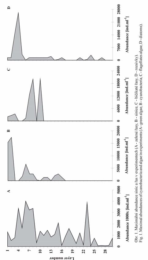
Results and discussion
Resting stages are thought to be the mechanisms whereby plankton
endures in deep, cold, dark, and often anoxic waters of lakes and oceans
(BURKHOLDER 1992).
Upper sediment layer can be characterized by significant irradiation and
aerobic-anaerobic gradient with the maximum of viable cyanobacteria and
algae in first few millimetres PADAN & COHEN (1982). Sediments at
fishpond Bílá Lhota were sampled under ice cover; light intensity and
oxygen concentrations were low (136 Hašler et al.: The survival and vitality
inoculum of later cyanobacterial growth. Differences in akinete
germination had been recorded previously by GIBSON & SMITH (1982).
Some of them germinate immediately (e.g. Anabaena, Aphanizomenon,
Gleotrichia) while other can germinate in the following years. Culture
conditions, for instance phosphate concentration, can influence akinete
germination (A. cylindrica; GIBSON & SMITH (1982). Although
cyanobacteria are successfully cultured in used Zehnder medium in culture
collections (LUKAVSKÝ et al. 1992), the phosphorus concentration is
probably lower than in natural conditions of the investigated fishpond (TP
in Zehnder medium = 16.54 µg.l-1; fishpond Bílá Lhota = annual average
of TP concentrations 2.06 mg.l-1)
However phytoplankton was dominated by P. agardhii throughout
last five years and its wintering in the form of hormogonia was observed
near the bottom, germination of P.agardhii was observed rarely (Table I/2)
at the end of the experiment (60 days of cultivation). GIBSON & FITZSIMONS
1982 found the wintering of other oscillatorean cyanobacteria in the water
column to be their main survival strategy. The germination of Oscillatoria
sp. (layer 3) and P. agardhii (layer 1,9) from sediment samples was
observed; therefore, we suppose that some resting bottom stages of these
cyanobacteria probably exist.
Diatoms
For some algae, physiological resting stages (as opposed to resting
spores or cysts, which are easily discernible with light microscopy) were
observed. SICKO-GOAD et al. (1989) identified resting cells in 18 diatom
species collected from aphotic sediments in the Laurentian Great Lakes.
These cells had all the cytoplasm condensed into one dense mass, which
usually laid in the centre of the frustule. They found that these resting cells
could rejuvenate within 2 hours of re-illumination.
Although the direct examination of sediment samples before the
cultivation established only the presence of empty diatom frustules,
diatoms were observed among first growing algae, already the 7th day of
experiment. Small Cyclotella sp. bunches among green algae occurred first.
These small diatoms (3 - 5 µm in diameter) were replaced later (after the
14th day) by pennate diatoms, e.g. Navicula, Pinnularia and Gomphonema.
After 45 days of cultivation, the majority of viable diatoms disappeared.
All frustules were thin, with indistinct stria pattern due to poor
silicification. The influence of light intensity, photoperiod and temperature
on diatom germination has been studied previously by MCQUOID et al.
(2002). They observed the germination even after the 1st day of cultivation
under optimum conditions (MCQUOID & HOBSON 1995, 1996).Czech Phycology, Olomouc, 4: 133-144 , 2004 137
Green algae and euglenophytes
Under unfavourable conditions, particularly desiccation, many
Chlorophyceae produce thick-walled resting cells (COLEMAN 1983 sec. cit.
SOUTH & WHITTICK 1995). LEMBI (1980) sec. cit. SOUTH & WHITTICK
(1995) recorded similar resting cells in unicellular volvocalean green algae
Akinetes are modified vegetative cells with a thickened wall. Hypnospores
and hypnozygotes also have thickened walls, but these are produced de
novo by protoplasts which had separated earlier from the walls of parental
cells. In laboratory, such cells may remain viable for periods exceeding 20
years (COLEMAN 1983 sec. cit. SOUTH & WHITTICK 1995).
The most frequent green algae were the representatives of genus
Scenedesmus. Tables 2/6, 7 present the development of their coenobia from
small resting cells. Similar stages had been previously recorded in
Scenedesmus abundans by HINDÁK (personal communication). Flagellates
were observed in the upper part of sediment, initially as pallmeloid stages
(Table III/6), later were identified as representatives of genus
Chlamydomonas (Table III/3,5). SUZUKI & JOHNSON (2002) studied the
photoperiodic control of germination in the unicellular Chlamydomonas.
They demonstrated that the occurrence of zygospore germination of this
alga is a genuine photoperiodic response. Germination efficiency is
enhanced in long days (15/9, 12/12, 11/13) compared to short days (9/15,
8/16).
Acknowledgement
This work was supported by the project FRVŠ 63/2003 of the
Ministry of Education of the Czech Republic GAČR No. 522/03/0323. The
authors express thanks to Mr. P. Hekele, the manager of the forest garden
Bílá Lhota, for his co-operation.
References
ANAGNOSTIDIS, K. & KOMÁREK, J. (1988): Modern approach to the classification
system of cyanophytes 3. Oscillatoriales. – Algological Studies 50–53: 327–472.
BAKER, P. D. (1999): Role of akinetes in the development of cyanobacterial populations
in the Lower Murray River, Australia. – Mar. Freshwater Res. 50: 265–279.
BELMONTE, G., MIGLIETTA, A., RUBINO, F. & BOERO, F. (1997): Morphological
convergence of resting stages of planktonic organisms: a review. –
Hydrobiologia 355: 159–165.
BURKHOLDER, J. M. (1992): Phytoplankton and episodic suspended sediment loading:
Phosphate partitioning and mechanisms for survival. – Limnol. Oceanogr. 37:
974–988.
ELLEGAARD, M., CHRITSENSEN, N. F. & MOESTRUP, Ø. (1994): Dinoflagellate cysts
from Recent Danish marine sediments. – Eur.J.Phycol. 29: 183–194.138 Hašler et al.: The survival and vitality
GIBSON, C. E. & FITZSIMONS, A. G. (1982): Periodicity and morphology of planktonic
blue–green algae in an unstratified lake. – Internat. Rev. ges. Hydrobiol. 67:
459–476.
GIBSON, C. E. & SMITH, R. V. (1982): Freshwater Plankton. – In: CARR, N.G. &
WHITTON, B. A. (eds): The Biology of Cyanobacteria, p. 463–490, Blackwell
Scientific Publications, Oxford.
HANSSON, L–A. (1996): Algal recruitment from lake sediments in relation to grazing,
sinking and dominance patterns in the phytoplankton community. – Limnology
and Oceanography 41(6): 1312–1323.
HARISTON, N. G., VAN BRUNT, R. A., KEARNS, C. M. & ENGSTROM, D. R. (1995): Age
and survioship of diapausing eggs in a sediment egg bank. – Ecology 76: 1706–
1711.
HAŠLER, P. & POULÍČKOVÁ, A. (2003): Diurnal changes in vertical distribution and
morphology of natural population of Planktothrix agardhii (GOM.) ANAGN.
& KOM. (Cyanophyta). – Hydrobiologia 506–509: 195–201.
KARLSSON, I. (2003): Benthic growth of Gloeotrichia echinulata Cyanobacteria. –
Hydrobiologia, in press.
KOMÁREK, J. & ANAGNOSTIDIS, K. (1989): Modern approach to the classification
system of cyanophytes 4. Nostocales. – Algological Studies 56: 247–345.
LUKAVSKÝ, J., CEPÁK, V., KOMÁREK, J., KAŠPÁRKOVÁ, M. & TAKÁČOVÁ, M. (1992):
Catalogue of algal and cyanobacterial strains of Culture Collection of
Autotrophic Organisms at Trebon. – Algological Studies 63: 59–112.
MCQUOID, M. R., GODHE, A. & NORDBERG, K. (2002): Viability of phytoplankton
resting stages in the sediments of a costal Swedish fjord. – Eur.J.Phycol. 37:
191–201.
MCQUOID, M. R. & HOBSON, L. A. (1995): Importance of resting stages in diatom
seasonal succession. – J.Phycol. 31: 44–50.
MCQUOID, M. R. & HOBSON, L. A. (1996): Diatom resting stages. – J.Phycol. 32:
889–902.
PADAN, E. & COHEN, Y. (1982): Anoxygenic Photosynthesis. – In: CARR, N.G. &
WHITTON, B.A. (eds): The Biology of Cyanobacteria, p. 215–236, Blackwell
Scientific Publications, Oxford.
PAERL, H. W. (1988): Nuisance phytoplankton blooms in coastal, estuarine and inland
waters. – Limnology and Oceanography 33: 823–847.
POKORNÝ, J. & HAUSER, V. (1996): Restoration of lakes by sediment removal –
fishpond Vajgar, Czech Republic. – In: EISELTOVÁ, M. (ed.): Restoration of lake
ecosystems – a holistic approach, 182 pp., Nature Conservation Bureau,
Berkshire.
POULÍČKOVÁ, A., HAŠLER, P. & KITNER, M. (in press): Annual cycle of Planktothrix
agardhii (GOM.) ANAGN. & KOM. nature population. – Int. Rev. Hydrobiol.
SICKO–GOAD, L., STOERMER, E. F. & KOCIOLEK, J. P. (1989): Diatom resting cell
rejuvenation and formation: Time course, species records and distribution. – J.
Plankton Res. 11: 375–389.
SOUTH, G. R. & WHITTICK, A. (1995): Introduction to phycology. – 293 pp., Blackwell
Scientific Publications, Oxford.
STARMACH, K. (1966): Cyanophyta–sinice, Glaucophyta – glaukofity. – In: STARMACH,
K. (ed.): Flora slodkow.Polski 2, 753 pp., PAN, Pañstw.Wyd. Nauk, Warszawa.
STAUB, R. (1961): Ernährungsphysiologisch-autökologische Untersuchungen an der
planktonischen Blaualge Oscillatoria rubescens DC. – Schweiz. Z . Hydrol. 23:
82–198.Czech Phycology, Olomouc, 4: 133-144 , 2004 139
SUZUKI, L. & JOHNSON, C. H. (2002): Photoperiodic control of germination in the
unicell Chlamydomonas. – Naturwissenschaften 89: 214–220.
VAN GEEL, B. (2001): Non–pollen palynomorphs. – In: SMOL, J.P., BIRKS, J.B. & W.M.
LAST (eds): Tracking Environmental Change Using Lake Sediments. Volume3:
Terrrestrial, Algal and Siliceous Indicators, p. 99–119, Kluwer Academic
Publishers, Dordrecht.
VAN GEEL, B., MUR, L. R., RALSKA–JASIEWICZOWA, M & GOSLAR, T. (1994): Fossil
akinetes of Aphanizomenon and Anabaena as indicators for medieval
phosphate–eutrophication of Lake Gosciaz (Central Poland). – Review of
Palaeobotany and Palynology 83: 97–105.
Table 1: List of cyanobacteria and algae found in sediment of fishpond Bílá Lhota
Species Found in layer number:
Cyanobacteria
Anabaena flos-aquae BRÉBISSON ex BORNET et FLAHAULT 12
Anabaena inaequalis BORNET et FLAHAULT 2, 7, 11
Anabaena cf. sphaerica BORNET et FLAHAULT 8, 12, 14
Anabaena sp. 5
Anabaena sp. 9
Nostoc linckia (ROTH) BORNET ET FLAHAULT 2, 6, 9
Nostoc sp. 7
Nostoc sp. 11
Nostoc sp. 15
Oscillatoria sp. 3
Planktothrix agardhii (GOMONT) ANAGNOSTIDIS et KOMÁREK 1, 9
Bacillariophyceae
Cocconeis placentula EHRENBERG 7, 12
Cyclotella atomus HUSTEDT 1-3, 5, 7-10, 13, 15
Cyclotella cf. meneghiniana KÜTZING 27
Cyclotella sp. 4
Fragilaria sp. 7
Gomphonema parvulum (KÜTZING) KÜTZING 7
Navicula rhynchocephala KÜTZING 7, 9, 10
Navicula sp. 11
Navicula sp. 12
Navicula sp. 15
Nitzschia communis RABENHORST 7
Nitzschia cf. obtusa 11
Pinnularia cf. viridis (NITZSCH) EHRENBERG 13
Pinnularia sp. 10
Pinnularia sp. 21
Synedra ulna (NITZSCH) EHRENBERG 3140 Hašler et al.: The survival and vitality
Euglenophyta
Euglena acus EHRENBERG 9, 11
Chlorophyta
Actinastrum hantzschii LANGERHEIM 4
Chlamydomonas debaryana GOROSH. 3, 7, 8, 10
Chlamydomonas sp. 1
Chlamydomonas sp. 2
Chlamydomonas sp. 4
Chlamydomonas sp. 6
Chlamydomonas sp. 8
Chlamydomonas sp. 10
Chlorella homosphaera SKUJA 1, 6, 10, 14, 18
Chlorella luteoviridis CHODAT 3
Chlorella vulgaris BEIJERINCK 2, 4, 5, 8-11, 20, 25, 30
Chlorogonium euchlorum EHRENBERG
Monoraphidium contortum (THURET in BRÉBISSON) KOMÁRKOVÁ- 2, 23
LEGNEROVÁ
Monoraphidium convolutum (CORDA) KOMÁRKOVÁ-LEGNEROVÁ 7, 25
Monoraphidium griffithii (BERKELEY) KOMÁRKOVÁ-LEGNEROVÁ 1-5
Scenedesmus abundans (LANGERHEIM) CHODAT 1-5, 7-9, 11-14, 16, 17
Scenedesmus acuminatus (LANGERHEIM) CHODAT 1, 3, 30
Scenedesmus alternans REINSCH 6, 20
Scenedesmus ecornis (RALFS) CHODAT 1-3, 6
Scenedesmus intermedius CHODAT 4, 8, 12, 15, 17, 23
Scenedesmus sp. 16Czech Phycology, Olomouc, 4: 133-144 , 2004 141
142                                           Hašler et al.: The survival and vitality

Fig. 2: 1 - Anabaena inaequalis, 2 - Planktothrix agardhii, 3 - Nostoc linckia, 4 - N.
linckia (hormogonium), 5 - N. linckia (akinetes formation), 6 - N. linckia (young
growing filaments)
Czech Phycology, Olomouc, 4: 133-144 , 2004                                           143

Fig. 3: 1 - Scenedesmus acutus, 2 - S. linearis, 3 - S. denticulatus, 4 - S. quadricauda, 5
- Actinastrum hantzschii, 6 - 7 - developmental stages of Scenedesmus
144                                           Hašler et al.: The survival and vitality

Fig. 4: 1 - Chlorella homosphaera, 2 - Ch. vulgaris, 3 - Chlamydomonas debaryana, 4 -
Chlorogonium euchlorum, 5 - Chlamydomonas sp., 6 - Chlamydomonas (palmelloid
stage), 7 - Monoraphidium griffithi, 8 - M. contortum
You can also read