TINEA INVERSICOLOR: A RARE DISTRIBUTION OF A COMMON ERUPTION - CUREUS
←
→
Page content transcription
If your browser does not render page correctly, please read the page content below
Open Access Case
                                         Report                                                DOI: 10.7759/cureus.6689

                                         Tinea InVersicolor: A Rare Distribution of a
                                         Common Eruption
                                         Matthew Ferry 1 , Lydia Shedlofsky 2 , Andrew Newman 2 , Yebabe Mengesha 2 , Brooke Blumetti
                                         2

                                         1. Dermatology, Ohio University Heritage College of Osteopathic Medicine, Athens, USA 2. Dermatology,
                                         Affiliated Dermatology, Scottsdale, USA

                                         Corresponding author: Matthew Ferry, mf579015@ohio.edu

                                         Abstract
                                         Tinea versicolor (TV), or pityriasis versicolor, is one of the most commonly occurring
                                         superficial mycoses. Typically, this condition is characterized by fine scaly hyper or
                                         hypopigmented macules and patches distributed on the trunk and upper extremities. Diagnosis
                                         is often based on clinical presentation. A Wood’s lamp examination or potassium hydroxide
                                         (KOH) preparation test is performed for confirmation. To date, numerous morphologic variants
                                         of this condition have been described. Here we present an inverse papular variant that, to our
                                         knowledge, has only been previously reported once. This case represents another unique
                                         presentation of TV and serves to highlight the clinical variety of this common mycosis.

                                         Categories: Dermatology, Infectious Disease
                                         Keywords: dermatology, tinea versicolor, pityriasis versicolor, malasseiza, mycosis, inverse

                                         Introduction
                                         Malassezia yeasts have been known to cause dermatologic disease for over 150 years [1]. While
                                         it is part of the normal human skin flora, this genus has been pathologically implicated in
                                         several skin conditions. One such condition is tinea versicolor (TV) [2]. TV, or pityriasis
                                         versicolor, is one of the most commonly occurring superficial mycoses. While it has a
                                         worldwide distribution, the highest incidence can be found in tropical climates where rates can
                                         be as high as 50% [3]. Generally, TV presents with characteristic fine scaly hyper or
                                         hypopigmented macules and patches located on the trunk, neck, and upper extremities. The
                                         diagnosis often entails a clinical assessment and confirmation via skin scraping with potassium
                                         hydroxide (KOH) preparation [4]. While most cases of TV follow a classic pattern, numerous
                                         varieties of TV have been described. In recent years, atrophic, papular, imbricate, and
                                         folliculocentric forms of TV have been reported [2,5-8]. This report depicts a rare presentation
                                         of this common disease, which the authors have termed “Tinea InVersicolor”.
Received 12/12/2019
Review began 12/23/2019
Review ended 01/13/2020
Published 01/17/2020
                                         Case Presentation
                                         A 47-year-old Caucasian female with psoriasis presented to an Arizona outpatient dermatology
© Copyright 2020
Ferry et al. This is an open access      clinic with concerns of a rash in the bilateral underarms with extension to the underside of
article distributed under the terms of   both breasts. The patient described her condition as red, bumpy, and associated with an
the Creative Commons Attribution         occasional burning sensation and pruritus. Her symptoms had persisted for three months. She
License CC-BY 3.0., which permits
                                         was initially treated with a short burst of clobetasol ointment and ciclopirox 0.77% gel. The
unrestricted use, distribution, and
                                         patient reported worsening of the rash with ciclopirox and partial improvement with
reproduction in any medium, provided
the original author and source are       clobetasol. While the patient denied the use of new skin-care products, medications, or recent
credited.                                travel prior to her rash onset, she did admit to the use of “crystal deodorant”, coconut oil, and
                                         hemp oil. Physical exam revealed a well-demarcated, sporadically configured, and bright red

                                         How to cite this article
                                         Ferry M, Shedlofsky L, Newman A, et al. (January 17, 2020) Tinea InVersicolor: A Rare Distribution of a
                                         Common Eruption. Cureus 12(1): e6689. DOI 10.7759/cureus.6689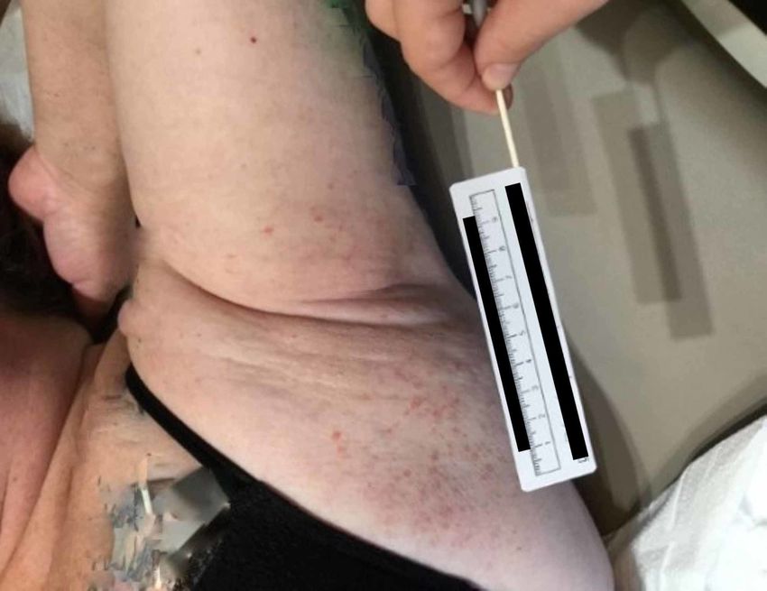
papular rash with minimal scale located in the bilateral axilla, which extended to the bilateral
inframammary skin (Figures 1 and 2). The rest of the physical exam including examination of
the hair and nails was unremarkable.
Based on history and visual morphology, a Wood’s lamp examination and shave biopsy were
performed. Upon Wood’s Lamp examination, the involved areas appeared to fluoresce a red-
orange color. Shave biopsy revealed stubby hyphae and round spores within the cornified layer,
consistent with the diagnosis of TV.
The patient was placed on fluconazole 300 mg orally every week for four weeks. At a follow-up
visit three weeks after starting fluconazole, the patient reported a drastic improvement in her
condition. Notably, the patient reported that pruritus had decreased and the rash had begun to
clear within 7-10 days of the first administration of the oral anti-fungal medication.
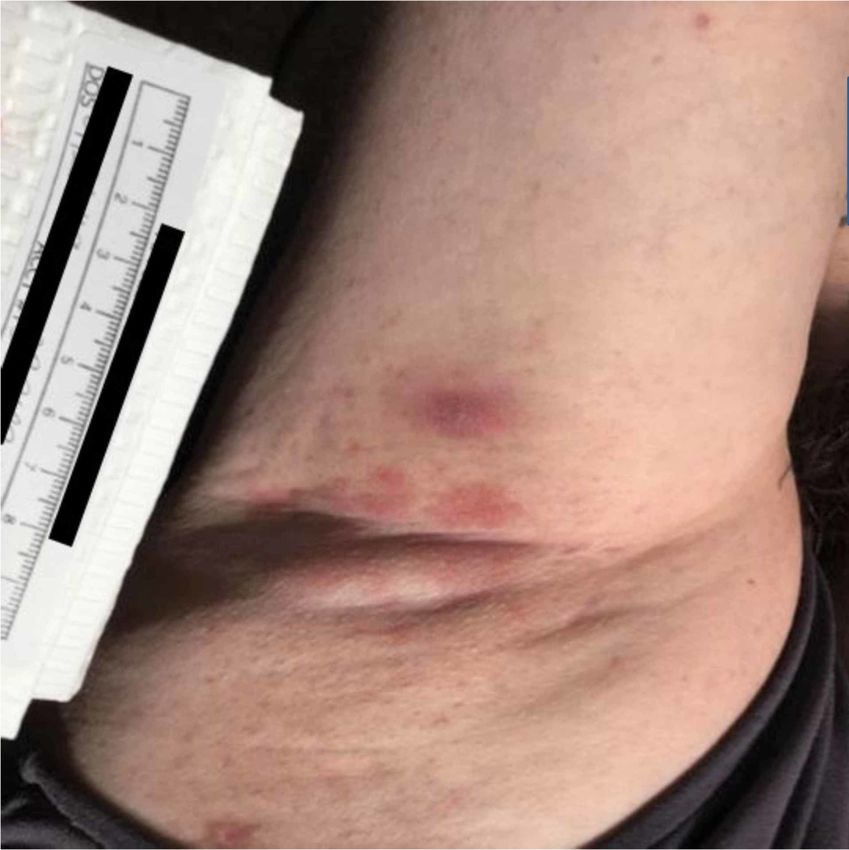
FIGURE 1: Patient’s right axilla demonstrates a well-
demarcated erythematous and papular rash with minimal scale
2020 Ferry et al. Cureus 12(1): e6689. DOI 10.7759/cureus.6689 2 of 6FIGURE 2: Patient’s left axilla demonstrates the papular
eruption extending from her axillary vault to her inframammary
tissue (patient’s tattoos have been digitally altered to maintain
anonymity)
Discussion
The building of the differential diagnosis for this case started with a consideration of the
papulosquamous reaction pattern described by Grooper in 2001 [9]. Given the erythematous,
mildly scaly papules defining our patient’s rash, conditions such as infectious intertrigo (i.e.
erythrasma, candidiasis, tinea), psoriasis, contact dermatitis, inverse pityriasis rosea, Darier
disease, and Grover disease were considered. Due to patient body habitus and intertriginous
distribution, an infective intertrigo was initially favored [10]. A lack of response to ciclopirox
gel and mild improvement with clobetasol ointment supported a bacterial etiology, such as
erythrasma, over candidiasis or dermatophytosis. A review of the patient’s skin-care regimen
revealed the use of numerous products including several natural remedies.
In their 2018 review of contact allergens, Uter et al. discuss the rise in contact dermatoses
associated with both cosmetic and natural skin-care products [11]. Such evidence, in addition
to the observable rash morphology, supports both allergic and irritant contact dermatoses as
plausible differentials. While our patient did reveal a history of psoriasis, her psoriasis was not
known to present with this morphology or inverse pattern, making this diagnosis less likely.
Though infectious intertrigo and contact dermatitis were highest on our differential, neither
Darier nor Grover disease could be ruled out by history and physical exam alone. Darier disease,
a keratinocyte adhesion disorder, commonly presents on the chest within intertriginous areas,
and Grover’s disease presents in a similar pattern with associated pruritus [12,13]. To further
distinguish our top differentials, clinical and histological tests were performed.
2020 Ferry et al. Cureus 12(1): e6689. DOI 10.7759/cureus.6689 3 of 6The patient had initially been referred from an allergist for consultation after her skin-prick
testing had revealed environmental allergens severe enough to qualify for immunotherapy.
Subsequently, the patient had undergone patch testing with 80 allergens, which had been
negative. Her history revealed that the timeline lent credence to a more chronic condition, and
so inverse pityriasis rosea was dropped to a lower position on the list of differentials. During
the initial office visit, a Wood’s lamp examination and shave biopsy were performed to
distinguish among the differentials. The Wood’s lamp, first described in 1903, is a source of
ultraviolet light in which all other wavelengths of the visible spectrum are filtered out [14].
False-positive and negative results with the Wood’s lamp, attributed to external factors
including skin-care products, have been described in the literature [15]. Our patient's
investigation included the Wood’s lamp, which was used to identify a possible superficial skin
infection such as erythrasma or TV. These conditions normally fluoresce coral red and
yellow/orange respectively under the lamp. With the exam room lights off during the test, the
involved areas of our patient’s rash appeared to illuminate red-orange. Additionally, a shave
biopsy was performed, which revealed stubby hyphae and round spores within the cornified
layer, consistent with the diagnosis of TV.
Malassezia, a genus of fungi known previously as Pitryosporum, comprises 14 species that are
predominately lipophilic, and several of these species are known to be pathogenic [16].
Malassezia globosa is the most common etiology of TV [17]. While these eukaryotic organisms
generally serve a commensal role on human skin, rapid proliferation and transformation to
their hyphal form are pathognomonic for the lesions of TV. While the exact reasons for
pathogenic transformation remain unknown, several environmental and biologically inherent
influences have been reported to serve as risk factors for the disease. From an environmental
perspective, the prevalence of TV in tropical regions approaches 50% [3]. Sunlight and the
associated increase in pilosebaceous secretions have been shown to support Malassezia
proliferation and likely account for the robust prevalence in tropical areas [17]. According to
Metin et al., other important risk factors for the disease include the use of oil-containing skin-
care products and the application of corticosteroids [2]. Generally, these substances provide a
substrate for yeast metabolism and create a nurturing microenvironment for yeast overgrowth
[2,17]. In hindsight, our patient exhibited several risk factors for disease. The warm Arizona
climate, use of multiple skin-care products, and initial treatment with topical corticosteroids
might have all contributed to the development and persistent nature of TV in our patient. In
considering the elicited history, the reported improvement with topical steroids directed
clinical thinking away from a mycotic etiology. The lack of improvement with topical ciclopirox
is likely secondary to a lack of compliance with treatment. The reported improvement with
topical steroids is a clinical outlier, rooted in the reduction of Malassezia-induced inflammatory
cytokines [17]. This case should encourage physicians to consider TV as a differential in
situations where topical steroids have elicited a partial treatment response.
Most commonly, TV lesions present as hyperpigmented or hypopigmented macules and
patches distributed on the trunk and upper extremities [2,4,5,17,18]. Generally, these lesions
exhibit a superficial scale and lack both clinical and histologic signs of inflammation. Atypical
variants of TV have been previously reported [2,5-8]. To date, variations of both clinical
distribution and morphology have been described. In their discussion of anatomic distributions
of TV, Varada et al. state that the trunk and upper extremities may be involved in up to 95% of
the cases. The most common non-classic distributions include the scalp and groin. Interestingly
enough, Varada et al. [6] also report that the flexural creases, namely the axilla and inguinal
folds, are commonly spared in cases of widespread infection. Our patient's anatomic
involvement was limited to the intertriginous areas of the trunk, including bilateral axilla and
inferior breast tissue. To our knowledge, only two cases of TV presenting with a predominantly
inverse anatomic distribution have been reported [7]. Rudolph et al. describe two cases of TV
with an inverse presentation. The first case involves a 32-year-old male with TV distributed
solely in the bilateral axilla and inguinal folds, while the second case depicts a 23-year-old
2020 Ferry et al. Cureus 12(1): e6689. DOI 10.7759/cureus.6689 4 of 6pregnant female with TV distributed solely in her perineal region and superior thighs [7]. In line
with the findings of Rudolph et al., the authors herein support the idea of expanding the
differential of inversely distributed eruptions to include TV.
In addition to the unusual anatomic distribution, our patient exhibited an unusual TV
morphology: well-defined and sporadically configured papules with minimal scale. In recent
years, several morphologic variants of TV have been described. These include atrophic, papular,
imbricate, and folliculocentric forms [2,5-8]. In their discussion of a papular variant, Pouldar et
al. describe brown flat-topped papules coalescing into reticulated plaques in a 26-year-old
female [8]. The lesions seen in our patient remained as discrete papules, were erythematous in
appearance, and may represent a newly reported form of the condition. Given the inimitable
morphology and extensive list of differential diagnoses, shave biopsy, rather than potassium
hydroxide (KOH) preparation, was performed. It is likely that a KOH preparation would have
likely expedited the diagnosis. In fact, all the cases of abnormal TV reviewed for this
publication had a positive KOH preparation [5-8]. Furthermore, in their discussion of bedside
dermatologic tests, Wanat et al. report the sensitivity and specificity of KOH preparation to be
88% and 95% respectively for the diagnosis of TV [19]. Moving forward, the authors of this
article advise clinicians to keep the anatomic and morphologic variants of TV in mind and to
consider KOH preparation to speed up the diagnosis of this easily treatable condition.
Conclusions
In summary, this case showcases an unusual inverse presentation of the commonly occurring
superficial mycosis, TV. To our knowledge, TV displaying a similar type of papular eruption
with a predominantly intertriginous distribution has been reported only once before. The
authors favor labeling this TV variant as “Tinea InVersicolor” because it is simple yet
descriptive. Our discussion of “Tinea InVersicolor” will hopefully serve as a useful reminder of
the variability often seen with common skin diseases. Finally, we hope our findings will
encourage clinicians to lower their threshold for including TV in the differential diagnosis of an
inverse rash suspected to be erythrasma or another infectious intertrigo, contact dermatitis of
the underarms, or a truncal papular eruption like Darier disease or Grover disease.
Additional Information
Disclosures
Human subjects: Consent was obtained by all participants in this study. Conflicts of interest:
In compliance with the ICMJE uniform disclosure form, all authors declare the following:
Payment/services info: All authors have declared that no financial support was received from
any organization for the submitted work. Financial relationships: All authors have declared
that they have no financial relationships at present or within the previous three years with any
organizations that might have an interest in the submitted work. Other relationships: All
authors have declared that there are no other relationships or activities that could appear to
have influenced the submitted work.
References
1. Gaitanis G, Velegraki A, Mayser P, Bassukas ID: Skin diseases associated with Malassezia
yeasts: facts and controversies. Clin Dermatol. 2013, 31:455-463.
10.1016/j.clindermatol.2013.01.012
2. Metin A, Dilek N, Demiseven DD: Fungal infections of the folds (intertriginous areas) . Clin
Dermatol. 2015, 33:437-447. 10.1016/j.clindermatol.2015.04.005
3. Goldstein BG, Goldstein AO: Tinea versicolor (pityriasis versicolor) . Dellavalle RP, Levy ML,
Rosen T (ed): Wolters Kluwer, 2018.
4. Bonifaz A, Gómez-Daza F, Paredes V, Ponce RM: Tinea versicolor, tinea nigra, white piedra,
2020 Ferry et al. Cureus 12(1): e6689. DOI 10.7759/cureus.6689 5 of 6and black piedra. Clin Dermatol. 2010, 28:140-145. 10.1016/j.clindermatol.2009.12.004
5. Hudson A, Carroll B, Kim SJ: Folliculocentric tinea versicolor. Dermatol Online J. 2017, 23:1-
3. Accessed: January 17, 2020: https://escholarship.org/uc/item/5kj574bd.
6. Varada S, Dabade T, Loo DS: Uncommon presentations of tinea versicolor . Dermatol Pract
Concept. 2014, 4:93-96. Accessed: January 17, 2020:
https://www.ncbi.nlm.nih.gov/pubmed/25126470. 10.5826/dpc.0403a21
7. Rudolph R, Holzwanger J: Letter: inverse tinea versicolor. Arch Dermatol. 1975, 111:1213.
10.1001/archderm.1975.01630210129020
8. Pouldar D, Shive M, Gee SN: Papular eruption in a woman with Down syndrome . J Am Acad
Dermatol. 2017, 77:e95-e96. 10.1016/j.jaad.2017.05.049
9. Grooper CA: An approach to clinical dermatologic diagnosis based on morphologic reaction
patterns. Clin Cornerstone. 2001, 4:1-14. 10.1016/s1098-3597(01)90002-x
10. Uzuncakmak TK, Akdeniz N, Karadag AS: Cutaneous manifestations of obesity and the
metabolic syndrome. Clin Dermatol. 2018, 36:81-88. 10.1016/j.clindermatol.2017.09.014
11. Uter W, Werfel T, White IR, Johansen JD: Contact allergy: a review of current problems from a
clinical perspective. Int J Environ Res Public Health. 2018, 15:E1108. 10.3390/ijerph15061108
12. Engin B, Kutlubay Z, Erkan E, Tuzun Y: Darier disease: a fold (intertriginous) dermatosis . Clin
Dermatol. 2015, 33:448-451. 10.1016/j.clindermatol.2015.04.009
13. Gonzales-Ramirez RA, Ocampo-Candiani J, Welsh-Lozano O: Grover disease: a diagnostic and
therapeutic challenge. J Am Acad Dermatol. 2009, 60:106. 10.1016/j.jaad.2008.11.470
14. Ruocco E, Baroni A, Donnarumma G, Ruocco V: Diagnostic procedures in dermatology . Clin
Dermatol. 2011, 29:548-556. 10.1016/j.clindermatol.2010.09.023
15. Gebhard R: Wood’s light examination. Pfenninger and Fowler’s procedures for primary care.
Saunders (imprint), Philadelphia, Pennsylvania; 2020. 32:213-215.
16. Kang S: Fitzpatrick’s dermatology in general medicine . McGraw-Hill Education, New York;
2019.
17. Mendez-Tovar LJ: Pathogenesis of dermatophytosis and tinea versicolor . Clin Dermatol. 2010,
28:185-189. 10.1016/j.clindermatol.2009.12.015
18. Saleem M, Oussedik E, Picardo M, Schoch JJ: Acquired disorders with hypopigmentation: a
clinical approach to diagnosis and treatment. J Am Acad Dermatol. 2019, 80:1233-1250.
10.1016/j.jaad.2018.07.070
19. Wanat KA, Dominquez AR, Carter Z, Legua P, Bustamante B, Micheletti RG: Bedside
diagnostics in dermatology. J Am Acad Dermatol. 2017, 77:197-218.
10.1016/j.jaad.2016.06.034
2020 Ferry et al. Cureus 12(1): e6689. DOI 10.7759/cureus.6689 6 of 6You can also read