TWELVE LIVE BIRTHS AFTER UTERUS TRANSPLANTATION IN THE DALLAS UTERUS TRANSPLANT STUDY
←
→
Page content transcription
If your browser does not render page correctly, please read the page content below
Original Research
Twelve Live Births After Uterus
Transplantation in the Dallas UtErus
Transplant Study
Downloaded from http://journals.lww.com/greenjournal by +eHMTJgP5IuXmvrmuJmsBgkFnGpvNHeLQxtgEAtGY2XNUltJuPp9+zxsmSZUVeaivgzl7Zi+8H8gjGzg0koT771SOYOX4ovm9YKlKXRYPTCHR87l+cnpcqJGWP2vPEbBw2Dps19QMFh130VFBI7Taw== on 01/29/2021
Liza Johannesson, MD, PhD, Giuliano Testa, MD, MBA, J. Michael Putman, MD, Gregory J. McKenna, MD,
E. Colin Koon, MD, Jackie R. York, MD, Johanna Bayer, MD, Lilly Zhang, PhD, Zachary S. Rubeo, MD,
Robert T. Gunby, MD, and Anthony R. Gregg, MD, MBA
OBJECTIVE: To describe aggregated pregnancy out- 79%. Ten uteri were from nondirected living donors
comes after uterus transplantation from a single, expe- and one uterus was from a deceased donor. In vitro
rienced center. fertilization was performed to achieve pregnancy. Ten
METHODS: This prospective study reports on live births recipients delivered one neonate, and one recipient
among 20 women who received a uterus transplant from delivered two neonates. One organ rejection episode
2016 to 2019 at Baylor University Medical Center at was detected during pregnancy and was resolved with
Dallas. These live births occurred between November steroids. The median birth weight was 2,890 g (range
2017 and September 2020. The main measures were live 1,770–3,140 g [median 68th percentile]). Maternal weight
birth, maternal complications, and fetal and newborn gain was higher than Institute of Medicine recommenda-
outcomes. tions. Maternal medical complications were observed in
five recipients (elevated creatinine level, gestational dia-
RESULTS: There were six graft failures (four surgical
betes, gestational hypertension [n52], and preeclamp-
complications and two with poor perfusion postopera-
sia). In five recipients, maternal medical or obstetric
tively). Of the 14 technically successful transplants, at
complications led to an unplanned preterm delivery (ele-
least one live birth occurred in 11 patients. Thus far, the
vated creatinine level, preeclampsia; preterm labor
live birth rate per attempted transplant is 55%, and the
[n53]). The median gestational age at delivery was 36
live-birth rate per technically successful transplant is
6/7 weeks (range 30 6/7–38 weeks). All neonates were
liveborn, with Apgar scores of 8 or higher at 5 minutes.
From the Annette C. and Harold C. Simmons Transplant Institute, the Fertility
CONCLUSION: Over the first 3 years, our program
Center of Dallas, the Department of Obstetrics and Gynecology, and the
Department of Neonatology, Baylor University Medical Center, and the Fetal experienced a live-birth rate per attempted transplant
Care Center, Dallas, Texas; and the Department of Obstetrics and Gynecology, of 55% and a live-birth rate per technically successful
PRISMA Health—University of South Carolina School of Medicine, Columbia,
transplant of 79%. In our experience, uterus transplan-
South Carolina.
tation resulted in a third-trimester live birth in all cases in
This study was supported by the Baylor Scott & White Dallas Foundation. The
Foundation played no role in designing the study, interpreting the results, or which pregnancies reached 20 weeks of gestation.
writing the manuscript. Maternal medical and obstetric complications can occur;
The authors thank Shelby Babcock, Hoylan Fernandez, MD, MPH, Amar Gupta, however, these were manageable by applying principles
MD, Eric J. Martinez, MD, Nicholas Onaca, MD, Heather H. Pirtle, Richard M. of generally accepted obstetric practice.
Ruiz, MD, Anji Wall, MD, PhD, and Kristin R. Wallis for their clinical
contributions. CLINICAL TRIAL REGISTRATION: ClinicalTrials.gov,
Each author has confirmed compliance with the journal’s requirements for NCT02656550.
authorship. (Obstet Gynecol 2021;137:241–9)
Corresponding author: Liza Johannesson, MD, PhD, Annette C. and Harold C. DOI: 10.1097/AOG.0000000000004244
Simmons Transplant Institute, Baylor University, Medical Center, Dallas, TX;
M
email: liza.johannesson@bswhealth.org.
ayer-Rokitansky-Küster-Hauser syndrome is a
Financial Disclosure developmental abnormality that results in
The authors did not report any potential conflicts of interest.
absence of the uterus. Women with this condition are
© 2021 by the American College of Obstetricians and Gynecologists. Published
by Wolters Kluwer Health, Inc. All rights reserved. infertile but develop normal secondary sexual character-
ISSN: 0029-7844/21 istics because ovarian function is not affected. Uterus
VOL. 137, NO. 2, FEBRUARY 2021 OBSTETRICS & GYNECOLOGY 241
© 2021 by the American College of Obstetricians
and Gynecologists. Published by Wolters Kluwer Health, Inc.
Unauthorized reproduction of this article is prohibited.transplantation is the only treatment that restores repro- (n52). A uterine transplantation procedure was con-
duction and the ability to experience gestation in women sidered technically successful when there was a vital
with absolute uterine-factor infertility, such as Mayer-Ro- graft 3 months postoperatively.
kitansky-Küster-Hauser syndrome. Uterus transplanta- Before uterus transplantation, recipients under-
tion is costly and requires the recipient and donor to went in vitro fertilization. The DUETS protocol
undergo major surgery. Once our uterus transplantation required good-quality day 5–6 expanded blastocysts
protocol was approved, we received inquiries from close (Society for Assisted Reproductive Technology
to 1,500 potential recipients and 700 potential donors. [SART] grading). All recipients underwent single
Our protocol was approved for 20 recipients. The first embryo transfer of an expanded blastocyst. Early in
live birth to a woman after uterus transplantation was the program (cases 4, 5), embryo transfer was planned
reported in 2014.1 Since then, the number of uterus 6–8 months after uterus transplantation. In subse-
transplantation procedures has increased, and births quent cases, to minimize recipient graft time, which
have occurred in multiple centers.1–6 Of the 20 live we defined as time from uterus transplantation to hys-
births after uterus transplantation that were mentioned terectomy,11 we individualized the timing of embryo
in media outlets worldwide, only 10 were reported in the transfer after uterus transplantation based on postop-
medical literature.1–4,7 These reports often were incom- erative recovery, graft function (eg, established men-
plete. This limits conclusions regarding pregnancy out- ses), immunosuppression regimen, and infectious
comes and safety. Our aim was to describe the outcomes disease susceptibility.11 This allowed us to shorten
of the first 12 pregnancies that led to live births at our the time interval from uterus transplantation to
center. embryo transfer.
An induction regimen with thymoglobulin (total
METHODS dose 4.5 mg/kg), and 500–1,000 mg of methylpred-
DUETS (the Dallas UtErus Transplant Study) was a nisolone was used at the time of uterus transplanta-
prospective cohort study of the first 20 women tion. Maintenance immunosuppression was achieved
undergoing uterus transplantation at a single center. with oral tacrolimus. The goal of trough tacrolimus
The study was approved by the Baylor University levels was 9–11 ng/mL during months 1–3 post–
Medical Center ethics committee and institutional uterus transplantation and 4–8 ng/mL thereafter.
review board in 2015. It was listed on clinicaltrials.gov Drug levels were monitored, and doses adjusted at 1-
(NCT02656550) before the first transplant. to 2-week intervals after uterus transplantation and
Our initial experience ascertaining recipients throughout pregnancy. Mycophenolate mofetil was
and donors for uterus transplantation (n5272 and used for cases 4, 5, and 7, and embryo transfer was
79, respectively), the screening protocol, and demo- performed after a 3-month washout. After the ninth
graphic data were published previously.8 Forty-five recipient, mycophenolate mofetil was eliminated from
potential recipients and 36 potential donors were the regimen and replaced with azathioprine or ever-
evaluated.8–10 Potential living donors were olimus, which allowed us to consider embryo transfer
excluded if they had a history of obstetric compli- 3 months after induction therapy. All recipients
cations, including recurrent pregnancy loss, sponta- received low-dose aspirin (81 mg) daily from uterus
neous abortion in their most recent pregnancy, or transplantation throughout pregnancy and the post-
any prior dilation and curettage. Pregnancy history partum period.
information for deceased donors was incomplete. After uterus transplantation, patients were moni-
Potential recipients with preexisting hypertension tored according to our protocol. This included cervi-
requiring medications, diabetes requiring pharma- cal biopsies to monitor for rejection. For 3 months
cologic therapy, or any ongoing serious medical after uterus transplantation, biopsies were done at
complication were excluded from uterus transplan- approximately 14-day intervals. Afterwards, monthly
tation in our protocol.8 biopsies were performed until embryo transfer. Pre-
Our protocol allows for recipients to experience a natal visits occurred every 2 to 3 weeks. Routine care
maximum of two pregnancies, and then a graft included fetal growth, biophysical profiles, and umbil-
hysterectomy is performed. The hysterectomy is ical artery Doppler studies. Rejection episodes were
performed after either the first or second delivery, managed with intravenous SoluMedrol, 1 g on day 1
depending on the patient’s wishes or medical and and 500 mg on days 2 and 3. A repeat cervical biopsy
obstetric considerations. Between September 2016 was done 1 week after rejection treatment. During
and August 2019, 20 women were transplanted with pregnancy, cervical biopsies were performed twice
uteri from living donors (n518) or deceased donors (12–14 and 24–26 weeks of gestation).
242 Johannesson et al Live Births After Uterus Transplantation OBSTETRICS & GYNECOLOGY
© 2021 by the American College of Obstetricians
and Gynecologists. Published by Wolters Kluwer Health, Inc.
Unauthorized reproduction of this article is prohibited.Uterus transplantation is performed at multiple complication when it spanned two trimesters (trimes-
centers around the world, and the delivery modality is ters: 14 weeks of gestation or less, 14 1/7 weeks to 28
universally cesarean delivery owing to concern that weeks or less, and 28 1/7 weeks to delivery). This
the vaginal anastomosis could dehisce during labor. In decision was made a priori, because bleeding in two
addition, a circumferential stricture at the anastomosis consecutive trimesters has been associated with
site is common, and this creates a small diameter of adverse pregnancy outcomes.16 A severe obstetric
the upper vagina. To prevent descent of the fetal head, complication was one that led to a surgical procedure
many centers plan for a preterm delivery before the or delivery before the planned date.
onset of labor. Our protocol allowed for delivery Fetal complications included congenital anoma-
timing to be at the discretion of the obstetrician and lies or abnormal fetal growth. Fetal growth abnormal-
maternal-fetal medicine subspecialist. Our initial ities were defined as either biometric measures or
protocol was to deliver patients at 35–36 weeks of estimated fetal weight less than the 10th percentile or
gestation. However, as we gained experience, our pro- greater than the 90th percentile. Doppler flow was
tocol was modified to deliver at 37–38 weeks of ges- considered abnormal when there was absent or
tation. In all cases in which there was a maternal reverse end diastolic velocity in any umbilical artery.
medical or obstetric complication, delivery time was A fetal echocardiogram was performed between 22
individualized. weeks and 24 weeks of gestation in all pregnancies.
The primary outcomes were live birth, pregnancy Apgar scores, umbilical artery pH, and birth weight
complications, fetal growth and development, and were recorded at delivery. Birth weight was adjusted
newborn weight. Pregnancy complications were for age and neonatal sex using Fenton growth charts
divided into three categories: maternal medical, for preterm births17,18 and World Health Organiza-
obstetric, and fetal. Maternal medical complications tion charts for full-term infants.19
included preeclampsia with and without severe fea-
tures and gestational diabetes. Preeclampsia with and RESULTS
without severe features were defined according to Of the 20 uterine transplants performed (Fig. 1), 14
standard American College of Obstetricians and were technically successful.20 Graft failures were
Gynecologists’ criteria.12 All patients were screened attributed to surgical complications (four cases) and
for gestational diabetes between 26 and 28 weeks of poor graft perfusion (two cases). The majority of the
gestation with a 50-g oral glucose load (140 mg/dL or graft losses were seen among the first 10 uterus trans-
higher); a 3-hour diagnostic test (100 g) was per- plants (cases 1–3, 8, 10). Of the 14 technically success-
formed if needed (95 mg/dL or less, 180 mg/dL or ful transplants, at least one live birth occurred in 11
less, 155 mg/dL or less, and 140 mg/dL or less at patients. At the time of this report, the success rate (ie,
fasting, 1 hour, 2 hours, and 3 hours, respectively). live birth) per attempted transplant is 55%, and the
A diagnosis of gestational diabetes was made with live birth rate per technically successful transplant is
two or more abnormal values on the 3-hour test.13 79%.
Maternal weight was recorded at each obstetric visit. With the 10 most recent transplants, the technical
At the end of pregnancy, maternal weight gain was success rate was 90%, and the live-birth rate per
compared with the Institute of Medicine’s (now technically successful transplantation is currently 67%
known as the National Academy of Medicine) recom- among these women. There was also one patient that
mendation for weight gain during pregnancy based on at preparation of this article was carrying a pregnancy
starting body mass index (BMI, calculated as weight (delivered November 2020). There also is one patient
in kilograms divided by height in meters squared).14 carrying a second pregnancy in this cohort (case 11),
Obstetric complications included preterm birth and two patients are proceeding with additional
(less than 37 weeks of gestation), conditions of embryo transfers (cases 12 and 17).
abnormal placentation after 28 weeks, cervical short- Among the 11 recipients with live births, the
ening (24 mm or less before 28 weeks), and abnormal indication for uterus transplantation was absolute
amniotic fluid volume as determined by either an uterine-factor infertility (Mayer-Rokitansky-Küster-
amniotic fluid index15 or a single deepest pocket of Hauser syndrome) in 10 and hysterectomy in early
less than 2 cm or more than 8 cm on a biophysical adulthood owing to a leiomyomatous uterus in one
profile. The amniotic fluid index was defined as (case 15). At the time of uterus transplantation, partic-
abnormal when the value was less than the 5th percen- ipants had a median age of 30 years (range 20–36
tile or more than the 95th percentile for gestational years) and a median BMI of 25 (range 21–31)
age.15 Vaginal bleeding was considered an obstetric (Table 1). One recipient (case 9) with Mayer-
VOL. 137, NO. 2, FEBRUARY 2021 Johannesson et al Live Births After Uterus Transplantation 243
© 2021 by the American College of Obstetricians
and Gynecologists. Published by Wolters Kluwer Health, Inc.
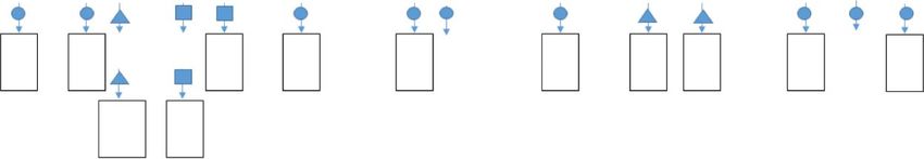
Unauthorized reproduction of this article is prohibited.Fig. 1. Schematic overview of the 14 technically successful uterus transplantations in DUETS (the Dallas UtErus Transplant
Study). Numbers are recipient case numbers. Live births, embryo transfers, time from uterus transplant to live birth, and graft
hysterectomy are shown. Patient numbers not shown are those with a nonvital graft at 3 months after transplant. Patients 12
and 17 have thus far had multiple embryo transfers and no live births; neither patient has proceeded beyond 20 weeks of
gestation. Patient 6 was a deceased donor. Numbers beneath live birth (LB) indicate completed months from uterine
transplant or *previous live birth. MC, miscarriage; P, ongoing pregnancy; C-H cesarean hysterectomy; H, delayed hys-
terectomy.
Johannesson. Live Births After Uterus Transplantation. Obstet Gynecol 2021.
Rokitansky-Küster-Hauser syndrome had a single kid- istration as described previously. One recipient had a
ney. The median serum creatinine level before uterus rejection episode that was detected during pregnancy
transplantation was 0.76 mg/dL (range 0.63–0.99 (case 20). The rejection episode resolved after steroid
mg/dL). Ten of the donors were living nondirected treatment.
(eg, the organ is intended for an individual neither In this cohort (Fig. 1), seven patients achieved a
named nor specified by the donor), and one was live birth after their first embryo transfer and two
deceased. The donors each had at least one full-term patients after two embryo transfers; two patients
live birth (median 3, range 1–7) and not more than required multiple embryo transfers to achieve live
two cesarean deliveries (Table 1). Three donors (cases birth. One patient had a second live birth (case 5).
D7, D11, D15) had prior cesarean deliveries. One After the first delivery, she miscarried after two
donor was a grand multipara (case D9). embryo transfers and subsequently achieved a second
Four recipients had a single episode of mild live birth after two additional embryo transfers.
rejection before pregnancy (cases 4, 5, 11, 13). In Seven patients had weight gain during pregnancy
three of these four patients, the rejection episodes exceeding the Institute of Medicine’s recommenda-
occurred with hormonal priming of the uterus before tions.21 The average weight gain during gestation
embryo transfer. In each case the rejection episode was a 12.6 kg (range 4.5–19.5). Six patients had BMIs
resolved before embryo transfer, using steroid admin- at delivery in a nonobese category.
Table 1. Demographics and Baseline Values for the 11 Recipients and Donors at Uterus Transplantation
Recipient Donor
No. of
Deliveries
Case Indication for Age BMI (kg/ SCr Level Renal Age (Vaginal/
No. UTx (y) m 2) (mg/dL) Malformation Type (y) Gravidity Parity Cesarean)
4 MRKH 30 25 0.88 None LNDD 34 2 2 2/0
5 MRKH 28 23 0.72 None LNDD 36 4 4 4/0
6 MRKH 36 21 0.8 None DD 33 3 3 NA
7 MRKH 25 31 0.67 None LNDD 39 331/2
9 MRKH 24 25 0.85 Single kidney LNDD 35 7 7 7/0
11 MRKH 20 25 0.99 None LNDD 32 4 5 3/2
13 MRKH 31 30 0.76 None LNDD 39 6 4 3/0*
15 Hysterectomy 31 29 0.82 None LNDD 43 2 2 0/2
16 MRKH 31 26 0.73 None LNDD 30 1 1 1/0
18 MRKH 33 21 0.63 None LNDD 38 2 2 2/0
20 MRKH 30 21 0.66 None LNDD 38 4 2 2/0
Median 30 25 0.76 36 3 3 2/0
UTx, uterus transplantation; BMI, body mass index; SCr, serum creatinine; MRKH, Mayer-Rokitansky-Küster-Hauser syndrome; LNDD,
living nondirected donor; DD, deceased donor.
* One twin delivery included.
244 Johannesson et al Live Births After Uterus Transplantation OBSTETRICS & GYNECOLOGY
© 2021 by the American College of Obstetricians
and Gynecologists. Published by Wolters Kluwer Health, Inc.
Unauthorized reproduction of this article is prohibited.When all categories of pregnancy complications The median umbilical artery pH was 7.26 (range
(maternal medical, obstetric, and fetal) were considered, 7.16–7.29) (Table 3).
8 of 12 (67%) pregnancies were noted to have at least All pregnancies concluded with cesarean delivery
one complication (Table 2). Five patients had at least per protocol. In four cases, the uterine graft was
one maternal medical complication, and seven had at removed at the time of the first delivery and in one
least one obstetric complication. None had a fetal case at the time of the second delivery (Fig. 1). Hys-
complication. terectomy and discontinuation of tacrolimus resulted
Three recipients had a blood pressure measure- in resolution of serum creatinine elevation in one
ment of 140 mm Hg or higher systolic or 90 mm Hg patient (case 4).3 Four patients had delayed postpar-
or higher diastolic. One of these three had an tum hysterectomies. The delay of hysterectomy was
unplanned delivery owing to preeclampsia without due to patient preference; in some cases, consider-
severe features, which was diagnosed the same day as ation was given to having a second pregnancy, as
delivery. The recipients, in aggregate, showed a blood allowed by our protocol. All hysterectomies were per-
pressure decline during the end of the second tri- formed without complications.
mester of pregnancy (Fig. 2). Tacrolimus levels were The median estimated blood loss at delivery for the
kept at target trough levels of 4–7 ng/mL in all par- entire cohort was 943 mL (range 500–2,600 mL). Esti-
ticipants. The serum creatinine concentrations in this mated blood loss for the patients who underwent cesar-
cohort remained stable during pregnancy. However, ean hysterectomy was 1,040 mL (range 500–2,600 mL).
one recipient had elevated prepregnancy serum creat- One patient (case 20) received a transfusion (2 units of
inine levels, and these remained above the median packed red blood cells) postoperatively owing to heavy
value throughout pregnancy (1.1–1.4 mg/dL) (case bleeding before cesarean hysterectomy.
4). This recipient had an unplanned delivery owing
to acute kidney injury attributed to exposure to DISCUSSION
tacrolimus. More than half of the first 20 uterus transplant
One recipient was diagnosed with gestational participants at our center have now had successful
diabetes. This was managed with diet and metformin live births. We searched PubMed using the following
starting at 30 weeks of gestation. Insulin was started at terms: uterus transplantation OR uterine transplanta-
32 weeks of gestation owing to side effects from tion AND live birth OR pregnancy, on November 11,
metformin. Delivery timing was not altered by this 2020. Our search generated 136 citations (starting
maternal medical complication. timeframe 1990) that included original cohort studies,
Preterm birth occurred as planned in two women case reports, and reviews. To our knowledge, this is
(17%), but delivery timing was unplanned in five the first detailed report of a series of live births after
cases (42%) (Table 3). In two cases (cases 4 and 15), uterus transplantation from a single center. All recip-
the unplanned preterm delivery was indicated owing ients underwent uterus transplantation owing to
to maternal medical complications, and in three absolute uterine-factor infertility caused by congenital
cases the cause was preterm labor. One of these agenesis of the uterus (Mayer-Rokitansky-Küster-
had an examination-indicated cerclage at 23 4/7 Hauser syndrome) or hysterectomy in early adult-
weeks of gestation for suspected cervical insuffi- hood. This is consistent with worldwide experience.5
ciency. The patient delivered at 30 6/7 weeks of ges- Before uterus transplantation, patients with Mayer-
tation owing to spontaneous preterm labor. The Rokitansky-Küster-Hauser syndrome and other causes
median gestational age for the entire cohort was 36 of absolute uterine-factor infertility had only two options
6/7 weeks of gestation (range 30 6/7–38 weeks) for reproduction: adoption and surrogacy. Gestational
(Table 3). surrogacy laws are complex. In some countries, there
All participants had estimated fetal weight by is complete prohibition; in the United States, state laws
ultrasonography across pregnancy between the 10th are not uniform, with some states prohibiting surrogacy.
and 90th percentiles. Umbilical artery Doppler flow Although uterus transplantation is costly and can require
was normal across the pregnancy for all patients. major surgery for two patients (donor and recipient),
The median time interval from uterus transplan- women with absolute uterine-factor infertility partici-
tation to delivery was 417 days (range 345–760). Neo- pated in our protocol.
nates had a median birth weight of 2,890 g (range In our cohort, uterus transplantation resulted in
1,770–3,140 g), which was between the 18th and third-trimester live births once pregnancies reached
88th percentile (Table 3). All neonates were liveborn, 20 weeks of gestation. All live births concluded with a
with Apgar scores of 8 or higher at 5 minutes (Table 3). delivery at or beyond 30 6 /7 weeks of gestation. All
VOL. 137, NO. 2, FEBRUARY 2021 Johannesson et al Live Births After Uterus Transplantation 245
© 2021 by the American College of Obstetricians
and Gynecologists. Published by Wolters Kluwer Health, Inc.
Unauthorized reproduction of this article is prohibited.Table 2. Maternal, Obstetric, and Fetal Complications in the 11 Uterus Transplant Recipients With Live
Births*
Maternal Medical Complications
Gestational Diabetes
Case Gestational Hypertension Preeclampsia Mellitus
4
5 (1st pregnancy)
5 (2ndpregnancy)
6 ✓
7 ✓
9
11
13
15 ✓
16 ✓
18
20
* Renal complications such as rising serum creatinine level, poor creatinine clearance, or proteinuria.
†
Vaginal bleeding spanning at least two trimesters (less than 14 weeks of gestation, 14–28 weeks, more than 28 weeks).
‡
Delivery for a maternal medical, obstetric, or fetal indication.
neonates were born without congenital anomalies. We Prior obstetric history was considered in the donor
had two maternal medical complications that led to selection process. Examination-indicated cerclage was
unplanned early deliveries. We attribute one (case 4) performed in one patient (case 9) who went on to deliver
to exposure to immunosuppressive agents and the spontaneously preterm (30 6/7 weeks of gestation). This
other (case 15) to preeclampsia. We consider both patient delivered earliest among the patients in our
maternal medical complications to be unavoidable cohort. Importantly, the live uterus donor was a grand
but easily recognized, followed, and managed. multipara but had no history of cesarean delivery,
Fig. 2. Maternal outcomes during pregnancy. Aggregated systolic blood pressure (mm Hg) (A), creatinine level (mg/dL) (B),
diastolic blood pressure (mm Hg) (C), and measured tacrolimus blood levels (ng/mL) (D). Values shown as median (trend
line) and range (vertical line).
Johannesson. Live Births After Uterus Transplantation. Obstet Gynecol 2021.
246 Johannesson et al Live Births After Uterus Transplantation OBSTETRICS & GYNECOLOGY
© 2021 by the American College of Obstetricians
and Gynecologists. Published by Wolters Kluwer Health, Inc.
Unauthorized reproduction of this article is prohibited.Obstetric Complications
Vaginal Placenta Cervical Preterm Preterm
Renal* Bleeding† Previa Insufficiency Polyhydramnios Oligohydramnios Labor Delivery‡
✓ ✓ ✓
✓
✓ ✓ ✓
✓
✓
✓ ✓ ✓ ✓
✓ ✓
cervical insufficiency, or prior preterm births. It is protein kinase inhibitors, or corticosteroids.2,5,22–24
unclear whether relaxing the selection process would Because the use of mycophenolate mofetil has to be
result in less favorable obstetric outcomes for the stopped before embryo transfer owing to its terato-
recipients. genic nature, we opted to abandon use of mycophe-
Most reports of uterus transplantation have used nolate mofetil. Instead, tacrolimus and azathioprine
an immunosuppression regimen based on tacrolimus were used as maintenance therapy. This alteration
maintenance therapy as a single agent or in combina- enabled earlier embryo transfer, which decreases
tion with mycophenolate mofetil, antimetabolites, cumulative immunosuppression exposure.
Table 3. Newborn Outcomes Among Live Births to Uterus Transplant Recipients
Birth
Recipient Age Weight Gestational Birth Weight Apgar Score Umbilical Indication
Case at Delivery (y) (g) Age (wk) Percentile(s)* (1-min/5-min) Artery (pH) Sex for Delivery
4 31 1,995 33 1/7 44 8/9 7.26 Male Elevated SCr
level
5 (1st 29 2,920 36 6/7 76 9/9 7.26 Female Per protocol
pregnancy)
5 (2nd 31 3,370 38 0/7 74/54 9/9 Not done Female Per protocol
pregnancy)
6 39 3,470 37 6/7 82/62 9/9 Not done Female Per protocol
7 28 2,860 35 6/7 74 8/8 7.29 Female Per protocol
9 25 1,770 30 6/7 83 7/8 7.29 Female Preterm
labor
11 21 3,140 37 2/7 62/30 8/8 7.25 Male Per protocol
13 32 2,960 37 0/7 51/23 8/9 7.24 Male Per protocol
15 32 2,400 36 6/7 18 8/9 7.26 Female Preeclampsia
16 32 3,025 37 0/7 57/23 4/8 Not done Male Per protocol
18 34 2,350 32 4/7 88 7/8 7.16 Male Preterm
labor
20 31 2,325 35 6/7 29 8/8 7.27 Female Preterm
labor
Median 31 2,890 36 6/7 68 8/9 7.26
Range 21–39 1,770– 30 6/7–38 0/ 18–88 4–9/8–9 7.16–7.29
3,140 7
SCr, serum creatinine.
* Measured birth weight for age. Sex-specific percentiles are derived from Fenton growth charts for preterm births and the World Health
Organization charts for full-term infants.17–19
VOL. 137, NO. 2, FEBRUARY 2021 Johannesson et al Live Births After Uterus Transplantation 247
© 2021 by the American College of Obstetricians
and Gynecologists. Published by Wolters Kluwer Health, Inc.
Unauthorized reproduction of this article is prohibited.Studies of female solid organ transplant recipients obstetric practice. Uterus transplantation only should
(not uterus) during pregnancy have shown an associa- be performed at centers with the capability of assem-
tion with preterm delivery and small-for-gestational-age bling a multidisciplinary team that can identify and
neonates.25,26 In our protocol, delivery was initially rec- respond to unforeseen complications during pregnancy.
ommended at 35–36 weeks of gestation. As we gained
experience, we delivered patients at later gestational REFERENCES
ages and observed a correspondingly greater birth 1. Brännström M, Johannesson L, Bokström H, Kvarnström N,
weight. The median gestational age in our study (36 6/ Mölne J, Dahm-Kähler P, et al. Livebirth after uterus transplan-
7 weeks) was higher than the world experience reported tation. Lancet 2015;385:607–16. doi: 10.1016/S0140-6736(14)
after uterus transplantation (34 5/7 weeks).1–5 Our mean 61728-1
birth weight (2,890 g, 68th percentile) compares favor- 2. Ejzenberg D, Andraus W, Baratelli Carelli Mendes LR, Ducatti
L, Song A, Tanigawa R, et al. Livebirth after uterus transplan-
ably to that reported by others (2,890 g, 52nd percentile, tation from a deceased donor in a recipient with uterine infer-
vs 2,576 g, 31st percentile, respectively).1–5 Umbilical tility. Lancet 2019;392:2697–704. doi: 10.1016/S0140-6736(18)
artery Dopplers were normal throughout pregnancy in 31766-5
all recipients. Similarly, all neonates were appropriate 3. Testa G, McKenna GJ, Gunby RT, Jr, Anthony T, Koon EC,
for gestational age at delivery. Low birth weight after Warren AM, et al. First live birth after uterus transplantation in
the United States. Am J Transpl 2018;18:1270–4. doi: 10.
uterus transplantation is likely due to preterm delivery 1111/ajt.14737
and not poor fetal growth after exposure to immunosup-
4. Castellón LAR, Amador MIG, González RED, Eduardo MSJ,
pressive agents or uteroplacental insufficiency. Díaz-García C, Kvarnström N, et al. The history behind suc-
In normal pregnancies, serum creatinine level cessful uterine transplantation in humans. JBRA Assist Reprod
declines in the second trimester and slowly rises 2017;21:126–34. doi: 10.5935/1518-0557.20170028
during the third trimester.27 Serum creatinine level 5. Jones BP, Saso S, Bracewell-Milnes T, Thum MY, Nicopoullos
J, Diaz-Garcia C, et al. Human uterine transplantation: a review
as a surrogate for renal function in uterus transplanta-
of outcomes from the first 45 cases. BJOG 2019;126:1310–9.
tion recipients before and during pregnancy is of great doi: 10.1111/1471-0528.15863
importance, because it can affect the recipients’ peri- 6. Flyckt R, Falcone T, Quintini C, Perni U, Eghtesad B, Richards
natal and long-term health. We did not observe a EG, et al. First birth from a deceased donor uterus in the United
clinically significant change in serum creatinine level. States: from severe graft rejection to successful cesarean deliv-
ery. Am J Obstet Gynecol 2020;223:143–51. doi: 10.1016/j.
Rates of preeclampsia are reported to be
ajog.2020.03.001
increased after uterus transplantation.5 In these
7. Brännström M, Enskog A, Kvarnström N, Ayoubi JM, Dahm-
reports, preeclampsia led to unplanned delivery at Kähler P. Global results of human uterus transplantation and
31–35 weeks of gestation.5 These patients had type 2 strategies for pre-transplantation screening of donors. Fertil
Mayer-Rokitansky-Küster-Hauser syndrome, which Steril 2019;112:3–10. doi: 10.1016/j.fertnstert.2019.05.030
includes abnormalities in kidney development, and 8. Johannesson L, Wallis K, Koon EC, McKenna GJ, Anthony T,
one had a single kidney. One of our patients devel- Leffingwell SG, et al. Living uterus donation and transplanta-
tion: experience of interest and screening in a single center in
oped preeclampsia, and, interestingly, that was the the United States. Am J Obstet Gynecol 2018;218:331.e1–7.
only patient not diagnosed with Mayer-Rokitansky- doi: 10.1016/j.ajog.2017.11.594
Küster-Hauser syndrome. It may be that exposure to 9. Järvholm S, Warren AM, Jalmbrant M, Kvarnström N, Testa G,
daily low-dose aspirin contributed to the infrequent Johannesson L. Preoperative psychological evaluation of uterus
observation of preeclampsia in our cohort. transplant recipients, partners, and living donors: suggested
framework. Am J Transpl 2018;18:2641–6. doi: 10.1111/ajt.
The primary limitation of our study is its small 15039
sample size. Although we report on 12 live births after 10. Warren AM, Testa G, Anthony T, McKenna GJ, Klintmalm
uterus transplantation, this is the largest experience GB, Wallis K, et al. Live nondirected uterus donors: psycho-
from a single center. A major strength of our report is logical characteristics and motivation for donation. Am J
that it is detailed and offers a framework of clinical Transpl 2018;18:1122–8. doi: 10.1111/ajt.14670
care for others embarking on a uterus transplantation 11. Johannesson L, Wall A, Putman JM, Zhang L, Testa G, Diaz-
Garcia C. Rethinking the time interval to embryo transfer after
program. We hope presentation of our data can bring
uterus transplantation—DUETS (Dallas UtErus Transplant
uterus transplantation centers together for data shar- Study). BJOG 2019;126:1305–9. doi: 10.1111/1471-0528.
ing to improve patient care. 15860
In our experience, uterus transplantation resulted in 12. American College of Obstetricians and Gynecologists, Task
a third-trimester live birth once pregnancies reached 20 Force on Hypertension in Pregnancy. Hypertension in preg-
nancy. Report of the American College of Obstetricians and
weeks of gestation. Maternal medical and obstetric
Gynecologists’ Task Force on Hypertension in Pregnancy.
complications can occur; however, these were manage- 2013;122:1122–31. doi: 10.1097/01.AOG.0000437382.03963.
able by applying principles of generally accepted 88
248 Johannesson et al Live Births After Uterus Transplantation OBSTETRICS & GYNECOLOGY
© 2021 by the American College of Obstetricians
and Gynecologists. Published by Wolters Kluwer Health, Inc.
Unauthorized reproduction of this article is prohibited.13. Carpenter MW, Coustan DR. Criteria for screening tests for tion: lessons learned and future perspectives. J Obstet Gynaecol
gestational diabetes. Am J Obstet Gynecol 1982;144:768–73. Res 2019;45:1458–65. doi: 10.1111/jog.13992
doi: 10.1016/0002-9378(82)90349-0
24. Johannesson L, Kvarnström N, Mölne J, Dahm-Kähler P, En-
14. Institute of Medicine and National Research Council Commit- skog A, Diaz-Garcia C, et al. Uterus transplantation trial: 1-year
tee to Reexamine IOM Pregnancy Weight Guidelines. Rasmus- outcome. Fertil Steril 2015;103:199–204. doi: 10.1016/j.fertn-
sen KM, Yaktine AL, editors. Weight gain during pregnancy: stert.2014.09.024
reexamining the guidelines. National Academies Press; 2009
25. Kainz A, Harabacz I, Cowlrick IS, Gadgil SD, Hagiwara D.
15. Moore TR, Cayle JE. The amniotic fluid index in normal Review of the course and outcome of 100 pregnancies in 84
human pregnancy. Am J Obstet Gynecol 1990;162:1168–73. women treated with tacrolimus. Transplantation 2000;70:1718–
doi: 10.1016/0002-9378(90)90009-V 21. doi: 10.1097/00007890-200012270-00010
16. Hossain R, Harris T, Lohsoonthorn V, Williams MA. Risk of 26. Nevers W, Pupco A, Koren G, Bozzo P. Safety of tacrolimus in
preterm delivery in relation to vaginal bleeding in early preg- pregnancy. Can Fam Physician 2014;60:905–6.
nancy. Eur J Obstet Gynecol Reprod Biol 2007;135:158–63.
doi: 10.1016/j.ejogrb.2006.12.003 27. Harel Z, McArthur E, Hladunewich M, Dirk JS, Wald R, Garg
AX, et al. Serum creatinine levels before, during, and after
17. Fenton TR, Kim JH. A systematic review and meta-analysis to
pregnancy. JAMA 2019;321:205–7. doi: 10.1001/jama.2018.
revise the Fenton growth chart for preterm infants. BMC Pe-
17948
diatr 2013;13:59. doi: 10.1186/1471-2431-13-59
18. Chou JH, Roumiantsev S, Singh R. PediTools electronic
growth chart calculators: applications in clinical care, research,
and quality improvement. J Med Internet Res 2020;22:e16204. Authors’ Data Sharing Statement
doi: 10.2196/16204
Will individual participant data be available (including
19. World Health Organization. Birthweight percentiles for full-term data dictionaries)? No.
infants. Accessed November 10, 2020. https://www.infantchart.com/ What data in particular will be shared? Not available.
20. Testa G, Koon EC, Johannesson L, McKenna GJ, Anthony T, What other documents will be available? Not
Klintmalm GB, et al. Living donor uterus transplantation: a available.
single center’s observations and lessons learned from early set- When will data be available (start and end dates)? Not
backs to technical success. Am J Transpl 2017;17:2901–10. doi: applicable.
10.1111/ajt.14326
By what access criteria will data be shared (including
21. Weight gain during pregnancy. Committee Opinion No. 548. with whom, for what types of analyses, and by what
American College of Obstetricians and Gynecologists. Obstet mechanism)? Not applicable.
Gynecol 2013;121:210–2. doi: 10.1097/01.AOG.0000425668.
87506.4c
22. Chmel R, Novackova M, Janousek L, Matecha J, Pastor Z,
Maluskova J, et al. Revaluation and lessons learned from the
first 9 cases of a Czech uterus transplantation trial: four
deceased donor and 5 living donor uterus transplantations. PEER REVIEW HISTORY
Am J Transpl 2019;19:855–64. doi: 10.1111/ajt.15096 Received October 5, 2020. Received in revised form October 30,
23. Chmel R, Pastor Z, Novackova M, Matecha J, Cekal M, Fronek 2020. Accepted November 5, 2020. Peer reviews and author corre-
J. Clinical pregnancy after deceased donor uterus transplanta- spondence are available at http://links.lww.com/AOG/C175.
VOL. 137, NO. 2, FEBRUARY 2021 Johannesson et al Live Births After Uterus Transplantation 249
© 2021 by the American College of Obstetricians
and Gynecologists. Published by Wolters Kluwer Health, Inc.
Unauthorized reproduction of this article is prohibited.You can also read