UC Davis Dermatology Online Journal - eScholarship
←
→
Page content transcription
If your browser does not render page correctly, please read the page content below
UC Davis
Dermatology Online Journal

Title
Gefitinib-associated lichen planus pigmentosus-like eruption

Permalink
https://escholarship.org/uc/item/1x9966rj

Journal
Dermatology Online Journal, 27(5)

Authors
Chuenwipasakul, Donlaporn
Wititsuwannakul, Jade
Asawanonda, Pravit
et al.

Publication Date
2021

DOI
10.5070/D327553616

Copyright Information
Copyright 2021 by the author(s).This work is made available under the terms of a Creative
Commons Attribution-NonCommercial-NoDerivatives License, available at
https://creativecommons.org/licenses/by-nc-nd/4.0/

Peer reviewed

 eScholarship.org                                 Powered by the California Digital Library
                                                                  University of California
Volume 27 Number 5| May 2021
Dermatology Online Journal || Case Presentation 27(5):9
Gefitinib-associated lichen planus pigmentosus-like
eruption
Donlaporn Chuenwipasakul MD, Jade Wititsuwannakul MD, Pravit Asawanonda MD DSc, Pawinee Rerknimitr
MD MSc
Affiliations: Division of Dermatology, Department of Internal Medicine, Faculty of Medicine, Skin and Allergy Research Unit,
Chulalongkorn University, Bangkok, Thailand
Corresponding Author: Pawinee Rerknimitr MD MSc, Division of Dermatology, Department of Medicine, Faculty of Medicine, Skin and
Allergy, Research Unit, Chulalongkorn University, 1873 Rama IV Road, Pathumwan, Bangkok 10330, Thailand, Tel & Fax: 66-2-2564253,
Email: pawinee.r@chula.ac.th
Case Synopsis
Abstract A 62-year-old woman presented with darkening of
The epidermal growth factor receptor (EGFR) the skin on the face and neck, eight months into a
signaling pathway is one of the oncogenic pathways course of gefitinib. Later, the rash gradually spread to
in non-small cell lung cancer. Gefitinib is classified as the neck and torso. The hyperpigmentation was not
a first-generation EGFR-tyrosine kinase inhibitor (TKI). preceded by any inflammatory changes. Her current
A variety of cutaneous adverse effects related to the medications were pravastatin, codeine, and
drug has been reported. Cutaneous betahistine. Pravastatin had been started 5 years
hyperpigmentation is a rare side effect of EGFR before the onset of the cutaneous symptoms.
inhibitor (EGFRi). Herein, we report a 62-year-old Codeine and betahistine had been used irregularly
woman with non-small cell lung carcinoma who for four months. Her topical products included 1%
presented with symmetrical, slate-gray-to-brownish- clindamycin lotion, sunscreen on the face and 10%
black macular pigmentation on sun-exposed and urea cream. No hair dye had been used for more than
non-sun-exposed areas after eight months of 10 years.
gefitinib administration. The clinical features were
consistent with lichen planus pigmentosus. This case On examination, multiple symmetrical, discrete and
highlights the unusual hyperpigmented condition confluent, greyish, small and large macules and
occurring in patients taking EGFR-TKIs. patches involving her forehead, lateral and posterior
neck, v-area of the chest, upper back, and antecubital
fossae were seen (Figure 1). Laboratory results were
Keywords: epidermal growth factor receptor, gefitinib,
unremarkable. Skin biopsy specimen taken from the
hyperpigmentation, inhibitor, lichen planus pigmentosus
lesion at posterior neck showed band-like
lymphohistiocytic infiltration and prominent
pigmentary incontinence in the upper dermis. The
overlying epidermis exhibited atrophy with focal
Introduction vacuolar alteration (Figure 2).
Cutaneous hyperpigmentation is a rare adverse
effect of epidermal growth factor receptor (EGFR)-
tyrosine kinase inhibitors (TKIs). Herein, we describe Case Discussion
a 62-year-old woman with slate gray Gefitinib is classified as a first-generation EGFR-TKI.
hyperpigmentation after an eight-month course of The mechanisms of action include binding to the
gefitinib for non-small cell lung carcinoma EGFR, inhibiting the intracellular phosphorylation of
-1-Volume 27 Number 5| May 2021
Dermatology Online Journal || Case Presentation 27(5):9
A
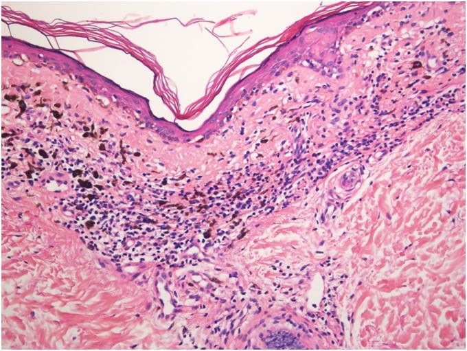
Figure 2. Biopsy specimen from a lesion on the neck shows band-
like lymphohistiocytic infiltration and prominent pigmentary
incontinence in the upper dermis. There is atrophy in overlying
epidermis with vacuolar alteration. H&E, 200×.
tyrosine kinases, and then blocking the cancer
growth [1]. Reported gefitinib side effects consist of
a variety of cutaneous eruptions. However,
hyperpigmentation is rarely reported [2]. We
recently reported a case of ashy dermatosis (AD)-like
hyperpigmentation induced by osimertinib, a
selective inhibitor against EGFR with the T790M
mutation [3].
Interestingly, besides cutaneous involvement,
hyperpigmentation of the hair and eyebrows in a
patient 7 months into gefitinib treatment has also
B been described [4]. In this case, dermoscopy
revealed brown scalp hyperpigmentation in the
affected hairy area. Moreover, reflectance confocal
microscopy showed hair bulb bigeminy with
pigmented keratinocytes. The authors speculated
that the interrupt of SCF/c-kit signaling pathway by
EGFR-TKIs might underlie these findings [4]. A
summary of the previous reports of EGFR-TKI-
induced pigmentary changes is presented in Table
1.
The clinical and histopathologic features of our
patient fit well into the proposed global consensus
for the diagnosis of lichen planus pigmentosus [5].
C
Pigmentary lesions gradually developed 8 months
Figure 1. Multiple slate-gray macules and patches were found on after the onset of gefitinib administration. The time
A) antecubital fossa, B) face and C) upper trunk. interval is compatible with the average time of onset
of drug-induced hyperpigmentation, that is 6.5
-2-Volume 27 Number 5| May 2021
Dermatology Online Journal || Case Presentation 27(5):9
Table 1. Summary of reported cases of EGFR inhibitor-induced pigmentary changes.
Onset
after the
Age, drug Morphology and
Author, year gender Cancer Drug initiation distribution Histopathology
Hyperpigmentation
on the face, trunk, and
Non-small Basal layer
Chang et al., 43, legs occurred
cell lung Gefitinib Months hyperpigmentation and
2004 [8] female following the
carcinoma the dermal macrophages
preceding acneiform
eruptions
Multifocal
Non-small hyperpigmentation
Chang et al., 60, A few
cell lung Gefitinib occurred following No data
2004 [8] female months
carcinoma the preceding
acneiform eruptions
Cosio et al., 77, Basal cell Hyperpigmentation of
Gefitinib 7 months No data
2020 [4] female carcinoma hair and eyebrows
Slate grey
Pigmentary incontinence
hyperpigmentation,
Non-small in the upper dermis and
Lertpichitkul et 71, ashy dermatosis-like
cell lung Osimertinib 6 months vacuolar degeneration at
al., 2020 [3] female eruptions on the
carcinoma the dermoepidermal
chest, buttocks, and
junction
forearms
Band-like
Multiple slate grey
lymphohistiocytic
Non-small macules and patches
62, infiltration and
Our case cell lung Gefitinib 8 months on the antecubital
female pigmentary incontinence
carcinoma fossa, face and upper
in the upper dermis with
trunk
focal vacuolar alteration
months [3,4]. The other current systemic drugs were [6,7]. It is characterized by diffuse, symmetrical, slate
used irregularly for a much longer time and despite gray-to-brownish-black macular pigmentation. The
stopping these drugs, there was further lesions occur on sun-exposed and non-sun-exposed
development of hyperpigmentation. Although areas and the head and neck regions are the
patch and photo-patch tests were not done, there common areas of involvement [5]. The
was no history of suspected allergens including hair histopathology exhibits a lichenoid infiltration and
dye. Moreover, the lesions were not limited to the melanophages on the superficial dermis. The first
areas in contact with the topical medications. line treatment of lichen planus pigmentosus are
Therefore, it is less likely for the contact substances topical corticosteroids and calcineurin inhibitors [6].
to be a cause. Since gefitinib was required, it was not
stopped. The hyperpigmentation was ongoing. A
trial of topical 2% kojic acid cream produced a Conclusion
modest improvement. We herein reported an unusual case of gefitinib-
Lichen planus pigmentosus is a rare entity that associated lichen planus pigmentosus-like
exhibits an acquired macular pigmentation of hyperpigmentation. It is interesting to note that the
uncertain etiology (MPUE). Risk factors for lichen adverse events in the group of MPUE, namely AD and
planus pigmentosus include viral infections lichen planus pigmentosus, have been reported
(hepatitis C) and medications such as methadone, mainly in Asians, South Asians and Africans [5].
henna dyes, mustard oil, and homeopathic remedies Dermatologists should be aware of this unusual
-3-Volume 27 Number 5| May 2021
Dermatology Online Journal || Case Presentation 27(5):9
hyperpigmented condition occurring in patients support, and Dr. Nattaya Teeyapun, medical
taking EGFR-TKIs. oncologist, who cares for this patient.
Acknowledgements Potential conflicts of interest
The authors graciously thank the Skin and Allergy The authors declare no conflicts of interest.
Research Unit, Chulalongkorn University, for their
References
1. Segaert S, Van Cutsem E. Clinical signs, pathophysiology and 32021862].
management of skin toxicity during therapy with epidermal 5. Kumarasinghe SPW, Pandya A, Chandran V, et al. A global
growth factor receptor inhibitors. Ann Oncol. 2005;16:1425-33. consensus statement on ashy dermatosis, erythema
[PMID: 16012181]. dyschromicum perstans, lichen planus pigmentosus, idiopathic
2. Hsu PC, Jablons DM, Yang CT, You L. Epidermal Growth Factor eruptive macular pigmentation, and Riehl's melanosis. Int J
Receptor (EGFR) Pathway, Yes-Associated Protein (YAP) and the Dermatol. 2019;58:263-72. [PMID: 30176055].
Regulation of Programmed Death-Ligand one (PDL1) in Non- 6. Weston G, Payette M. Update on lichen planus and its clinical
Small Cell Lung Cancer (NSCLC). Int J Mol Sci. 2019;20. [PMID: variants. Int J Womens Dermatol. 2015;1:140-9. [PMID: 28491978].
31387256]. 7. Vollono L, Bianchi L, Mazzilli S, et al. Drug-induced lichen planus
3. Lertpichitkul P, Wititsuwannakul J, Asawanonda P, Rerknimitr P. pigmentosus: Do supportive and complementary drugs count?
Osimertinib-associated ashy dermatosis-like hyperpigmentation. Dermatol Ther. 2019;32:e12871. [PMID: 30843626].
JAAD Case Rep. 2020;6:86-8. [PMID: 32051836]. 8. Chang GC, Yang TY, Chen KC, et al. Complications of therapy in
4. Cosio T, Mazzilli S, Bianchi L, Campione E. The Dark Side of cancer patients: Case 1. Paronychia and skin hyperpigmentation
Gefitinib: Reflectance Confocal Microscopy Applied to Hair induced by gefitinib in advanced non-small-cell lung cancer. J Clin
Hyperpigmentation. Skin Appendage Disord. 2020;6:44-7. [PMID: Oncol. 2004;22:4646-8. [PMID: 15542815].
-4-You can also read