Disseminated histiocytic sarcoma in Asian palm civet (Paradoxurus hermaphroditus)
←
→
Page content transcription
If your browser does not render page correctly, please read the page content below
NOTE
                                 Pathology

                                 Disseminated histiocytic sarcoma in Asian
                                 palm civet (Paradoxurus hermaphroditus)
                                 Kittikorn BOONSRI1)*, Sakorn DECHKAJORN2), Kornravee PHOTICHAI1),
                                 Saralee SRIVORAKUL1), Hassadin BOONSRIROJ3), Atigan THONGTHARB4,5) and
                                 Kidsadagon PRINGPROA1,6)
                                 1)VeterinaryDiagnostic Center, Chiang Mai University Animal Hospital, Faculty of Veterinary Medicine,
                                   Chiang Mai University, Chiang Mai 50100, Thailand
                                 2)Chiang Mai Night Safari, Chiang Mai 50230, Thailand
                                 3)Department of Pathology, Faculty of Veterinary Medicine, Mahanakon University of Technology,

                                   Bangkok 10530, Thailand
                                 4)Department of Companion Animal and Wildlife Clinic, Faculty of Veterinary Medicine, Chiang Mai University,

                                   Chiang Mai 50100, Thailand
                                 5)Small Animal Hospital, Chiang Mai University Animal Hospital, Faculty of Veterinary Medicine,

                                   Chiang Mai University, Chiang Mai 50200, Thailand
                                 6)Department of Veterinary Biosciences and Veterinary Public Health, Faculty of Veterinary Medicine,

                                   Chiang Mai University, Chiang Mai 50100, Thailand

                                    ABSTRACT. This case study had focused on a male, 7-year-old Asian palm civet (Paradoxurus
                                    hermaphroditus) with a history of biting its tail and the development of skin masses around
                                    its inguinal area, prior to its death. Macroscopically, multiple firm white nodular masses of
J. Vet. Med. Sci.                   0.5–5 cm in diameter were found in the subcutis of the inguinal area, and in the lungs, spleen
83(1): 108–111, 2021                and liver. Microscopically, masses in the skin, lungs and spleen were composed of neoplastic
                                    spindle cells admixed with mononuclear cells and multinucleated giant cells. The neoplastic
doi: 10.1292/jvms.20-0403           cells were arranged in a sheet pattern. Immunohistochemically, the neoplastic cells were
                                    immunohistochemically positive for vimentin, Iba-1, CD 204 and Human leukocyte antigen
                                    (HLA)-DR, while the cells were negative for cytokeratin and smooth muscle actin. Based on the
Received: 2 July 2020
                                    histopathological and immunohistochemical results, disseminated histiocytic sarcoma was
Accepted: 13 November 2020          diagnosed.
Advanced Epub:
                                    KEY WORDS: civet, disseminated histiocytic sarcoma
          24 November 2020

   Canine and feline histiocytic diseases have been specifically characterized and classified as histiocytoma, cutaneous Langerhans
cell histiocytosis, pulmonary Langerhans cell histiocytosis, cutaneous histiocytosis, systemic histiocytosis, histiocytic sarcoma,
histiocytic sarcoma-hemophagocytic, feline progressive histiocytosis and dendritic cell leukemia [6]. Histiocytic sarcoma (HS) is
categorized as being localized if it originates at a single site or organ; however, if the neoplasms occur as multiple lesions in and/or
on many organs, the term disseminated HS is used [6, 7]. Localized and disseminated cases of HS have been observed in dogs and
cats [1, 6, 7] but have rarely been reported among zoo animals. The Asian palm civet (Paradoxurus hermaphroditus) belongs to the
family viverridae along with other civets which are naturally found in South and Southeast Asia [2]. To the best of our knowledge,
no published reports exist concerning HS in Asian palm civets. Here, we describe the pathological characteristics of disseminated
HS in an Asian palm civet and summarize our findings in comparison to related literatures.
   A male 7-year-old Asian palm civet born at Chiang Mai Night Safari, Chiang Mai, Thailand was kept within the exhibition
zone and routinely fed with tropical fruit and chicken bones. The civet had a history of biting its tail for 1 year and had skin
masses around its inguinal area about 1 month prior to being found dead by zookeepers. A necropsy was performed at the zoo, and
sampled tissues were submitted for further histopathological investigation. Macroscopically, multiple firm white nodular masses
measuring 5 × 4 × 2 cm were present in the subcutis of the inguinal area (Fig. 1A). Similar masses of 0.5–1 cm in diameter were
distributed throughout the lungs (Fig. 1B), while masses of 0.2–0.3 cm in diameter were randomly located in the liver parenchyma
(Fig. 1C). Furthermore, the spleen contained a large mass (2 cm in diameter) and a small mass (0.5 cm in diameter) (Fig. 1D).
   The animal’s skin, heart, lungs, liver, kidneys and spleen were fixed in 10% neutral buffered formalin and processed for
hematoxylin and eosin (H&E) staining at the Veterinary Diagnostic Center, Faculty of Veterinary Medicine, Chiang Mai University,

*Correspondence to: Boonsri, K.: kittikorn.boonsri@cmu.ac.th
©2021 The Japanese Society of Veterinary Science
                  This is an open-access article distributed under the terms of the Creative Commons Attribution Non-Commercial No Derivatives (by-nc-nd)
                  License. (CC-BY-NC-ND 4.0: https://creativecommons.org/licenses/by-nc-nd/4.0/)

                                                                                                                                                   108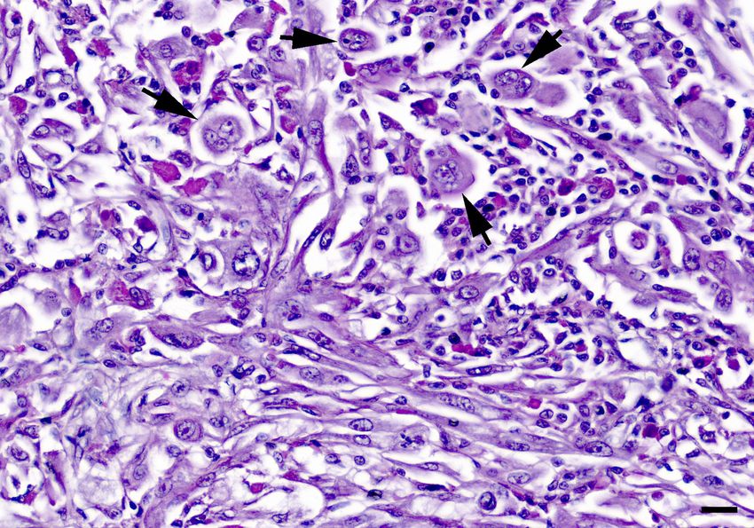
HISTIOCYTIC SARCOMA IN CIVET

Thailand. Immunohistochemistry was performed using the Avidin-Biotin complex (ABC) method as has been previously described
[9]. The using primary antibodies were listed in Table 1.
   Under microscopic examination, an unencapsulated, poorly circumscribed subcutaneous mass was characterized by sheets
of pleomorphic spindle and polygonal cells with distinct cell borders (Fig. 2). Furthermore, a number of lymphocytes and
multinucleated giant cells were distributed in the subcutis. The neoplastic cells were observed to possess oval and bizarre
hyperchromatic nuclei that contained prominent nucleoli and abundant eosinophilic cytoplasm. Mitotic figures were comprised of 7
cells/10 as observed under high power field (HPF). Extensive necrosis was present in the center of the mass. Neoplastic cells with
similar morphology were observed in the spleen with a high mitotic index (13 cells/10HPF). The pulmonary mass was composed
of spindle neoplastic cells. Mitotic figures were comprised of 4 cells/10 HPF, while multinucleated giant cells were comprised of 7
cells/10HPF. Focal necrosis was found in the liver along with aggregation of round cells that were similar to the histiocytic cells.
The other organs showed no remarkable lesions.

  Fig. 1. Gross findings of tumor in a male 7-year-old Asian palm civet (Paradoxurus hermaphroditus) with disseminated histiocytic sarcoma.
     Skin masses at the inguinal area (A). Multifocal nodular masses (arrows) in the lungs (B). White foci (arrows) in the liver (C). Nodular
     masses (arrows) in the spleen (D). Bars=1 cm.

Table 1. Primary antibodies used in the present case
                                                                                                                 Positive control tissue (Asian
        Antibody        Clone      Dilution               Source                      Antigen retrieval
                                                                                                                          palm civet)
Pan-Cytokeratin       AE1/AE3       1:300     Abcam, Cambridge, MA, USA       Microwave (100°C) for 15 min in   Epidermis and adnexa of the skin
                                                                              citrate buffer, pH 6.0
Vimentin                 V9         1:300     Abcam                           Microwave (100°C) for 15 min in   Macrophages and smooth muscle
                                                                              citrate buffer, pH 6.0            in the lung
Iba-1                 20A12.1       1:400     Millipore, Billerica, MA, USA   Microwave (100°C) for 15 min in   Alveolar macrophages in the lung
                                                                              citrate buffer, pH 6.0
CD 204                Polyclonal    1:300     Abcam                           Microwave (100°C) for 20 min in   Alveolar macrophages in the lung
                       antibody                                               citrate buffer, pH 6.0
HLA-DR                  LN3         1:300     Novocastra, Chicago, IL, USA    Microwave (100°C) for 30 min in   Subcutaneous macrophages
                                                                              citrate buffer, pH 6.0
Smooth muscle actin     1A4         1:300     Cell Marque, St. Louis, MO,     Microwave (100°C) for 20 min in   Smooth muscle of the bronchioles
                                              USA                             citrate buffer, pH 6.0

J. Vet. Med. Sci. 83(1): 108–111, 2021                                                                                                      109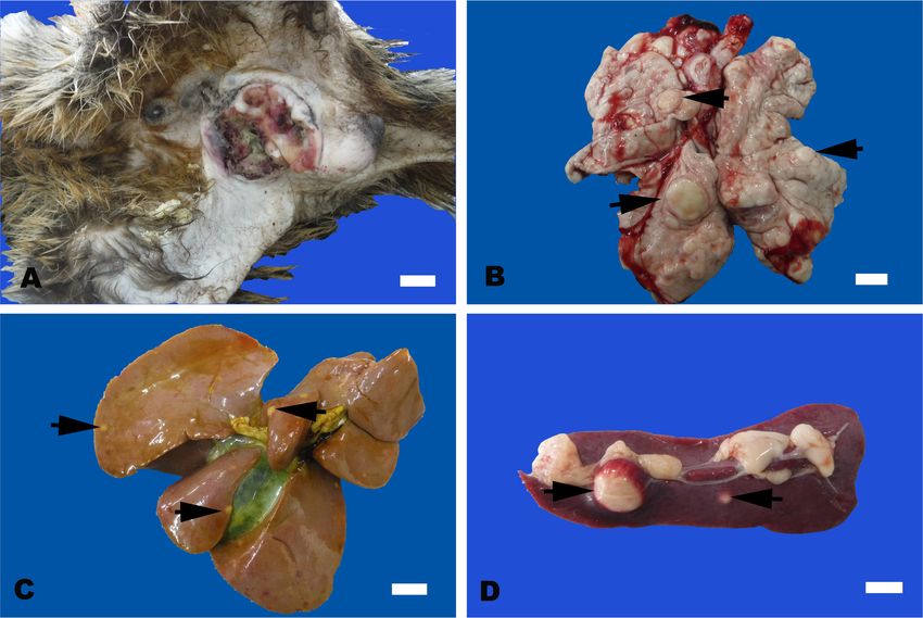
K. BOONSRI ET AL.
Fig. 2. Histological findings of disseminated histiocytic sarcoma in a male 7-year-old Asian palm civet (Paradoxurus hermaphroditus); subcu-
taneous mass. Neoplastic spindle cells are notably oval and bizarre with hyperchromatic nuclei containing prominent nucleoli and abundant
eosinophilic cytoplasm. Many multinucleated giant neoplastic cells are also observed (arrows). Hematoxylin and eosin. Bar=20 µm.
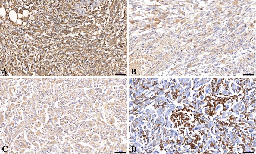
Fig. 3. Immunohistopathological findings of disseminated histiocytic sarcoma in a male, 7-year-old Asian palm civet (Paradoxurus hermaph-
roditus). Neoplastic cells in the subcutaneous mass are positive for vimentin (A), major histocompatibility complex (MHC) class II (B),
CD204 (C) and Iba1 (D). Bars=50 µm.
Immunohistochemically, the neoplastic spindle cells and multinucleated giant cells were positive for vimentin, major
histocompatibility complex (MHC) class II and CD 204 (Fig. 3A–C). Neoplastic spindle cells were strongly positive for Ionised
calcium-binding adaptor molecule-1 (Iba-1) while multinucleated giant cells were negative (Fig. 3D). Neoplastic cells were
negative for AE1/AE3 cytokeratin and smooth muscle actin. Based on the histopathologic and immunohistochemical results, the
tumor in the present case was diagnosed as disseminated HS.
HS is the most aggressive syndrome among histiocytic tumors [3]. HS lesions can occur in or on the spleen, lymph nodes, lungs,
J. Vet. Med. Sci. 83(1): 108–111, 2021 110HISTIOCYTIC SARCOMA IN CIVET
bone marrow, skin and subcutis, brain and articular tissue of the appendicular joints [6]. In this case, the lesions were present
subcutaneously and on multiple organs, such as the lung, spleen and liver. This form of the disease is termed disseminated HS [3,
6, 7]. The spleen, liver, bone marrow, lungs and lymph nodes are the most frequently targeted areas of tissue in disseminated HS
[1]. Predominant tumor cells are large, round and spindle-shaped with ovoid nuclei and abundant eosinophilic cytoplasm [3, 6].
Most neoplastic cells in this case were spindle cells. Spindle cells as the prominent form of HS present a challenge because this
form mimics other spindle cell tumors, such as fibroblastic sarcoma, anaplastic sarcoma ad leiomyosarcoma [6]. For fibrosarcoma,
the tumor is known to be infiltrative and produces collagen. Smooth muscle tumors are characterized by the expression of smooth
muscle actin [3]. In this instance, an immunohistochemical examination was recommended to confirm the immunophenotype
of the neoplastic cells. [1]. In this present study, immunohistochemistry indicated that the cytoplasm of neoplastic cells was
positive for Iba-1, vimentin, CD 204 and MHC class II. These outcomes were applied to determine their interstitial dendritic cell
immunophenotype [6]. Recommended immunohistochemical histiocytic sarcoma markers in dogs, cats, ferrets and four-toed
hedgehogs are CD1a, CD11c/CD18, CD204, Human leukocyte antigen-DR (HLA-DR) or MHC class II and Iba-1 [6–8, 10]. The
etiology and pathology of histiocytic sarcoma is largely unknown. HS has also been reported in exotic pets such as four-toed
hedgehogs [5, 8], rabbits [4], capybaras [9] and ferrets [10]. In conclusion, this is the first report to describe the pathological
characteristics of disseminated HS in an Asian palm civet.
POTENTIAL CONFLICTS OF INTEREST. The author(s) declared no conflicts of interest with respect to the authorship and/or
publication of this article.
REFERENCES
1. Affolter, V. K. and Moore, P. F. 2002. Localized and disseminated histiocytic sarcoma of dendritic cell origin in dogs. Vet. Pathol. 39: 74–83.
[Medline] [CrossRef]
2. Denver, M. 2003. Procyonidae and viverridae. pp. 516–523. In: Zoo and Wild Animal Medicine (Fowler M. E. and Miller R. E. eds.), W.B.
Saunders, Philadelphia.
3. Hendrick, M. J. 2017. Mesenchymal tumors of the skin and soft tissues. pp. 142–175. In: Tumors in Domestic Animals, 5th ed. (Donald, J. M. rd.),
John Wiley & Sons, Ames.
4. Ishimori, M., Michishita, M., Yoshimura, H., Azakami, D., Ochiai, K., Ishiwata, T. and Takahashi, K. 2017. Disseminated histiocytic sarcoma with
hemophagocytosis in a rabbit. J. Vet. Med. Sci. 79: 1503–1506. [Medline] [CrossRef]
5. Koizumi, I. and Kondo, H. 2019. Clinical management and outcome of four-toed hedgehogs (Atelerix albiventris) with histiocytic sarcoma. J. Vet.
Med. Sci. 81: 545–550. [Medline] [CrossRef]
6. Moore, P. F. 2014. A review of histiocytic diseases of dogs and cats. Vet. Pathol. 51: 167–184. [Medline] [CrossRef]
7. Moore, P. F. 2017. Canine and feline histiocytic disease. pp. 322–336. In: Tumors in Domestic Animals, 5th ed. (Donald, J. M. ed.), John Wiley &
Sons Inc., Ames.
8. Son, N. V., Chambers, J. K., Dung, L. T., Kishimoto, T. E., Nishimura, M., Kita, C., Takada, Y., Miwa, Y., Nakayama, H. and Uchida, K. 2020.
Histological and immunohistochemical features of normal histiocytes and Langerhans cells, and histiocytic sarcomas in four-toed hedgehogs
(Atelerix albiventris). J. Comp. Pathol. 178: 32–40. [Medline] [CrossRef]
9. Srivorakul, S., Boonsri, K., Vechmanus, T., Boonthong, P., O’Sullivan, M. G. and Pringproa, K. 2017. Localized histiocytic sarcoma in a captive
capybara (Hydrochoerus hydrochaeris). Thai. J. Vet. Med. 47: 131–135.
10. Thongtharb, A., Uchida, K., Chambers, J. K., Miwa, Y., Murata, Y. and Nakayama, H. 2016. Histological and immunohistochemical features of
histiocytic sarcoma in four domestic ferrets (Mustela putorius furo). J. Vet. Diagn. Invest. 28: 165–170. [Medline] [CrossRef]
J. Vet. Med. Sci. 83(1): 108–111, 2021 111You can also read