Usefulness of Ultrasound Devices in Facial Plastic Surgery
←
→
Page content transcription
If your browser does not render page correctly, please read the page content below
pISSN 2733-4813 • eISSN 2733-631X

                                                                                                                                                 Review Article
                                                      Aesthet 2021;2(1):1-6
                                                      https://doi.org/10.46738/Aesthetics.2021.2.1.1

Usefulness of Ultrasound Devices in Facial Plastic Surgery

Won Lee, MD, PhD1                                            The use of ultrasound technology in aesthetic fields is common. The proper
Wook Oh, MD2                                                 use thereof can help plastic surgeons achieve satisfactory outcomes. The
Hyun Jun Park, MD2                                           study was designed to outline the use of ultrasound technology devices in
                                                             plastic surgery. The PubMed database was searched for articles reporting on
1
                                                             the use of diagnostic ultrasound, microfocused ultrasound technology, and
 Yonsei E1 Plastic Surgery Clinic, Anyang, Korea
2
 Maylin Clinic, Seoul, Korea
                                                             non-focused ultrasound technology. Doppler ultrasound technology has
                                                             found use in detecting arteries of the face. Emitting thermal energy, microfo-
                                                             cused ultrasound has been used to elicit skin tightening and fat reduction ef-
                                                             fects. The use of non-focused ultrasound technology to achieve cellular level
                                                             effects has garnered greater interest. Understanding of different applications
                                                             of ultrasound technology could help with improving treatment results in fa-
                                                             cial plastic surgery.

                                                             Key words
                                                             Ultrasound technology, Microfocused ultrasound, Non-focused ultrasound,
                                                             HIFU, Hyaluronic acid filler

Received April 6, 2021
Accepted April 19, 2021

Correspondence
Won Lee
Yonsei E1 Plastic Surgery Clinic, Anyang
14072, Korea
E-mail: e1clinic@daum.net
https://orcid.org/0000-0001-7411-0198

C   Korean Society for Laser, Dermatology and Trichology

CC This is an open access article distributed under

the terms of the Creative Commons Attribution Non-
Commercial License (http://creativecommons.org/
licenses/by-nc/4.0) which permits unrestricted non-
commercial use, distribution, and reproduction in any
medium, provided the original work is properly cited.

                                                                                                                     www.theaesthetics.org   1
INTRODUCTION RESULTS
In recent years, energy-based devices have been increas- 1. Ultrasound technology for visualization
ingly used in fields of dermatology, aesthetic medicine, and For ultrasound devices, 1 Hz means 1 cycle/sec and indi-
plastic surgery. In plastic surgery, laser and non-surgical cates how many pulses are made during 1 second. For tis-
fat removal devices are popular [1]. Among these devices, sue visualization, settings of 8 MHz to 20 MHz are typically
those emitting ultrasound waves are most widely used used, and the use of 8 MHz provides a depth of visualization
because of their safety: ultrasound devices emit high fre- of approximately 50 mm. At 20 MHz, a depth of 10 mm can
quency sound waves that pose no harm to facial tissues. Re- be visualized (Fig. 1).
cently, research on the use of ultrasound in plastic surgery Upon emitting ultrasound energy to tissue, waves are
has focused on the detection of facial features beneath the reflected to the probe at 90 degrees. During the procedure,
surface of the skin, especially when performing filler injec- however, the energy can be absorbed by the tissue or scat-
tions and thread lifting, which are considered blind tech- tered. When the attenuated energy, such as absorbed or
niques. High-intensity focused ultrasound technology has scattered energy, is high, images are not obtainable. Ultra-
been used to achieve skin tightening and face contouring. sound at a relatively high frequency leads to more attenu-
Non-focused ultrasound technology has also found use. In ation, and as such, a lower frequency is needed to detect
this study, we aimed to review the literature on the use of deeper structures.
ultrasound technology and to propose possible application The mechanism of the treatment effects elicited by ul-
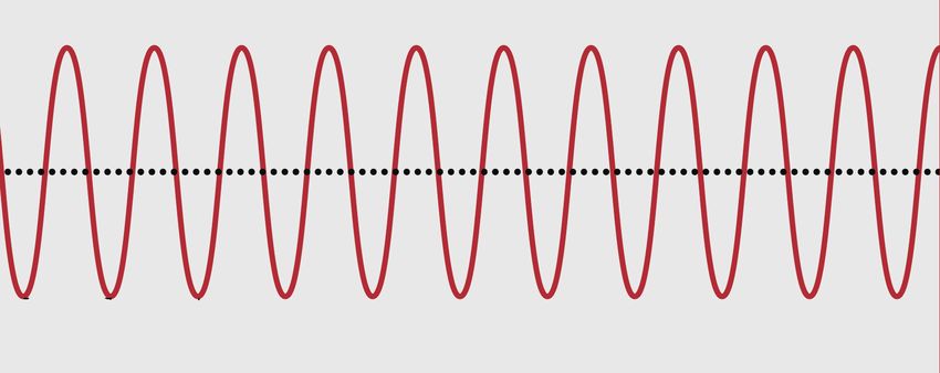
thereof in plastic surgery. trasound energy relies on repetitive electric stimulations.
These stimulations exert compression and rarefaction pres-
MATERIALS AND METHODS sure, due to piezoelectric effects (Fig. 2). When ultrasound
energy interacts with human tissue, attenuation of ultra-
The PubMed database was searched for the following sound energy induces thermal changes. Ultrasound devices
terms in April 2021: “ultrasound hyaluronic acid filler”, “ul- for diagnosis generate minimal thermal energy and pose
trasound thread lifting,” “microfocused ultrasound,” and no harm to human tissue. However, some devices, such
“non-focused ultrasound.” We excluded articles not written as high-intensity focused ultrasound devices, can emit an
in English, that described the use of ultrasound devices for intensity up to 5,000 W/cm2 and can elicit tissue tempera-
non-aesthetic purposes, that used ultrasound to merely tures up to 60℃. The mechanism of diagnostic ultrasound
confirm the presence of filler, that were experimental in is seen in Fig. 3.
design, that focused on the hands and neck, and that com-
prised ophthalmic ultrasound studies. In total, 338 articles 1) Vascular mapping for injections
were found, and after applying the exclusion criteria, 23 The most tragic complications of filler injections, includ-
articles were finally reviewed. ing ocular complications, are related to the vascularity in
8 MHz 10 MHz 20 MHz
Penetration
10 mm
Penetration
35 mm
Penetration
50 mm A B C
Fig. 1. Different depths of visualization with different ultrasound frequencies.
2 www.theaesthetics.org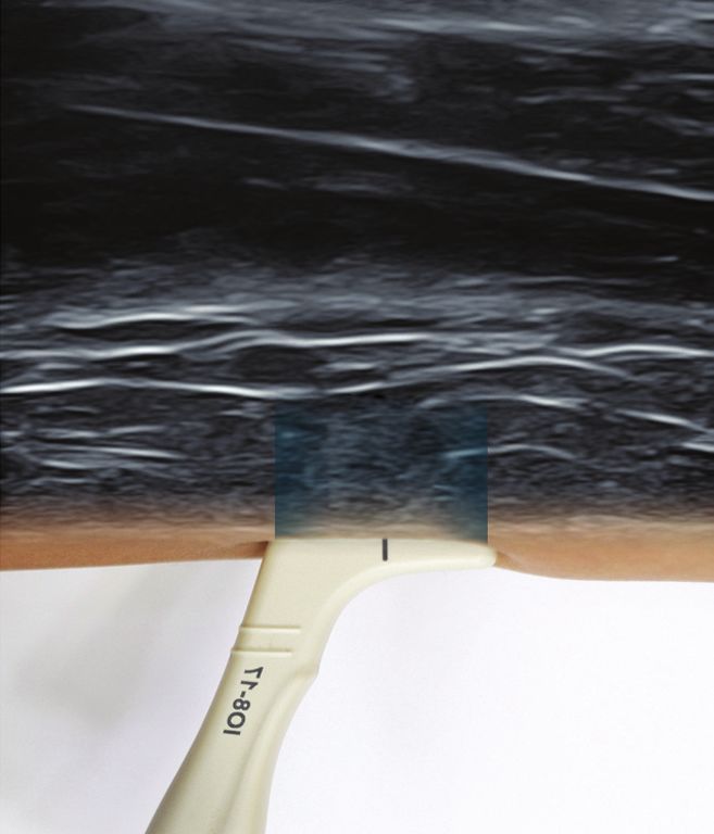
Ultrasound Technology in Plastic Surgery
                                                                                                                              Won Lee, et al.

                                                                                                                                                        Review Article
                                       Higher frequency

                      Period
                       (T)
                 P
Pressure

           0


                                                                                                              10 MHz

                                       Lower frequency

                               T
Pressure

           0
                                                                                                             Penetration
                                                                                                               35 mm


               Time                                                           Fig. 3. Mechanism of diagnostic ultrasound. An ultrasound probe
                                                                              sends signals at a right angle and receives signals after the energy is
                P: Pressure        : Wavelength   f: Frequency   T: Period   reflected. The energy is attenuated (i.e., scattered or absorbed) dur-
                                                                              ing energy transfer, eliciting thermal effects.
Fig. 2. Depiction of ultrasound frequencies. High frequencies are bet-
ter for detecting dermal layers; low frequencies are better for detect-
ing deeper layers. Repetitive compression pressure and rarefaction
pressure occur with ultrasound energy.
                                                                              with minimal damage to surrounding structures. MFU is
                                                                              able to effectively disrupt tissue by exploiting the transfor-
                                                                              mation of acoustic energy into thermal damage. MFU can
the face. During injection procedures, Doppler ultrasound                     be focused on subcutaneous tissue, where temperatures
can be used to detect arteries and vessels: Facial arteries                   briefly increase beyond 60℃, producing small thermal co-
can be detected during nasolabial fold correction [2]. The                    agulation points to a depth of up to 5 mm within the mid-
supratrochlear artery can be detected during correction of                    to-deep reticular layer of the dermis and subdermis [13].
glabellar frown lines [3]. The frontal branch of the superfi-                 These thermal changes shrink the tissue for skin tightening
cial temporal artery also can be detected [4]. Additionally,                  effects. The target layer of the face for skin tightening would
the dorsal nasal artery can also be detected during nose                      be the musculocutaneous layer [14]. When applied at the
augmentation by filler injections [5]. The use of Doppler ul-                 subcutaneous fat layer, ultrasound thermal energy can be
trasound to locate vessels that could be potentially harmed                   applied to elicit adipose cell destruction [15]. Histologic
during filler injections can help prevent vascular complica-                  reports have shown that MFU targets subcutaneous adi-
tions [6].                                                                    pose tissue [16]. The mechanism of MFU is outlined in Fig.
                                                                              4. Dual-depth focused ultrasound can also be used for skin
   2) Detection of filler complications                                       tightening [17].
   Doppler ultrasound can also be used to detect complica-
tions from filler injections. Vascular complications, such as                 3. Non-focused ultrasound technology
those associated with compression [7] or embolism [8], can                        Non-focused ultrasound can also be used in facial plas-
be diagnosed upon vessel detection in all areas of the face,                  tic surgery, although reports thereof in the literature are
such as the tear trough, nose, temple, and lips [9]. Ultra-                   scarce. Whereas high frequency ultrasound can reach a
sound can also be used to detect palpable nodules [10] and                    depth of more than 50 mm for diagnosis and vascular visu-
to determine which kinds of filler have been injected [11].                   alization and high-intensity focused ultrasound can be used
Research has shown that the amount, location, and depth                       to generate thermal changes in target tissue, non-focused
of previous injected filler can be identified for precise intral-             ultrasound can be used to target the cellular level. Matrix
esional delivery of hyaluronidase to dissolve filler [12].                    metalloproteinases (MMPs) are known to be important
                                                                              in both chronological and photo-induced skin ageing [18],
2. Microfocused ultrasound technology                                         and heat shock proteins (HSPs) are important in fibroblast
   Microfocused ultrasound (MFU) technology facilitates                       activity and collagen production [19]. Interestingly, decreas-
the precise delivery of acoustic energy to targeted tissue,                   ing the production of HSPs has been found to be a possible

                                                                                                                 Aesthet Vol. 2 No. 1 April 2021   3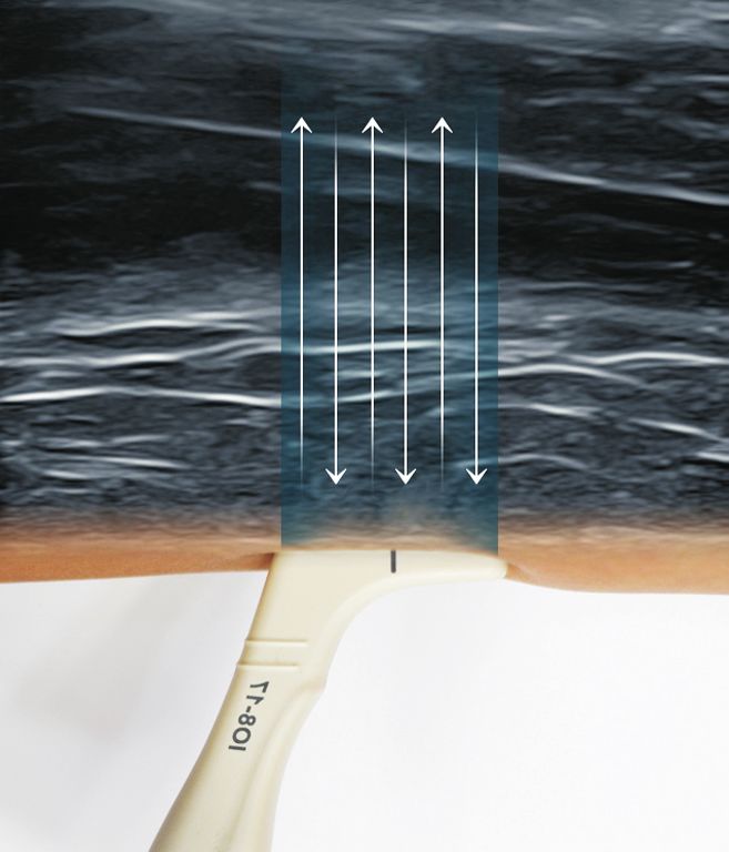
4 MHz 7 MHz
Penetration Penetration Fig. 4. Use of microfocused ultrasound
4.5 mm 4.5 mm on the skin. Microfocused ultrasound
technology generates thermal coagula-
tion in the musculocutaneous layer for
skin tightening effects or destroys fat
cells in the subcutaneous layer for face
contouring. Ultrasound energy at 7 MHz
reaches a shallower depth than 4 MHz,
and thus, a higher intensity is needed to
reach the same depth.
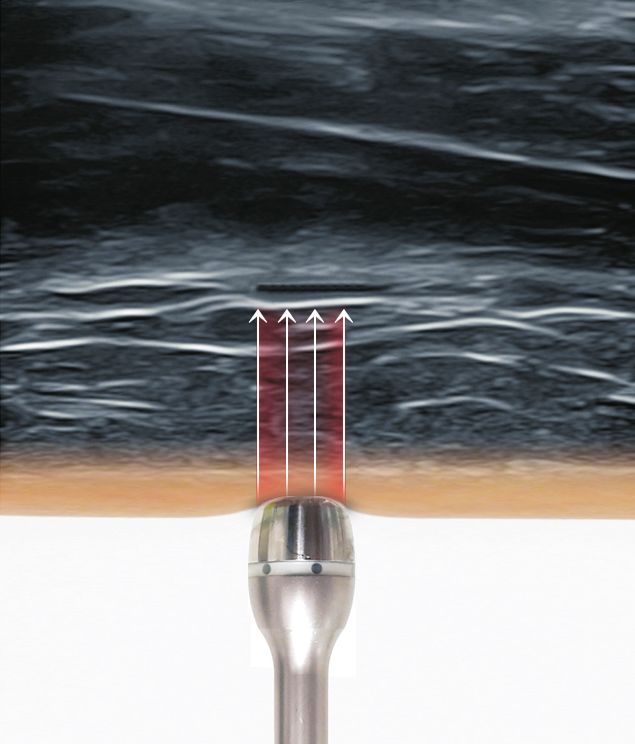
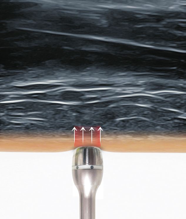
1 MHz 3 MHz 10 MHz
Penetration
3 mm
Penetration
10 mm
Penetration
30 mm
Fig. 5. The use of non-focused ultrasound energy to generate tissue changes. The probe (1 MHz, 3 MHz, 10 MHz) is from Intense Ultra (Ionto
Comed, Germany). Energy at 10 MHz emits a large number of acoustic waves, but the depth is relatively shallow. Therefore, high intensity is re-
quired to overcome energy attenuation from scattering and absorption.
target for reducing the signs of ageing [20]. Modulation of One potential use for ultrasound technology is in three-
both MMPs and HSPs could be possible with non-focused dimensional restoration of the facial vasculature. Although
ultrasound technology [21]. The use of non-focused ultra- doppler ultrasound can detect arteries of the face, it is quite
sound energy is outlined in Fig. 5. difficult to learn how to use ultrasound at the face because
of its two-dimensional images. Future technology could
DISCUSSION make three-dimensional images more accessible and in real
time for safer injection procedures. Another potential use is
Ultrasound technology is being widely used in aesthetic for microfocused ultrasound in procedures to reduce sub-
fields for detecting arteries of the face, for detecting pos- mental fat. Ultrasound energy is transferred to water con-
sible filler complications, for eliciting therapeutic thermal tent easily. Applying MFU in combination with tumescent
damage to the musculoaponeurotic and subcutaneous solution and/or other solutions, such as fat dissolving solu-
layers, and for anti-ageing by stimulating connective tissue tions, can induce larger thermal injury zones and potentially
restoration. Several other potential uses for ultrasound greater fat reductions [22]. Thus, for patients desiring less
technology are proposed below. invasive techniques, MFU for fat reduction could prove pref-
4 www.theaesthetics.org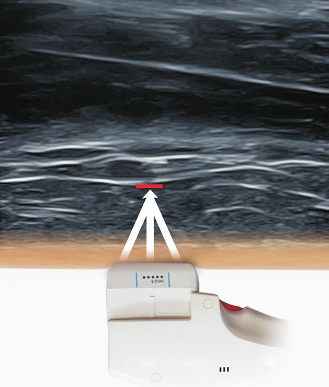
Ultrasound Technology in Plastic Surgery
                                                                                                                      Won Lee, et al.

                                                                                                                                              Review Article
erable over conventional liposuction. A third potential use                 sjaa197
is in post-operative swelling and preoperative treatment.              4.   Lee W, Moon HJ, Kim JS, Chan BL, Yang EJ. Doppler ultra-
Stimulating connective tissue restoration, non-focused ul-                  sound-guided thread lifting. J Cosmet Dermatol 2020;19:
trasound technology might help reduce inflammation and                      1921-7.
post-operative swelling. This could potentially be applied to          5.   Moon HJ, Lee W, Do Kim H, Lee IH, Kim SW. Doppler ultraso-
conventional face lift operations, thread lifting, and microfat             nographic anatomy of the midline nasal dorsum. Aesthetic
grafting. Further studies are warranted to assess postopera-                Plast Surg. Forthcoming 2020. https://doi.org/10.1007/
tive swelling at the cellular level. Ultrasound also can be                 s00266-020-02025-1
used for preoperative treatment to restore connective tis-             6.   Lee W. Prevention of hyaluronic acid filler-induced blindness.
sue balance. Use of 3-MHz non-focused ultrasound energy                     Dermatol Ther 2020;33:e13657.
can reach the subcutaneous fatty layer and might help fa-              7.   Lima VGF, Regattieri NAT, Pompeu MF, Costa IMC. External
cilitate smoother injections or surgical dissection. One final              vascular compression by hyaluronic acid filler documented
potential use for ultrasound technology is in the elimination               with high-frequency ultrasound. J Cosmet Dermatol 2019;18:
of cellulite. Cellulite is a common condition caused by ab-                 1629-31.
normal tethering of fat strands to the subcutaneous tissues.           8.   Schelke LW, Velthuis P, Kadouch J, Swift A. Early ultrasound
Research has shown that high-intensity focused ultrasound                   for diagnosis and treatment of vascular adverse events with
cavitation causes architectural changes in subcutaneous                     hyaluronic acid fillers. J Am Acad Dermatol. Forthcoming
fat tissue, disrupting the cellulite [23]. Another interesting              2019. https://doi.org/10.1016/j.jaad.2019.07.032
report mentioned the use of shock wave treatment for                   9.   Jaguś D, Skrzypek E, Migda B, Woźniak W, Mlosek RK. Useful-
cellulite [24]: Acoustic wave therapy originates from shock                 ness of Doppler sonography in aesthetic medicine. J Ultrason
wave technology successfully used for over 30 years in urol-                2021;20:e268-72.
ogy. Ultrasound devices, such as MFU or non-focused ultra-            10.   Mlosek RK, Migda B, Skrzypek E, Słoboda K, Migda M. The
sound, might also find use for treating cellulite and other                 use of high-frequency ultrasonography for the diagnosis of
conditions, such as lipedema, striae, or capsular fibrosis                  palpable nodules after the administration of dermal fillers. J
after breast operations.                                                    Ultrason 2021;20:e248-53.
                                                                      11.   Urdiales-Gálvez F, De Cabo-Francés FM, Bové I. Ultrasound
CONCLUSION                                                                  patterns of different dermal filler materials used in aesthet-
                                                                            ics. J Cosmet Dermatol. Forthcoming 2021. https://doi.org/
   Ultrasound technology is widely used in aesthetic fields,                10.1111/jocd.14032
and many possible applications thereof in several plastic             12.   Schelke LW, Decates TS, Velthuis PJ. Ultrasound to improve
surgery fields await study. Understanding of action mecha-                  the safety of hyaluronic acid filler treatments. J Cosmet Der-
nisms for ultrasound technology should be established to                    matol 2018;17:1019-24.
use better surgical and/or minimal invasive results.                  13.   Fabi SG. Noninvasive skin tightening: focus on new ultra-
                                                                            sound techniques. Clin Cosmet Investig Dermatol 2015;8:47-
CONFLICT OF INTEREST                                                        52.
                                                                      14.   White WM, Makin IR, Barthe PG, Slayton MH, Gliklich RE.
  No potential conflict of interest relevant to this article                Selective creation of thermal injury zones in the superficial
was reported.                                                               musculoaponeurotic system using intense ultrasound ther-
                                                                            apy: a new target for noninvasive facial rejuvenation. Arch
REFERENCES                                                                  Facial Plast Surg 2007;9:22-9.
                                                                      15.   Kwon TR, Im S, Jang YJ, Oh CT, Choi EJ, Jung SJ, et al. Im-
 1. The Aesthetic Society’s Cosmetic Surgery National Data                  proved methods for evaluating pre-clinical and histological
    Bank: Statistics 2019. Aesthet Surg J 2020;40(Suppl 1):1-26.            effects of subcutaneous fat reduction using high-intensity fo-
 2. Lee W, Kim JS, Moon HJ, Yang EJ. A safe Doppler ultrasound-             cused ultrasound in a porcine model. Skin Res Technol 2017;
    guided method for nasolabial fold correction with hyaluronic            23:194-201.
    acid filler. Aesthet Surg J. Forthcoming 2020. https://doi.org/   16.   Gadsden E, Aguilar MT, Smoller BR, Jewell ML. Evaluation of
    10.1093/asj/sjaa153                                                     a novel high-intensity focused ultrasound device for ablating
 3. Lee W, Moon HJ, Kim JS, Yang EJ. Safe glabellar wrinkle cor-            subcutaneous adipose tissue for noninvasive body contour-
    rection with soft tissue filler using Doppler ultrasound. Aes-          ing: safety studies in human volunteers. Aesthet Surg J 2011;
    thet Surg J. Forthcoming 2020. https://doi.org/10.1093/asj/             31:401-10.

                                                                                                        Aesthet Vol. 2 No. 1 April 2021   5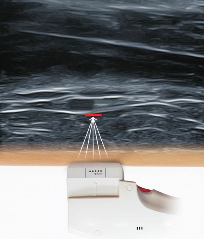
17. Baumann L, Zelickson B. Evaluation of micro-focused ultra-       21. Kruglikov IL, Sontag W. Ultrasound of 10 MHz frequency as a
    sound for lifting and tightening neck laxity. J Drugs Dermatol       novel strategy for skin anti-aging therapy. Med Hypotheses
    2016;15:607-14.                                                      2010;74:620-1.
18. Fisher GJ, Kang S, Varani J, Bata-Csorgo Z, Wan Y, Datta S, et   22. Lee S, Kim HJ, Park HJ, Kim HM, Lee SH, Cho SB. Morpho-
    al. Mechanisms of photoaging and chronological skin aging.           metric analysis of high-intensity focused ultrasound-induced
    Arch Dermatol 2002;138:1462-70.                                      lipolysis on cadaveric abdominal and thigh skin. Lasers Med
19. Dams SD, de Liefde-van Beest M, Nuijs AM, Oomens CW,                 Sci 2017;32:1143-51.
    Baaijens FP. Heat shocks enhance procollagen type I and          23. Moravvej H, Akbari Z, Mohammadian S, Razzaghi Z. Focused
    III expression in fibroblasts in ex vivo human skin. Skin Res        ultrasound lipolysis in the treatment of abdominal cellulite:
    Technol 2011;17:167-80.                                              an open-label study. J Lasers Med Sci 2015;6:102-5.
20. Calderwood SK, Murshid A, Prince T. The shock of aging: mo-      24. Siems W, Grune T, Voss P, Brenke R. Anti-fibrosclerotic effects
    lecular chaperones and the heat shock response in longevity          of shock wave therapy in lipedema and cellulite. Biofactors
    and aging--a mini-review. Gerontology 2009;55:550-8.                 2005;24:275-82.

6    www.theaesthetics.org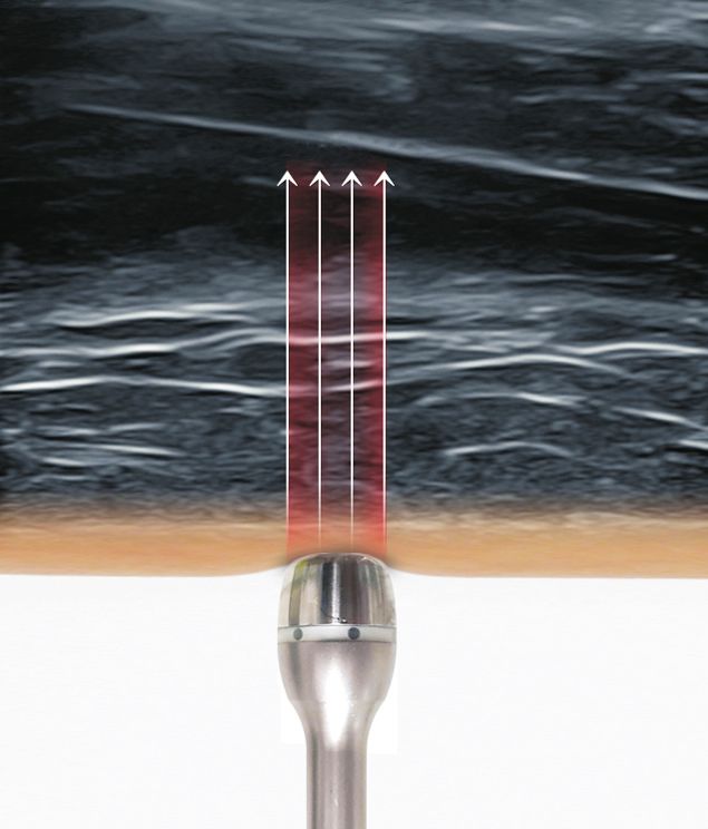
You can also read