Usefulness of virtual reality based training to diagnose strabismus
←
→
Page content transcription
If your browser does not render page correctly, please read the page content below
www.nature.com/scientificreports
OPEN Usefulness of virtual reality‑based
training to diagnose strabismus
Hyun Sik Moon1, Hyeon Jeong Yoon1, Sang Woo Park1, Chae Yeon Kim2, Mu Seok Jeong2,
Sung Min Lim2, Jee Heon Ryu2 & Hwan Heo1*
To study the usefulness of virtual reality (VR)-based training for diagnosing strabismus. Fourteen
residents in ophthalmology performed at least 30 VR training sessions to diagnose esotropia and
exotropia. Examinations of real patients with esotropia or exotropia before and after the VR training
were video-recorded and presented to a strabismus expert to assess accuracy and performance
scores for measuring the deviation angle and diagnosing strabismus with anonymization. A feedback
survey regarding the usefulness and ease of use of the VR application was conducted for participants.
The mean age of the 14 ophthalmology residents (10 men and 4 women), was 29.7 years. Before
VR training, participants showed a mean accuracy score of 14.50 ± 5.45 and a performance score
of 9.64 ± 4.67 for measuring the deviation angle and diagnosing strabismus in real patients with
strabismus. After VR training, they showed a significantly improved accuracy score of 22.14 ± 4.37
(p = 0.012) and a performance score of 15.50 ± 1.99 (p = 0.011). According to the survey, most
participants agreed on the usefulness of VR applications. This study suggests that VR-based training
improved ophthalmology residents’ clinical diagnostic skills for strabismus in a short period.
Strabismus is not uncommon in children, and without appropriate approach and treatment, exacerbation of
strabismus and subsequent sequelae, such as amblyopia, could occur. The ability to diagnose strabismus is an
essential skill for ophthalmology specialists, and proper education and extensive clinical experience are required
during residency training.
To date, methods such as practice-based learning, problem-based learning, team-based learning, and e-learn-
ing have been used to train medical specialists. In recent years, virtual reality (VR) and augmented reality (AR)
simulation training have also been utilized, focusing on various medical fields. Advancements in VR represent
some of the newest modalities being integrated into ophthalmologic practice and resident education1–3. The
importance of incorporating simulation in the education of the residents and skills assessment is increasingly
emphasized4. The 2019 coronavirus (COVID-19) pandemic also affected the medical curriculum; although
junior doctors had to potentially serve as frontliners in this situation, they only had limited experience and
education, both medically and surgically5. Therefore, the need for alternative educational strategies, such as VR
simulation, has e merged6,7.
Therefore, we developed a VR application for diagnostic training of strabismus that could measure the devia-
tion angle and diagnose strabismus using the head-mounted display (HMD) and VR technology. This study was
aimed at investigating the usefulness of VR application as a training tool for ophthalmology trainees in the field
of clinical strabismus.
Methods
VR application for diagnostic training of strabismus. The VR application was created based on the
Oculus Rift (Oculus VR, LLC, Irvine, USA), the VR HMD, Oculus Touch (Oculus VR, LLC, Irvine, USA), wire-
less haptic controllers, and a computer running Windows 10 (Microsoft Corporation, Redmond, WA, USA).
The Oculus Rift HMD device was equipped with a liquid–crystal display (5.7″ diagonal, resolution of
1280 × 1440 pixels per eye), with a 110° field of view, mounted with an accelerometer, a gyroscope, and a mag-
netometer sensor for the positional tracking system. Oculus Touch controllers were peripheral accessories of the
Oculus Rift and were employed to track the user’s hand position and orientation in a three-dimensional space.
The HMD device and controllers were connected to a PC system.
A simple user manual was prepared to enable the usage of the VR application, and tutorials were also included
in the application (Fig. 1). Users could use controllers to select and set the desired type of strabismus in the
1
Department of Ophthalmology, Chonnam National University Medical School and Hospital, 42 Jebongro,
Dong‑gu, Gwangju 61469, Republic of Korea. 2Department of Education, College of Education, Chonnam National
University, Gwangju, Republic of Korea. *email: hwanheo@jnu.ac.kr
Scientific Reports | (2021) 11:5891 | https://doi.org/10.1038/s41598-021-85265-8 1
Vol.:(0123456789)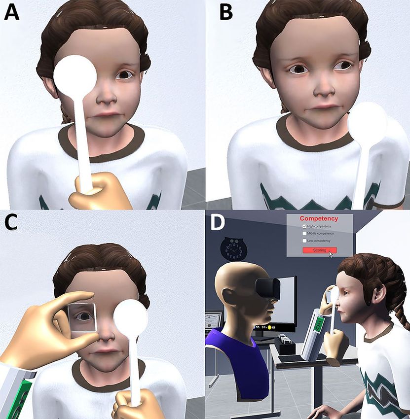
www.nature.com/scientificreports/
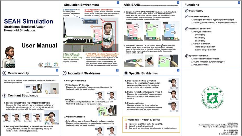
Figure 1. The user manual of the virtual reality application for diagnostic training of strabismus. The manual,
which contains 8 pages, describes the simulation environment and the user interface implemented on the left
wrist. The user interface provides lists of examinations that can be performed through simulation and how to
carry them out.
virtual environment. Thus, they could perform clinical tests and observe ocular motility in the various types of
strabismus, such as horizontal, vertical, paralytic, and specific strabismus.
In a VR environment, similar to an outpatient clinic, the strabismus patient was sited looking at a distant
object, and the user was facing the patient from the right side. Furthermore, a virtual occluder and prisms with
a spacing of five prism diopters were placed step by step on a virtually implemented table. The users could use
the controllers to operate the virtual instrument. We also implemented cover-uncover, alternate cover, and prism
cover tests to allow users to measure the patient’s deviation angle (Fig. 2). In virtual patients with exotropia and
esotropia, the non-dominant eye moves for fixation when the dominant eye is covered. In contrast, the dominant
eye does not move when the non-dominant eye is covered. In the alternate cover test, an ocular movement is
made, such that the type of strabismus could be distinguished. Furthermore, When the users perform the prism
cover test with a smaller prism than the patient’s deviation angle, strabismus decreases but persists. In contrast,
when a prism larger than the virtual patient’s deviation angle is applied, the eye moves in the opposite direction.
When using a prism matching the patient’s deviation angle, no ocular movement is ascertained upon use (see
Supplementary Video S1 online).
Study design, setting, and participant. All participants provided written informed consent. The
study complied with the tenets of the Declaration of Helsinki. The Institutional Review Board of the Chon-
nam National University Hospital approved the study protocol. This study followed the Standards for Qual-
ity Improvement Reporting Excellence Strengthening the Reporting (SQUIRE) guidelines for pre-post quality
improvement study.
A total of 14 ophthalmology residents volunteered to participate in the study from the Chonnam national
university hospital of South Korea between March 30, 2020, and July 31, 2020. Exclusion criteria were strabis-
mus, previous ocular surgery, and ocular media’s opacification, including cataract, and active ocular disease. The
same person (H.S.M.) instructed participants with a brief standardized instruction on using this VR application,
including a manual, tutorials of each module, and information about controlling the virtual occluder and prisms.
Participants carried out examinations on 3 actual patients with esotropia or exotropia, within 2 weeks before
and after 30 sessions of VR training. The interval between the actual patient examination and before and after
VR training was limited to two weeks. Each VR training session lasted at least 5 min, and participants performed
at least one ocular motility examination, cover-uncover test, alternate cover test, and prism cover test on virtual
patients. We implemented the application to count sessions wherein the completion requirement was met, and
we recommended and confirmed them to run 30 sessions in 2 weeks. The virtual patient’s strabismus settings
were limited to esotropia and exotropia between 20–40 prism diopter. We designed the study to ensure that each
participant’s pediatric ophthalmic training was not duplicated during the study period. They were given a little
opportunity to see strabismus patients outside the pediatric ophthalmology clinic. We designed it such that each
participants’ pediatric ophthalmic training did not overlap with the study period.
Scientific Reports | (2021) 11:5891 | https://doi.org/10.1038/s41598-021-85265-8 2
Vol:.(1234567890)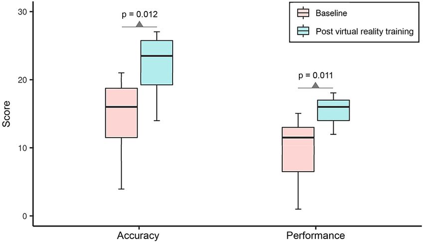
www.nature.com/scientificreports/

                                  Figure 2.  The virtual examination of virtual patients using an occluder and prisms implemented in virtual
                                  reality. (A,B) The cover-uncover test in a patient with left exotropia. (C) The prism cover test. (D) A user and a
                                  patient from the perspective of a third party.

                                   Procedures                    Items to check                                                                         Score
                                                                 Using the instrument (1)
                                   Ocular motility examination   Specifying the target (2)                                                              5
                                                                 Checking the oblique muscles (2)
                                   Cover–uncover test            Occlusion skill (5)                                                                    5
                                   Alternate cover test          Occlusion skill (5)                                                                    5
                                                                 Prism-applying skill (3)
                                   Prism cover test                                                                                                     5
                                                                 Occlusion skill (2)
                                                                 Accuracy of identifying the dominant and non-dominant eye/direction of deviation (3)
                                   Diagnosis report              Measured deviation angle (3)                                                           10
                                                                 Strabismus diagnosis (4)
                                   Total score                                                                                                          30

                                  Table 1.  Accuracy score rating standard of the diagnostic procedure for strabismus.

                                  Evaluation. Examinations of actual patients were video-recorded and anonymous. After completing the
                                  process, videos were blinded as to which video recorded exams were conducted pre- versus post-VR training
                                  and presented to the strabismus specialist (H.H.) in a random order. The primary outcome measure was par-
                                  ticipants’ accuracy and performance scores, measured by a predetermined scoring standard, which comprises
                                  specific procedures. To rate accuracy and performance, scores for 4 procedures, consisting of the ocular motility
                                  exam, cover-uncover test, alternate cover test, and prism cover test, were evaluated. Table 1 shows the accuracy
                                  score, with a total of 30 points, which was rated by scoring detail items for each procedure and diagnostic report.
                                  The performance score consisted of a total of 20 points, evaluated on a scale of 1 to 5 for mastery and natural
                                  performance for each item, where 1 means “poorly performed” and 5 means “performed well” (Table 2). Then,
                                  the scores of all participants before and after VR training were compared.
                                      Subsequently, participants completed a feedback survey on the usefulness and ease of use of the VR applica-
                                  tion. Participants were asked to quantitatively rate their perceptions of the VR application on a scale of 1 to 5,
                                  where “1” meant “Disagree” and “5” meant “Agree” (Table 3)8.

                                  Statistical analysis. Statistical analysis was performed using SPSS Statistics for Windows version 18.0
                                  (IBM Corp., Armonk, NY, USA). Data are presented as mean ± standard deviation. The paired t-test was used to
                                  compare each participant’s mean accuracy and performance scores before and after the VR application training.
                                  A p-value < 0.05 was considered to be statistically significant.

Scientific Reports |   (2021) 11:5891 |                   https://doi.org/10.1038/s41598-021-85265-8                                                                 3

                                                                                                                                                                Vol.:(0123456789)
www.nature.com/scientificreports/
Grade
Procedures Poorly performed Performed with some errors or hesitation Performed well
Ocular motility examination 1 2 3 4 5
Cover–uncover test 1 2 3 4 5
Alternate cover test 1 2 3 4 5
Prism cover test 1 2 3 4 5
Total score 20
Table 2. Performance score rating standard of the diagnostic procedure for strabismus. The performance
score was evaluated on a scale of 1 to 5, where 1 meant “Poorly performed” and 5 meant “Performed well”.
Questions Mean value
Perceived usefulness
The application improves my understanding of the processes involved in the diagnosis of strabis-
4.43 ± 0.49
mus
The application improves my strabismus inspection ability 3.86 ± 0.64
The application made it easier to observe the anomalies related to ocular position and movement 4.14 ± 0.35
The application will give me the confidence to perform this task on someone in the future 3.86 ± 0.64
Average mean score 4.07
Perceived ease of use
Learning to use the application would be easy for me 3.57 ± 1.18
I find it easy to control virtual examination tools 3.71 ± 0.70
Average mean score 3.64
Table 3. Results of the virtual reality application experience survey from participants. Participants’ perception
of virtual reality applications was quantitatively evaluated on a scale of 1 to 5, where 1 meant “Disagree” and 5
meant “Agree”. Data were expressed as mean ± standard deviation unless otherwise indicated.
Results
A total of 14 ophthalmology residents, including 10 men and 4 women, with a mean age of 29.7 years (range,
26–34 years), were enrolled in this study. No participants were excluded. The mean corrected visual acuity of
participants was 0.00 LogMAR, and the mean stereoacuity with the Titmus Stereo test (Stereo Optical Co., Inc.,
Chicago, IL, USA) was 40 s of arc. When examining three real patients with strabismus before VR training, par-
ticipants had a mean accuracy score of 14.50 ± 5.45 and a performance score of 9.64 ± 4.67. After 30 sessions of
VR training for 2 weeks, participants again examined three real patients with strabismus and showed an accuracy
score that had improved by 53% to 22.14 ± 4.37 (p = 0.012) and a performance score that had improved by 61%
to 15.50 ± 1.99 (p = 0.011) (Fig. 3).
On a 5‑point rating scale, the overall score was 4.07 for the usefulness of the VR application and 3.64 for
the ease of using the device and VR application (Table 3). Most participants agreed on the usefulness of the VR
application, while some participants had a neutral opinion.
Discussion
VR application for diagnostic training for strabismus could improve the accuracy and performance of resi-
dents’ skills to diagnose strabismus after performing only 30 sessions for 2 weeks. The analysis of the recording
of participants’ examination on real patients with strabismus revealed that the participants’ skills of holding
prisms, placing the occluder and performing cover-uncover and alternating occlusion improved after VR train-
ing. Participants were trained by applying a virtual occluder and prism to virtual patients, such as in the cover-
uncover, alternate cover, and prism cover tests, which are essential tests to diagnose strabismus. Thus, users
could determine both the type of strabismus and the angle of deviation. Although our VR application includes
other vertical, incomitant, or specific strabismus. Our study was limited to participants performing VR training
with virtual patients with exotropia and esotropia, which were implemented with various angles of deviation to
assess the accuracy of the measured angle.
Studies regarding VR simulators and programs related to ophthalmic examination, treatment, and education
have been reported. Tsapakis et al.9 developed a VR-based visual field (VF) test and reported that the results
significantly correlated for glaucoma patients. Nakanishi et al.10 developed a VF test device by combining an
electroencephalogram and electrooculogram with a VR device. As for the treatment, VR spectacles that could
expand peripheral VF, HMD devices for low-vision rehabilitation, and vision enhancement were r eported11,12.
Besides, several studies on amblyopia and binocular vision using VR technology have also been conducted. A
study about the improvement of visual acuity in patients with amblyopia through dichoptic t raining2, showed
Scientific Reports | (2021) 11:5891 | https://doi.org/10.1038/s41598-021-85265-8 4
Vol:.(1234567890)www.nature.com/scientificreports/
Figure 3. Comparisons of accuracy and performance scores before and after virtual reality training. After
30 sessions of virtual reality training for 2 weeks, participants’ accuracy and performance scores significantly
improved (total accuracy and performance scores are 30 and 20 points, respectively; the paired t-test was used to
compare changes in scores).
improvements in stereopsis in patients with intermittent exotropia3, and reported the assessment of ocular
misalignment of strabismus patients through a dissociative test13. We have developed a system using HMD,
combining the eye-tracking technology and VR, to measure the angle of strabismus with the principle of cover-
uncover and alternate cover tests. We found that this system could identify ocular deviation with high accuracy
and efficiency14.
Most reports on ophthalmology education using VR technology involved cataract surgery training using
VR simulators. Thomsen et al.1 reported that clinically relevant cataract surgical skills could improve with pro-
ficiency-based training using a VR simulator, particularly for novices and intermediate-level surgeons. McCan-
nel et al.15 presented that VR simulation could reduce the rate of errant capsulorhexis. Ng et al.16 reported that
residents who had completed the VR training had greater confidence in performing phacoemulsification. In
addition to surgical training, Wilson et al.8 reported that the VR ophthalmoscope training could successfully
simulate performing eye examinations.
Residency training has not been easy or straightforward in the strabismus field, consistent with other fields.
The process of accurately diagnosing strabismus is complicated and requires specialized skills.
During the current COVID-19 pandemic, the medical and surgical experience and education of residents
were limited5. Recent research demonstrated that the demand for alternative technical educational tools to tra-
ditional medical and surgical education had increased, along with its use in practice: for web-based teaching, tel-
ementoring telemedicine, self-directed learning, and in-person clinical encounters, including VR s imulators6,7,17.
In such situations, this VR application, as in other aforementioned educational programs, could play a role
in allowing residents to improve their skills free from the burdens of space and time limitations. Additionally,
residents developed the confidence to examine and diagnose children with strabismus. Although the VR applica-
tion in this study was aimed to help ophthalmology residents, it could be beneficial for trained ophthalmologists
to maintain or practice their skills.
There is a well-developed strabismus simulator to teach basic strabismus18. However, our VR program has
more advantages than the simulator. Examiners can inspect using prisms and occluder by controlling both hands’
haptics in the virtual clinic that was implemented like a real environment in a three-dimensional space. It makes
the examiners feel the real appearance of the examination of strabismus patients. And the virtual patients’ eye
movements could be observed not only from the front but also from various directions, and the movement of the
eyes covered by the occluder could be observed. Virtual patient’s eyes could accurately react to different virtual
prism powers. These characteristics could be applied to objective structured clinical examination or clinical
performance examination for the education of medical students. An additional advantage was being able to train
on the VR program without a specific simulator, such as for cataract surgery training, and only requiring a VR
headset and wireless haptic controllers. Thus, learners could train without time and space constraints.
A limitation of our study was the lack of a control group. It was not possible to conduct a randomized con-
trolled trial because of the small sample size. It would have been optimal to randomize one group of residents to
VR training while another group recieved no training or examined real patients with strabismus. The lack of a
control group led to the risk of confounding factors and bias. To address this risk, we applied the generalizability
theory to the before and after study design. The natural learning curve for regular education schedules for resi-
dents could affect the results as a confounding factor. To remove this element, we let them conduct VR training
only for 2 weeks. We also restricted their rotation in the pediatric ophthalmology clinics or seeing strabismus
patients during the study. Besides, there was one evaluator (H.H.) for rating participants’ examinations. This
could lead to the risk of subjective assessment. To compensate for objectivity, we prepared the detailed rating
Scientific Reports | (2021) 11:5891 | https://doi.org/10.1038/s41598-021-85265-8 5
Vol.:(0123456789)www.nature.com/scientificreports/
scales in advance. Further, the recorded videos were also processed as much as possible so that the evaluator
could not identify the participants. We provided the videos in random order to the evaluator so that it was not
possible to check when they were recorded. Additional limitations were the small number of participants and
the inclusion of horizontal strabismus alone. Including more participants and various types of strabismus would
have enabled multiple regression analysis, with the experience level and complexity of strabismus as variables.
Currently, the virtual patient model is being improved through a continuous update of the application and VR
environment to determine various strabismus types in addition to the disease entity. Moreover, a real-time
evaluation mode should be developed within the VR environment in order to create a proficiency-based educa-
tion program beyond repetitive and time-based education. Based on these, we intended to evaluate the efficacy
of training on the various types of strabismus and compare them among various groups and other strabismus
training simulators in the future.
In conclusion, the results of our study suggest that the VR application for diagnostic training for strabismus
could improve the accuracy and performance of examination skills of ophthalmology residents in a short term.
Data availability
The datasets generated during the current study are available from the corresponding author on reasonable
request.
Received: 17 January 2021; Accepted: 28 February 2021
References
1. Thomsen, A. S. S. et al. Operating room performance improves after proficiency-based virtual reality cataract surgery training.
Ophthalmology 124, 524–531 (2017).
2. Žiak, P., Holm, A., Halička, J., Mojžiš, P. & Piñero, D. P. Amblyopia treatment of adults with dichoptic training using the virtual
reality oculus rift head mounted display: preliminary results. BMC Ophthalmol. 17, 105 (2017).
3. Li, X. et al. Intermittent exotropia treatment with dichoptic visual training using a unique virtual reality platform. Cyberpsychol.
Behav. Soc. Netw. 22, 22–30 (2019).
4. Barsom, E., Graafland, M. & Schijven, M. Systematic review on the effectiveness of augmented reality applications in medical
training. Surg. Endosc. 30, 4174–4183 (2016).
5. Dedeilia, A. et al. Medical and surgical education challenges and innovations in the COVID-19 era: A systematic review. Vivo 34,
1603–1611 (2020).
6. Duong, A. T. et al. Medical education and path to residency in ophthalmology in the COVID-19 era: Perspective from medical
student educators. Ophthalmology 127, e95–e98 (2020).
7. Ferrara, M. et al. Reshaping ophthalmology training after COVID-19 pandemic. Eye 34, 2089–2097 (2020).
8. Wilson, A. S., O’Connor, J., Taylor, L. & Carruthers, D. A 3D virtual reality ophthalmoscopy trainer. Clin. Teach. 14, 427–431
(2017).
9. Tsapakis, S. et al. Visual field examination method using virtual reality glasses compared with the Humphrey perimeter. Clin.
Ophthalmol. Auckl. NZ 11, 1431–1443 (2017).
10. Nakanishi, M. et al. Detecting glaucoma with a portable brain-computer interface for objective assessment of visual function loss.
JAMA Ophthalmol. 135, 550–557 (2017).
11. Sayed, A. M. et al. Expansion of peripheral visual field with novel virtual reality digital spectacles. Am. J. Ophthalmol. 210, 125–135
(2020).
12. Ehrlich, J. R. et al. Head-mounted display technology for low-vision rehabilitation and vision enhancement. Am. J. Ophthalmol.
176, 26–32 (2017).
13. Nesaratnam, N., Thomas, P. & Vivian, A. Stepping into the virtual unknown: Feasibility study of a virtual reality-based test of
ocular misalignment. Eye 31, 1503–1506 (2017).
14. Miao, Y., Jeon, J. Y., Park, G., Park, S. W. & Heo, H. Virtual reality-based measurement of ocular deviation in strabismus. Comput.
Methods Programs Biomed. 185, 105132 (2020).
15. McCannel, C. A., Reed, D. C. & Goldman, D. R. Ophthalmic surgery simulator training improves resident performance of capsu-
lorhexis in the operating room. Ophthalmology 120, 2456–2461 (2013).
16. Ng, D.S.-C. et al. Impact of virtual reality simulation on learning barriers of phacoemulsification perceived by residents. Clin.
Ophthalmol. Auckl. NZ 12, 885–893 (2018).
17. Wong, T. Y. & Bandello, F. Academic ophthalmology during and after the COVID-19 pandemic. Ophthalmology 127, e51–e52
(2020).
18. Orge, F. H. Strabismus Simulator. https: //www.aao.org/intera ctive -tool/strabi smus- simula tor (American Academy of Ophthalmol-
ogy, 2015).
Acknowledgements
This research was supported by Basic Science Research Program through the National Research Foundation of
Korea (NRF) funded by the Ministry of Science, ICT & Future Planning (NRF-2017R1D1A3B03032579) and by
a grant (CRI 17031-1) from Chonnam National University Hospital Biomedical Research Institute.
Author contributions
H.S.M.: study design and conception, data acquisition, data analysis and interpretation, writing manuscript,
manuscript revision, final approval, and agreement to be accountable. H.J.Y.: study design and conception. S.W.P.:
study design and conception, manuscript revision. C.Y.K., M.S.J., S.M.L., J.H.R.: Administrative, technical, or
material support. H.H.: study design and conception, data analysis and interpretation, writing manuscript,
manuscript revision, final approval, and agreement to be accountable. All authors reviewed the manuscript.
Competing interests
The authors declare no competing interests.
Scientific Reports | (2021) 11:5891 | https://doi.org/10.1038/s41598-021-85265-8 6
Vol:.(1234567890)www.nature.com/scientificreports/
Additional information
Supplementary Information The online version contains supplementary material available at https://doi.
org/10.1038/s41598-021-85265-8.
Correspondence and requests for materials should be addressed to H.H.
Reprints and permissions information is available at www.nature.com/reprints.
Publisher’s note Springer Nature remains neutral with regard to jurisdictional claims in published maps and
institutional affiliations.
Open Access This article is licensed under a Creative Commons Attribution 4.0 International
License, which permits use, sharing, adaptation, distribution and reproduction in any medium or
format, as long as you give appropriate credit to the original author(s) and the source, provide a link to the
Creative Commons licence, and indicate if changes were made. The images or other third party material in this
article are included in the article’s Creative Commons licence, unless indicated otherwise in a credit line to the
material. If material is not included in the article’s Creative Commons licence and your intended use is not
permitted by statutory regulation or exceeds the permitted use, you will need to obtain permission directly from
the copyright holder. To view a copy of this licence, visit http://creativecommons.org/licenses/by/4.0/.
© The Author(s) 2021
Scientific Reports | (2021) 11:5891 | https://doi.org/10.1038/s41598-021-85265-8 7
Vol.:(0123456789)You can also read