VHL mosaicism: the added value of multi-tissue analysis
←
→
Page content transcription
If your browser does not render page correctly, please read the page content below
www.nature.com/npjgenmed

                  CASE REPORT                         OPEN

                  VHL mosaicism: the added value of multi-tissue analysis
                  Leslie E. Oldfield1, Jessica Grzybowski 2, Sylvie Grenier3, Elizabeth Chao2, Gregory S. Downs3, Kirsten M. Farncombe                                           4
                                                                                                                                                                                    ,
                  Tracy L. Stockley 3,5, Ozgur Mete1 and Raymond H. Kim 6 ✉

                      Von Hippel-Lindau disease (VHL) is an autosomal dominant, inherited syndrome with variants in the VHL gene causing
                      predisposition to multi-organ benign and malignant neoplasms. A germline VHL variant is identified in 95–100% of individuals with
                      a clinical diagnosis of VHL. Here, we present the case of an individual with a clinical diagnosis of VHL disease where peripheral
                      blood DNA analysis did not detect a VHL variant. Sequencing of four tumor tissues (ccRCC, pheochromocytoma, lung via sputum,
                      liver) revealed a VHL c.593 T > C (p.Leu198Pro) variant at varying allele fractions (range: 10–55%) in all tissues. Re-examination of the
                      peripheral blood sequencing data identified this variant at 6% allele fraction. Tumor analysis revealed characteristic
                      cytomorphological, immunohistochemical reactivity for alpha-inhibin, and CAIX, and reduced pVHL reactivity supported VHL-
                      related pseudohypoxia. This report of a rare case of VHL mosaicism highlights the value of tissue testing in VHL variant
                      negative cases.
                      npj Genomic Medicine (2022)7:21 ; https://doi.org/10.1038/s41525-022-00291-3
1234567890():,;

                  INTRODUCTION                                                                               reported. Several studies have attempted to quantify the rate of
                  Von Hippel-Lindau disease (VHL) is an autosomal dominant                                   mosaicism within VHL, the most notable being Sgambati et al
                  syndrome that predisposes individuals to benign and malignant                              (2000)16. This study found mosaic variants in ~5% (2/42) of
                  neoplasms in various organs1. The most common neoplasms                                    patients with a clinical diagnosis of VHL, but without a family
                  affecting individuals with VHL are hemangioblastoma of the                                 history. A recent study used Next Generation Sequencing (NGS) in
                  retina, hemangioblastoma of the central nervous system (CNS),                              combination with single mutation-specific PCR methods in a
                  endolymphatic sac tumor, clear cell renal cell carcinoma (ccRCC),                          cohort of gene-negative patients with either clinical VHL or
                  pheochromocytoma (PCC), paraganglioma, and pancreatic neu-                                 suspected VHL17. This combination method was able to detect
                  roendocrine neoplasm or cyst2. VHL is caused by a germline                                 mosaic variants in 8.5% (4/47) of their cohort17. Another paper by
                  pathogenic variant in the tumor suppressor gene VHL and has a                              the same group performed deep sequencing (>1000×) on eight
                  prevalence of ~1 in 36,0003. Tumorigenesis typically follows the                           patients suggestive of VHL, with two patients fulfilling clinical VHL
                  two-hit mechanism where the wild-type VHL allele becomes lost                              criteria. A pathogenic VHL variant was detected at 1.7% and 5.7%
                  or inactivated through multiple mechanisms, including somatic                              variant allele fractions (VAFs) in the two cases with clinical VHL18.
                  methylation of the VHL promoter region, point mutations, small                             Of note, tumor testing was performed in only one study where
                  insertions/deletions, or loss-of-heterozygosity (often through large                       half the patients (2/4) with mosaic variants had tumor tissue
                  deletions)4.                                                                               analyzed17. All studies defined mosaicism in terms of a variant
                     Patients with suspected VHL disease undergo confirmation                                 with a low VAF, with no specific cutoff provided.
                  through molecular genetic analysis of the unaffected DNA. A                                   Advances in NGS allow for high throughput and more sensitive
                  germline VHL variant (sequence or copy number variant) is                                  detection of variants at lower VAFs and more cases of mosaic VHL
                  identified in 95–100% of individuals who fulfill the clinical criteria                       may emerge19. To further ascertain and counsel these cases,
                                                                                                             tumor analysis of the VHL gene and tumor immunohistochemistry
                  for VHL5–8. A study of 945 VHL families found the majority of
                                                                                                             will become increasingly important. Here, we present a case of a
                  individuals have a missense variant (52%), followed by frameshift
                                                                                                             suspected mosaic individual who fulfilled clinical criteria for VHL
                  (13%), large deletion (11%), nonsense (11%), splice site (7%), and
                                                                                                             but initially received negative germline VHL results through
                  in-frame deletion/insertion (6%)9. It is typically believed that up to
                                                                                                             multigene panel testing on DNA extracted from peripheral blood
                  5% of individuals with a clinical diagnosis of VHL have negative
                                                                                                             leukocytes. We describe further evaluation with tumor tissue
                  genetic test results from germline DNA analysis6–8. However, a
                                                                                                             analysis as a complementary tool in the workup of such variant
                  national study in Denmark found no disease-causing VHL variant
                                                                                                             elusive VHL cases.
                  in 21% (15/71) assumed VHL patients that underwent genetic
                  testing, which included patients with a hemangioblastoma of the
                  CNS10. This may be due to postzygotic mosaicism, phenotypic
                  coincidence, methylation, alteration in another gene that results in                       RESULTS
                  a phenotype similar to VHL, or a cryptic variant in VHL not                                Patient characteristics
                  detected by conventional methods4,11,12.                                                   A 55-year-old male presented in the emergency room with
                     Only a few case studies13–15 have reported VHL mosaicism; the                           shortness of breath, chest pain, and coughing; a chest X-ray found
                  rate of mosaicism in VHL is largely unknown and may be under-                              a perihilar mass on the left lung. A CT scan of the chest confirmed

                  1
                   Princess Margaret Cancer Centre, Toronto, ON, Canada. 2Ambry Genetics, Aliso Viejo, CA, USA. 3Division of Clinical Laboratory Genetics, Laboratory Medicine Program, University
                  Health Network, Toronto, ON, Canada. 4Toronto General Hospital Research Institute, University Health Network, Toronto, ON, Canada. 5Department of Laboratory Medicine and
                  Pathobiology, University of Toronto, Toronto, ON, Canada. 6Princess Margaret Cancer Centre, University Health Network, Sinai Health System, Hospital for Sick Children,
                  Department of Medicine, University of Toronto, Toronto, ON, Canada. ✉email: raymond.kim@uhn.ca

                  Published in partnership with CEGMR, King Abdulaziz University
L.E. Oldfield et al.
2
1234567890():,;
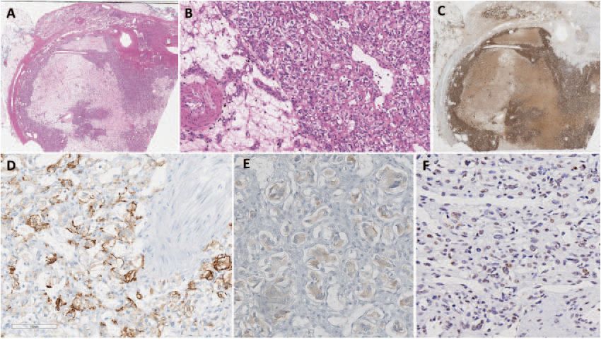
Fig. 1 Morphological and immunohistochemical findings of pheochromocytoma. Whole scanned images: The adrenalectomy specimen
(hematoxylin and eosin) shows an encapsulated pheochromocytoma with clear cell change, variable fibrohyaline, and myxoid stroma rich in
microvasculature (a, b). The tumor cells are diffusely positive for tyrosine hydroxylase (the rate limiting enzyme in catecholamine synthesis) (c).
The tumor is positive for carbonic anhydrase IX (d). Alpha-inhibin shows variable weak reactivity in the tumor cells (e). pVHL shows variable
loss or significantly reduced staining intensity in the tumor cells (f).
the mass on the left lung (measuring 3.1 × 1.8 cm) and noted a left family history, both daughters were enrolled in full VHL screening
renal lesion. A CT scan of the abdomen and pelvis revealed a large including ophthalmologic assessment, brain, spine, and abdominal
mass on the left kidney and right adrenal gland. An ultrasound MRI. They have no VHL manifestations at the ages of 25 and 30.
guided biopsy showed that the left kidney mass was a renal cell
carcinoma. The patient subsequently underwent a left-sided radical Morphological and immunohistochemical characteristics of
nephrectomy and right-sided adrenalectomy. The pathology results pheochromocytoma
confirmed ccRCC (measuring 12 × 11 × 9.5 cm) and identified a
The adrenal tumor tissue underwent detailed morphological
pheochromocytoma (measuring 6.7 × 5.8 × 5 cm). Biochemical eva-
assessment by an endocrine pathologist. The adrenal tumor was
luation of urine and plasma metanephrines and urine catechola-
an encapsulated pheochromocytoma with variable clear cell
mines were within normal limits. Two pieces of lung tissue orally
change and stromal degeneration with increased microvascula-
expelled from the patient further supported metastatic ccRCC. While
ture (Fig. 1a, b). For immunohistochemistry analysis, positivity for
spinal MRI and chest CT identified bone metastases, there were also
tyrosine hydroxylase (Fig. 1c) and GATA3 confirmed the diagnosis
two spinal lesions (measuring 2.2 mm and 14 mm) consistent with
of pheochromocytoma. The cytomorphological findings (e.g., clear
spinal hemangioblastomas. In addition, histologically confirmed liver
cell change, encapsulation with stromal changes) were highly
metastases from ccRCC were noted. The patient succumbed to his
suggestive of VHL-related pheochromocytoma20. While there was
disease 10 months after initial presentation. The patient’s family
no cyclinD1 overexpression, the tumor was positive for carbonic
history was unremarkable for VHL and no other VHL-associated
anhydrase IX (CAIX) and alpha-inhibin (variable and weak) (Fig. 1d,
manifestations were found on the patient’s subsequent imaging. A
1e). Alpha-inhibin expression21, along with CAIX expression21–23,
clinical diagnosis of Type 2B VHL was presumed, and blood was sent
for germline molecular genetic testing. was consistent with VHL-disease-related pseudohypoxia, espe-
cially in the background of morphological findings. Moreover,
variable loss of pVHL also supported an altered pseudohypoxia
Initial genetic testing results pathway in this tumor (Fig. 1f).
The patient underwent germline testing on DNA isolated from
peripheral blood leukocytes with a panel of genes targeted at Genetic testing of tumor specimens
hereditary renal cell cancer and a hereditary paraganglioma- Paraffin embedded tissue retrieved from the ccRCC, pheochro-
pheochromocytoma. The patient tested negative for any patho- mocytoma, expelled lung tissue (sputum), and liver biopsy were
genic, likely pathogenic, or uncertain DNA sequence or copy tested on a targeted hereditary cancer panel24. A likely pathogenic
number variants in FH, FLCN, MET, MITF, MLH1, MSH2, MSH6, PMS2, variant in exon 3 of VHL (NM_0000551.3: c.593 T > C; p.Leu198Pro)
PTEN, SDHA, SDHB, SDHC, SDHD, TP53, TSC1, TSC2, MAX, MEN1, NF1, was detected in all four tissues at varying VAFs (Fig. 2). This
RET, SDHAF2, TMEM127, and VHL. Variant allele threshold for missense variant is consistent with Type 2 VHL and has been
review on the germline panel was 10% or greater VAF. classified as likely pathogenic in the germline context25–27.
As a molecular genetic diagnosis of VHL was not identified, the Although no second hits in VHL were detected, copy number
daughters of the proband underwent germline molecular genetic calling revealed evidence of 3p loss-of-heterozygosity in all tumor
analysis of VHL and results were negative. However, given the tissues (kidney, adrenal, lung, and liver).
npj Genomic Medicine (2022) 21 Published in partnership with CEGMR, King Abdulaziz University
L.E. Oldfield et al.
                                                                                                                                                 3

Fig. 2 Visualization of VHL variant in tumor and germline DNA. Integrative Genomics Viewer display of the c.593 T > C (p.Leu198Pro) variant
in tissue from four tumor specimens (a) and peripheral blood leukocytes (b). The percentage of sequencing reads (gray bars) depicting the
variant are beside the biospecimen type. The blue bars depict a cytosine nucleotide instead of a thymine nucleotide at the variant loci. The
overall proportion of cytosine:thymine reads is represented by the blue:red bars at the top of each tissue panel. Tumor cellularity was
estimated by a pathologist to be 65% in the kidney tissue and 85% in the adrenal gland, lung, and liver tissue.

   The VHL p.Leu198Pro variant has been previously reported in          this was the expected lower VAF range for a germline hetero-
the germline of two families25–27 and tumor of a patient with           zygous variant on this panel. Variants present at reduced VAFs
sporadic pheochromocytoma28. In the first family, three members          may not be reported in all clinical labs. In addition, the lower limit
carried the variant and all three had bilateral pheochromocytomas       threshold for reporting variants is not a standard value for NGS
and one had an additional sympathetic paraganglioma25,27. A             panels whose intended use is to detect inherited germline variants
protein prediction research software (symphony) predicted that          expected to be heterozygous or homozygous. Communication
this missense variant affects a VHL surface protein residue25,29. In    with the testing laboratory, including clinical suspicions for a
the second family, one member presented with an early onset             specific genetic diagnosis, can be helpful in triggering re-
pancreatic neuroendocrine tumor, bilateral pheochromocytomas,           examination of low-level calls or raw data for potentially mosaic
and an optic nerve hemangioblastoma26.                                  results. In addition, negative genetic testing should not be
                                                                        considered as a definitive conclusion and further investigations,
Reanalysis of germline testing results                                  including of other tissue types, may be required to arrive at a
Re-examination of the NGS data from the proband’s initial               diagnostic molecular finding. Our VAF of the various affected
hereditary cancer testing identified the T allele at c.593 in the        tissues ranged from as low as 10% to as high as 55% despite
VHL gene in ~6% of the sequencing reads (77 reads of the total          adequate tumor cellularity. This variability may be due to tumor
1298 at this base pair position). This was below the typical            biology such as inflammatory cells causing normal tissue
reportable threshold for NGS multigene panel testing of 10%, and        contamination and other genomic events and is a reported
therefore was not initially reported in clinical genetic testing.       limitation of VAF from tumor sequencing30.
                                                                           Mosaicism results from the acquisition of a somatic mutation
                                                                        during embryogenesis, leading to variable mutant cell frequency
DISCUSSION                                                              throughout the body. While previously reported that individuals
Approximately 95–100% of individuals with clinical VHL receive a        with mosaic VHL are asymptomatic or present with a mild
positive result when they undergo standard genetic testing on DNA       phenotype13–15, Coppin et al. (2014) have reported a severe
extracted from blood or saliva. This case report explores the rare      phenotype in a mosaic VHL patient18. Here we presented an
event of an individual with typical VHL manifestations testing          individual with three VHL-associated manifestations, which
negative for a VHL variant on germline DNA extracted from peripheral    provides further evidence that mosaicism in VHL can present
blood leukocytes, with subsequent sequencing of four postmortem         with a severe phenotype. Cases of mosaicism in similar Mendelian
tumor tissue specimens revealing a VHL pathogenic variant in all four   conditions have been described previously with phenotypes less
tissues. Tumor analysis revealed protein markers consistent with VHL-   severe than might be expected from these autosomal dominant
driven tumorigenesis and pVHL. Reanalysis of germline genetic           conditions; however, given the incomplete penetrance and
testing results on the blood sample detected the variant at 6% VAF,     variable expressivity of these conditions themselves, it is difficult
confirming the strong likelihood of somatic mosaicism in this case.      to make specific phenotypic predictions31,32. A systematic review
   This case illustrates several factors to consider for conditions     of 111 articles reporting on 320 mosaic NF1 individuals found that
where somatic mosaicism is suspected based on the proband and           mosaic NF1 patients present with a milder phenotype and have
family history. Clinical genetic testing results are typically          fewer complications and manifestations31. A study on 39 Tuberous
anticipated to be categorical (wild-type, heterozygous, and             Sclerosis Complex patients with mosaicism (
L.E. Oldfield et al.
4
Notably, the two offspring of this proband had negative variants were filtered using Alissa software (Agilent, Santa Clara, CA) to
germline testing, which included analysis of VHL prior to the detect variants with a VAF > 10% and to remove benign variants. All
identification of this mosaic finding. The eventual identification of variants were manually reviewed in IGV (version 2.3)38. All genomic regions
a molecular cause for their father’s phenotype may have clinical achieved at least 25× coverage.
implications for these two daughters. However, given the findings
from this report, the risk of VHL disease to the daughters may be Immunohistochemistry
significantly decreased and may play a role in counseling them to Immunohistochemical staining for GATA3 (Biocare Medical, Pacjeco, CA;
continue to undergo intensive VHL surveillance. The current gold- catalog number: BC-CM405bl dilution: 1:100), tyrosine hydroxylase (Abcam,
standard for mosaic testing involves analysis of tissue from all Cambridge, UK; catalog number: ab75875-2; dilution: 1:500), cyclinD1 (Cell
three germ layers and may not be possible in many clinical Marque, Rocklin, CA; catalog number: CMQ-241R16; dilution: 1:250), alpha-
scenarios, including in rapidly deteriorating patients. This case inhibin (Cedarlane, Burlington, ON; catalog number: MCA951S; dilution:
1:100) and CAIX (Leica, Wetzlar, Germany; catalog number: CAIX-L-CE;
report shows the reality of mosaic testing and proves the value in dilution: 1:100) was performed in our clinical diagnostic immunohisto-
completing tissue testing on available tissue as the result provides chemistry laboratory using appropriate positive and negative controls.
valuable information that can be used to guide the surveillance pVHL immunohistochemistry was performed in an institutional research
and treatment of family members. laboratory according to the manufacturer’s instructions (OriGene, Rockville,
MD; catalog number: TA506222; dilution 1:500), along with appropriate
positive and negative controls.
METHODS
Ethics approval and clinical trial registration Reporting summary
Written informed consent for this study was obtained from the family of the Further information on research design is available in the Nature Research
proband following a clinical research protocol approved by the Research Ethics Reporting Summary linked to this article.
Board at University Health Network (#16-5831). This research study is registered
at clinicaltrials.gov (clinical trials registration number: NCT03857594).
DATA AVAILABILITY
Genetic analysis Tumor bam files from the four tissues that underwent targeted sequencing have
Genomic DNA extracted from peripheral blood leukocytes, and quantified been deposited in the European Genome-phenome Archive repository under
using a spectrophotometer, underwent two targeted NGS panels at a accession code EGAS00001005895. The germline data derived for this study are
commercial laboratory (Ambry Genetics Corporation, Aliso Viejo, California) not publicly available as they contain information that may compromise the research
for clinical testing. Ambry Genetics is a CLIA-certified, CAP accredited participant’s privacy but may be accessed by qualified researchers through R.H.K.
laboratory, which performs clinical genetic testing. The requested assays (raymond.kim@uhn.ca).
included NGS of all coding base pairs and 5 base pairs of intronic region
flanking the exons of 12 genes associated with hereditary renal cell cancer
and hereditary paraganglioma-pheochromocytoma predisposition. The CODE AVAILABILITY
genes and reference sequences used for analysis are: FH- NM_000143, No custom code was used to analyze the data and all tools used within the
MAX- NM_002382, MEN1-NM_130799.2, NF1- NM_001042492, RET- customized bioinformatics pipeline are included in the Methods section, with the
NM_020975, SDHA- NM_004168, SDHAF2-NM_017841, SDHB- version used.
NM_003000, SDHC- NM_003001, SDHD- NM_003002, TMEM127-
NM_017849, and VHL- NM_000551. Sequence enrichment of the targeted
Received: 8 July 2021; Accepted: 15 February 2022;
coding exons and adjacent intronic nucleotides was carried out by a bait-
capture methodology using long biotinylated oligonucleotide probes
followed by polymerase chain reaction and NGS. Sequence reads were
aligned to the reference human genome (GRCh37) using NovoAlign
(version 3.02.07; Novocraft Technologies, Selangor, Malaysia) and variant
calls generated using the Genome Analysis Toolkit (version 3.2.2; Broad REFERENCES
Institute, Cambridge, MA). Suspect variant calls, other than those classified 1. Kim, J. J., Rini, B. I. & Hansel, D. E. in Diseases of DNA Repair (ed. Ahmad, S. I.)
as “likely benign” or “benign”, were verified by Sanger sequencing. A 228–249 (Springer New York, 2010).
minimum coverage of 25×, Q score of 30 and VAF of >10% were required 2. Binderup, M. L. M. et al. Von Hippel-Lindau disease (vHL). National clinical
for candidate variants to pass quality control metrics for reporting. Any guideline for diagnosis and surveillance in Denmark. 3rd edition. Dan. Med. J. 60,
regions with an insufficient depth of coverage for heterozygous variant B4763 (2013).
calling were analyzed by Sanger sequencing. The sequence of copy 3. Maher, E. R. et al. Von Hippel-Lindau disease: a genetic study. J. Med. Genet. 28,
number variants which did not meet internal quality control metrics for 443–447 (1991).
confidence were confirmed by a secondary methodology, such as 4. Prowse, A. H. et al. Somatic inactivation of the VHL gene in Von Hippel-Lindau
chromosomal microarray or Sanger sequencing, prior to reporting. disease tumors. Am. J. Hum. Genet. 60, 765–771 (1997).
Paraffin embedded clinical blocks were retrieved postmortem and DNA 5. Maher, E. R., Neumann, H. P. & Richard, S. von Hippel-Lindau disease: a clinical
from four separate tissues (ccRCC, pheochromocytoma, expelled lung and scientific review. Eur. J. Hum. Genet. 19, 617–623 (2011).
tissue, and liver biopsy). The tissue underwent pathology review by 6. Cho, H.-J., Ki, C.-S. & Kim, J.-W. Improved detection of germline mutations in
experienced clinical pathologists who determined histology and tumor Korean VHL patients by multiple ligation-dependent probe amplification analysis.
cellularity. DNA was extracted from the four tissues and used in 59-gene J. Korean Med. Sci. 24, 77–83 (2009).
NGS panel (Clinical Genome Diagnostics Laboratory, University Health 7. Stolle, C. et al. Improved detection of germline mutations in the von Hippel-
Network, Toronto, Ontario) for detection of pathogenic variants associated Lindau disease tumor suppressor gene. Hum. Mutat. 12, 417–423 (1998).
with hereditary cancer syndromes, using methods previously described for 8. Hes, F. J. et al. Frequency of Von Hippel-Lindau germline mutations in classic and
analysis of tumor DNA from FFPE material24. For variant analysis, a custom non-classic Von Hippel-Lindau disease identified by DNA sequencing, Southern
bioinformatic pipeline was used with targeted sequence reads aligned to blot analysis and multiplex ligation-dependent probe amplification. Clin. Genet.
the reference human genome (hg19) using Burrows-Wheeler Aligner 72, 122–129 (2007).
(version 0.7.12)33. Duplicate reads were marked (Picard Mark Duplicates; 9. Nordstrom-O’Brien, M. et al. Genetic analysis of von Hippel-Lindau disease. Hum.
version 1.130) and the Genome Analysis Toolkit (version 3.3.0; Broad Mutat. 31, 521–537 (2010).
Institute, Cambridge, MA) best practices followed Base Quality Score 10. Binderup, M. L. M., Galanakis, M., Budtz-Jørgensen, E., Kosteljanetz, M. & Luise
Recalibration. Somatic single nucleotide variants and small insertion/ Bisgaard, M. Prevalence, birth incidence, and penetrance of von Hippel-Lindau
deletions were detected by Varscan2 (version 2.3.8) and copy number disease (vHL) in Denmark. Eur. J. Hum. Genet. 25, 301–307 (2017).
status was determined by CNVkit (version 0.7.11)34,35. Loss-of- 11. Ricketts, C. J. et al. A germline 1;3 translocation disrupting the VHL gene: a novel
heterozygosity was detected using pureCN (version 1.12.0) in R (version genetic cause for von Hippel-Lindau. J. Med. Genet. https://doi.org/10.1136/
3.5.0), with input data from CNVkit and MuTect (version 1.1.5)36,37. All jmedgenet-2020-107308 (2020).
npj Genomic Medicine (2022) 21 Published in partnership with CEGMR, King Abdulaziz UniversityL.E. Oldfield et al.
5
12. Lenglet, M. et al. Identification of a new VHL exon and complex splicing alterations 37. Cibulskis, K. et al. Sensitive detection of somatic point mutations in impure and
in familial erythrocytosis or von Hippel-Lindau disease. Blood 132, 469–483 (2018). heterogeneous cancer samples. Nat. Biotechnol. 31, 213 (2013).
13. Wu, P. et al. Mosaicism in von Hippel-Lindau disease with severe renal mani- 38. Thorvaldsdóttir, H., Robinson, J. T. & Mesirov, J. P. Integrative Genomics Viewer
festations. Clin. Genet. 84, 581–584 (2013). (IGV): high-performance genomics data visualization and exploration. Brief.
14. Murgia, A. et al. Somatic mosaicism in von Hippel-Lindau Disease. Hum. Mutat. Bioinform. 14, 178–192 (2013).
15, 114 (2000).
15. Santarpia, L. et al. Mosaicism in von Hippel-Lindau disease: an event important to
recognize. J. Cell. Mol. Med. 11, 1408–1415 (2007). ACKNOWLEDGEMENTS
16. Sgambati, M. T. et al. Mosaicism in von Hippel-Lindau disease: lessons from We would like to thank the patient and their family for allowing us to complete this
kindreds with germline mutations identified in offspring with mosaic parents. study. In addition, we would like to thank the Princess Margaret Applied Molecular
Am. J. Hum. Genet. 66, 84–91 (2000). Profiling—Drug Development Biomarker lab, Ming Tsao and Jing Xu for completing
17. Coppin, L. et al. Optimization of next-generation sequencing technologies for the pVHL immunohistochemical staining. Thank you to Charlotte Fung and Karen Ott
von Hippel Lindau (VHL) mosaic mutation detection and development of con- for reviewing the manuscript. This study was funded by the University of Toronto
firmation methods. J. Mol. Diagn. 21, 462–470 (2019). Division of Medical Oncology Strategic Innovation Fund and the Soper Kidney Cancer
18. Coppin, L. et al. VHL mosaicism can be detected by clinical next-generation Foundation. R.H.K. is supported by the Bhalwani Family Charitable Foundation.
sequencing and is not restricted to patients with a mild phenotype. Eur. J. Hum.
Genet. 22, 1149–1152 (2014).
19. Gajecka, M. Unrevealed mosaicism in the next-generation sequencing era. Mol.
AUTHOR CONTRIBUTIONS
Genet. Genomics 291, 513–530 (2016).
20. Mete O., Hannah-Shmouni F., Kim R., Stratakis C. A. in The Spectrum of Neu- R.H.K. devised the study, secured funding, and supervised the study. L.E.O. completed
roendocrine Neoplasia (eds. Asa, S. L., Rosa, S. L., Mete, O.) 409–459 (Springer, 2020). the analysis with input from J.G., S.G., T.S., and R.H.K. O.M. completed the
21. Mete, O., Pakbaz, S., Lerario, A. M., Giordano, T. J. & Asa, S. L. Significance of alpha- immunohistochemical staining and interpretation. L.E.O. and R.H.K. wrote the manu-
inhibin expression in pheochromocytomas and paragangliomas. Am. J. Surg. script with support from all authors. All authors read and approved the manuscript.
Pathol. https://doi.org/10.1097/PAS.0000000000001715 (2021).
22. Papathomas, T. G. et al. What have we learned from molecular biology of para-
gangliomas and pheochromocytomas? Endocr. Pathol. 32, 134–153 (2021). COMPETING INTERESTS
23. Juhlin, C. C. Challenges in paragangliomas and pheochromocytomas: from his- J.G. and E.C. are employed by Ambry Genetics. The remaining authors declare no
tology to molecular immunohistochemistry. Endocr. Pathol. https://doi.org/ competing interests.
10.1007/s12022-021-09675-0 (2021).
24. Care, M. et al. Tumor and germline next generation sequencing in high grade
serous cancer: experience from a large population-based testing program. Mol.
Oncol. https://doi.org/10.1002/1878-0261.12817 (2020). ADDITIONAL INFORMATION
25. Lomte, N. et al. Genotype phenotype correlation in Asian Indian von Hippel- Supplementary information The online version contains supplementary material
Lindau (VHL) syndrome patients with pheochromocytoma/paraganglioma. Fam. available at https://doi.org/10.1038/s41525-022-00291-3.
Cancer 17, 441–449 (2018).
26. Wittström, E., Nordling, M. & Andréasson, S. Genotype-phenotype correlations, Correspondence and requests for materials should be addressed to Raymond H. Kim.
and retinal function and structure in von Hippel-Lindau disease. Ophthalmic
Genet. 35, 91–106 (2014). Reprints and permission information is available at http://www.nature.com/
27. Pandit, R. et al. Germline mutations and genotype-phenotype correlation in Asian reprints
Indian patients with pheochromocytoma and paraganglioma. Eur. J. Endocrinol.
175, 311–323 (2016). Publisher’s note Springer Nature remains neutral with regard to jurisdictional claims
28. Dannenberg, H. et al. Von Hippel-Lindau gene alterations in sporadic benign and in published maps and institutional affiliations.
malignant pheochromocytomas. Int. J. Cancer 105, 190–195 (2003).
29. Gossage, L. et al. An integrated computational approach can classify VHL mis-
sense mutations according to risk of clear cell renal carcinoma. Hum. Mol. Genet.
23, 5976–5988 (2014).
30. Strom, S. P. Current practices and guidelines for clinical next-generation Open Access This article is licensed under a Creative Commons
sequencing oncology testing. Cancer Biol. Med. 13, 3–11 (2016). Attribution 4.0 International License, which permits use, sharing,
31. García-Romero, M. T., Parkin, P. & Lara-Corrales, I. Mosaic neurofibromatosis type adaptation, distribution and reproduction in any medium or format, as long as you give
1: a systematic review. Pediatr. Dermatol. 33, 9–17 (2016). appropriate credit to the original author(s) and the source, provide a link to the Creative
32. Giannikou, K. et al. Low-level mosaicism in tuberous sclerosis complex: prevalence, Commons license, and indicate if changes were made. The images or other third party
clinical features, and risk of disease transmission. Genet. Med. 21, 2639–2643 (2019). material in this article are included in the article’s Creative Commons license, unless
33. Li, H. & Durbin, R. Fast and accurate short read alignment with Burrows–Wheeler indicated otherwise in a credit line to the material. If material is not included in the
transform. Bioinformatics 25, 1754–1760 (2009). article’s Creative Commons license and your intended use is not permitted by statutory
34. Koboldt, D. C. et al. VarScan 2: somatic mutation and copy number alteration regulation or exceeds the permitted use, you will need to obtain permission directly
discovery in cancer by exome sequencing. Genome Res. 22, 568–576 (2012). from the copyright holder. To view a copy of this license, visit http://creativecommons.
35. Talevich, E., Hunter Shain, A., Botton, T. & Bastian, B. C. CNVkit: Genome-wide org/licenses/by/4.0/.
copy number detection and visualization from targeted DNA sequencing. PLoS
Comput. Biol. 12, e1004873 (2016).
36. Riester, M. et al. PureCN: copy number calling and SNV classification using tar- © The Author(s) 2022
geted short read sequencing. Source Code Biol. Med. 11, 13 (2016).
Published in partnership with CEGMR, King Abdulaziz University npj Genomic Medicine (2022) 21You can also read