Case Report: End-Stage Recurrent Glioblastoma Treated With a New Noninvasive Non-Contact Oncomagnetic Device - Frontiers
←
→
Page content transcription
If your browser does not render page correctly, please read the page content below
PERSPECTIVE
                                                                                                                                                 published: 22 July 2021
                                                                                                                                         doi: 10.3389/fonc.2021.708017

                                              Case Report: End-Stage Recurrent
                                              Glioblastoma Treated With a New
                                              Noninvasive Non-Contact
                                              Oncomagnetic Device
                                              David S. Baskin 1,2,3*, Martyn A. Sharpe 1,2, Lisa Nguyen 1,2 and Santosh A. Helekar 1,2,3
                                              1Kenneth R. Peak Brain and Pituitary Tumor Treatment Center, Department of Neurosurgery, Houston Methodist
                                              Neurological Institute, Houston, TX, United States, 2 Department of Neurosurgery, Houston Methodist Research Institute,
                                              Houston, TX, United States, 3 Department of Neurosurgery, Weill Cornell Medical College, New York, NY, United States

                                              Alternating electric field therapy has been approved for glioblastoma (GBM). We have
                                              preclinical evidence for anticancer effects in GBM cell cultures and mouse xenografts with
                                              an oscillating magnetic field (OMF) generating device. Here we report OMF treatment of
                                              end-stage recurrent glioblastoma in a 53-year-old man who had undergone radical
                        Edited by:
                  David Nathanson,
                                              surgical excision and chemoradiotherapy, and experimental gene therapy for a left
        UCLA David Geffen School of           frontal tumor. He experienced tumor recurrence and progressive enlargement with
            Medicine, United States           leptomeningeal involvement. OMF for 5 weeks was well tolerated, with 31% reduction
                      Reviewed by:            of contrast-enhanced tumor volume and reduction in abnormal T2-weighted Fluid-
                     Peter LaViolette,
        Medical College of Wisconsin,         Attenuated Inversion Recovery volume. Tumor shrinkage appeared to correlate with
                        United States         treatment dose. These findings suggest a powerful new noninvasive therapy
                Kenneth D. Swanson,
Beth Israel Deaconess Medical Center
                                              for glioblastoma.
         and Harvard Medical School,
                                              Keywords: magnetic resonance imaging, contrast enhanced tumor, compassionate use treatment, radiation-type
                        United States
                                              tumor necrosis 2, oscillating magnetic fields
                *Correspondence:
                    David S. Baskin
      dbaskin@houstonmethodist.org
                                              INTRODUCTION
                   Specialty section:
         This article was submitted to        For glioblastoma (GBM), the most common malignant tumor of the brain in adults, treatment
                  Neuro-Oncology and          outcome remains dismal. In over 40 years median survival has only shown modest improvement
             Neurosurgical Oncology,          (1), and standard of care treatment often has negative impact on quality of life (2). Treatment
               a section of the journal       including radiation and chemotherapy takes a heavy toll. Frequently patients cannot tolerate the
                 Frontiers in Oncology        completion of the prescribed chemotherapy cycles. Thus, there is a great unmet need for a
             Received: 11 May 2021            completely different therapeutic approach with better outcome and less toxicity.
            Accepted: 21 June 2021               A new FDA-approved treatment involving electric fields alternating at 200 kHz called Optune™
            Published: 22 July 2021           therapy is now available for recurrent GBM as monotherapy and in combination with
                            Citation:         temozolomide for newly diagnosed GBM (3, 4). It is also being tested in clinical trials for other
Baskin DS, Sharpe MA, Nguyen L and            cancers. Its hypothesized mechanism of action involves disruption of tubulin dimers, mitotic
Helekar SA (2021) Case Report: End-
                                              spindles, and cell division by electric field-induced dipole alignment and dielectrophoresis (5). It has
Stage Recurrent Glioblastoma Treated
             With a New Noninvasive
                                              a modest effect on survival, increasing median overall survival by 0.6 month in recurrent GBM (3),
 Non-Contact Oncomagnetic Device.             and in newly diagnosed GBM by 31% (4). Even this modest effect is encouraging for patients.
            Front. Oncol. 11:708017.             It has been shown that electromagnetic fields (EMF) produce anticancer effects in vitro (6, 7). We
     doi: 10.3389/fonc.2021.708017            have conducted preclinical experiments with a new noninvasive wearable device known as an

Frontiers in Oncology | www.frontiersin.org                                          1                                           July 2021 | Volume 11 | Article 708017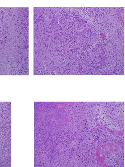
Helekar et al. Oncomagnetic Treatment of Recurrent Glioblastoma
Oncomagnetic device that generates oscillating magnetic fields care, he received concomitant radiation therapy and chemotherapy
(OMF) by rotating strong permanent magnets (8, 9). The OMF with temozolomide.
generating components (oncoscillators) of the device can be In August 2019, the patient presented with an area of contrast
attached to a helmet and treatment with the device does not enhancement on MRI scan along the left ventricle. At first this was
require shaving the head. Using the oncoscillators of the device thought to be a treatment effect. This area progressively enlarged.
and specially devised patterns of magnet rotations we have Evaluations done before OMF treatment initiation on January 16,
produced strong selective anticancer effects in patient derived March 3, and April 15, 2020, demonstrated a clear recurrence. The
GBM and xenografted mouse models without causing adverse tumor abutted the ventricle and there was evidence of
effects on cultured normal cells and normal mice (10–12). The leptomeningeal spread. The patient had already had radiation
mechanism of action of OMF differs from Optune™ and therapy and chemotherapy and the tumor was now progressing.
involves disruption of the electron transport in the The presence of leptomeningeal disease portends poor outcome,
mitochondrial respiratory chain causing elevation of reactive with median survival of 3.5 to 3.9 months (13).
oxygen species and caspase-dependent cancer cell death (10–12). Because of inadequacy of any standard of care options he was
Here we report evidence of treatment response in the first enrolled in an FDA-approved Expanded Access Program (EAP)
patient to ever receive this therapy with an untreatable left for compassionate use treatment with the Oncomagnetic device.
frontal GBM, treated with a wearable Oncomagnetic device in He signed an informed consent on April 15, 2020. The EAP
an FDA-approved Expanded Access Program. study was carried out under a protocol approved by the Houston
Methodist Research Institute Institutional Review Board.
METHODS Oncomagnetic Device
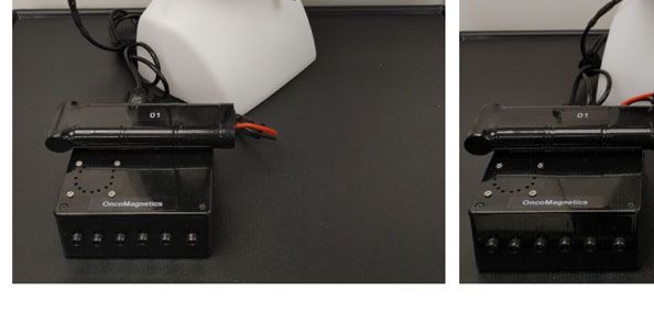
The Oncomagnetic device consists of 3 oncoscillators securely
Case Description attached to an acrylonitrile butadiene styrene helmet and
The patient is a 53-year-old man who first presented with altered connected to a microprocessor-based electronic controller
mental status in May 2018. Imaging studies documented a large operated by a rechargeable battery (Figure 1). Further details
tumor in the left frontal lobe extending across the midline into the regarding the device are given in the Supplementary Appendix.
right frontal lobe, with diffuse and extensive infiltration through the Based on a finite element model-based calculation of the spread of
corpus callosum. There was mass effect and severe edema. He was the field and the size and magnetization of the rotated diametrically
taken to the operating room on June 4, 2018, where he underwent magnetized neodymium magnets, we estimated that the combined
left frontal craniotomy and radical excision of the tumor. The tumor effective field (at least 1 mT in strength) of the 3 oncoscillators
was histopathologically confirmed as GBM. At the time of the covered the entire brain, including the upper part of the brain stem.
surgery, the excision extended across the midline into the right
frontal lobe. He was enrolled in a herpes simplex virus-thymidine Oscillating Magnetic Field Treatment
kinase gene therapy program and received viral injection during The treatment consists of intermittent application of an OMF
surgery per protocol. In addition, per protocol, and as standard of that needs to be generated by rotating permanent magnets in a
A B
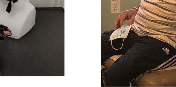
FIGURE 1 | Oncomagnetic Device. (A) Device helmet with 3 oncoscillators securely attached to it. The oncoscillators are connected to a controller box powered by
a rechargeable battery. (B) The patient wearing the device helmet with three oncoscillators attached.
Frontiers in Oncology | www.frontiersin.org 2 July 2021 | Volume 11 | Article 708017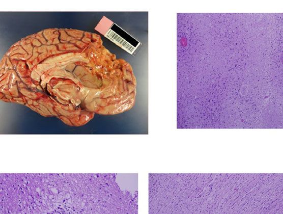
Helekar et al.                                                                                    Oncomagnetic Treatment of Recurrent Glioblastoma

specific frequency profile and timing pattern to be effective. The          probability of a decrease at each post-treatment initiation
patient received this treatment initially in the Peak Center clinic       time point.
under the supervision of the treating physician and the Principal
Investigator (DSB) of this study for the first 3 days. The dose was
escalated over this period as follows. On the first day, the
treatment was for 2 hours with a 5-min break between the first
                                                                          RESULTS
and the second hour. On the second and third days, it was                 The patient received OMF treatment with the Oncomagnetic
increased to 2 and 3 2-hour sessions, respectively, with 1-hour           device for 36 days. The treatment regimen was changed at
breaks between the sessions. The patient’s spouse was trained in          various times during this period based on the caregiver reports
the use and care of the device on these days. After this initial          and clinical findings, as described below.
supervised phase, the treatment was continued at home
unsupervised with the same regimen as on the third day,                   Clinical Findings
above. The spouse was instructed to maintain a daily log of the           After the initial 3 days of supervised treatment, the patient was
conduct and progress of treatment, and any observed treatment             seen again by the treating physician in the outpatient clinic on
and adverse effects.                                                      Day 7 from the start of treatment. Because of inattention at
                                                                          baseline, the patient was having difficulty with the length of
Clinical Evaluations and Neuroimaging                                     treatment sessions. They were reduced to 2 hours/day Monday
The patient was evaluated clinically by the treating physician on         through Friday with Saturday and Sunday off. The Day 16
each of the 3 days that he received treatment in the clinic and 7,        clinical examination revealed that he was tolerating the
16, 30 and 44 days after initiation of treatment. Magnetic                treatment sessions well, so they were increased to a total of 3
Resonance Imaging (MRI) scans were done on Days 1, 3, 7, 16,              hours/day (in one-hour increments with 5 min breaks) Monday
30 and 44. The Day 1 scan was done before initiation of                   through Friday and the weekends off. On Day 30 visit, the patient
treatment. All other scans were done after treatment initiation.          reported headaches related to transient hypertension for which
The treatment was paused on Day 37 because of an unfortunate              he was taking medication. The treating physician increased blood
but unrelated severe closed head injury (CHI). MRI scans were             pressure medication (Valsartan) with improvement. The
done on a Siemens Magnetom Terra 7T scanner. MRI scans                    treatment was paused on Day 36 because of a closed head
included T1 magnetization prepared rapid gradient echo scans              injury from a fall. Whether the fall was related to the
with and without gadolinium contrast, and T2-weighted Fluid-              treatment in any way is uncertain. It is worth noting, however,
Attenuated Inversion Recovery (FLAIR), T2-weighted Turbo                  that the patient had experienced several falls before initiation of
Spin Echo, Diffusion Weighted Imaging, Susceptibility                     treatment. At the last follow-up on Day 44 the patient was
Weighted Imaging, proton Magnetic Resonance spectroscopy                  admitted to the inpatient unit for evaluation of closed head
and Diffusion Tensor Imaging scans. Treatment effect on                   injury and underwent detailed assessment. There were no serious
contrast-enhanced tumor (CET) was evaluated according to the              adverse events reported during treatment. The patient’s
response assessment in neuro-oncology (RANO) criteria for                 caregivers reported subjective improvement in speech and
clinical trials (14). In addition, an automated software-based            cognitive function.
method developed in house was used to objectively calculate the
CET volume (see below and Supplementary Appendix).                        MRI Findings
                                                                          Evaluation of the T1 post-contrast clinical MRI scans obtained
Data Analysis                                                             before initiation of treatment showed progression in accordance
Post-contrast T1 anatomical and T2-FLAIR MRI scans at each of             with the RANO criteria (Figure 2A). All scans acquired during
the 6 time points were used to determine changes in contrast-             treatment showed stable disease, according to these criteria
enhanced tumor (CET) volume and non-enhanced tumor                        (Figure 2A). To obtain an objective quantitative assessment of
infiltration, respectively, before and after initiation of                 the CET volume we used an automated MATLAB software-
treatmen t. Inf ormation on image processin g, d ata                      based script. This analysis showed marked changes in CET
normalization and plotting are given in the Supplementary                 volume with treatment. Figure 2B shows a plot of the CET
Appendix. Values obtained from pre-treatment clinical scans               volume as a function of time before and after initiation of
taken at 2 time points over 3 months before enrollment of the             treatment. It reveals that there was substantial growth of the
patient were also plotted on the same graph. Because this is a            tumor volume over the 3 months before the treatment. Within
single patient case report, we could not perform any meaningful           the first 3 days of treatment the trend is reversed with the volume
statistical analysis. However, to obtain a semi-quantitative              steeply decreasing by ~10% on Day 7 and then less steeply by
assessment of the significance of the trend seen with treatment,           31% on Day 30. Based on a Bayesian-type assessment of the
we analyzed the changes in CET volume using Bayesian logic,               probability of a decrease in CET volume at each post-treatment
given the observed increasing trend at two pre-treatment time             initiation time point, the decrease at Day 30 is statistically
points. Accordingly, we assumed that the chance of increase,              significant at P = 0.036. The treatment was paused on Day 37.
decrease and no change in the rate of tumor growth was the same           After the pause we see another trend reversal and an increase in
at each time point after treatment initiation to calculate the            CET volume on Day 44.

Frontiers in Oncology | www.frontiersin.org                           3                                     July 2021 | Volume 11 | Article 708017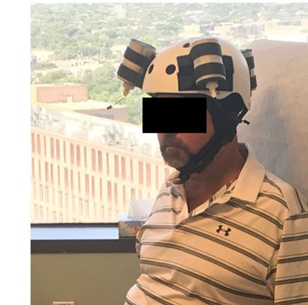
Helekar et al. Oncomagnetic Treatment of Recurrent Glioblastoma
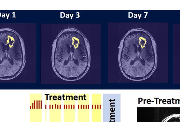
A
B
FIGURE 2 | Change in Contrast-Enhanced Tumor Volume. (A) T1-weighted axial post-contrast scans showing the contrast-enhanced tumor (CET) highlighted
with an overlayed automated computer program-generated light-yellow mask at different time points (B) Left – A graph showing the change in CET volume over
time. The treatment times and durations are shown as red bars and light-yellow highlights. The long pause in treatment is shown as a light-blue highlight. Right –
T1-weighted axial post-contrast scans showing CET at two levels along the dorso-ventral axis at Day 1 before treatment and Day 30 of treatment.
A B C
D E
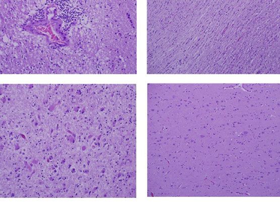
FIGURE 3 | Variation in Enhanced Intensity Volumes in T2-FLAIR MRI Scans and Autopsy Findings. (A) Top – Bar plots of the volumes of T2-FLAIR intensity
enhancement in the whole brain at different time points. Overall, there was up to 11% decrease in T2 FLAIR volume over the course of treatment. Bottom –
Representative T2-FLAIR images are shown. (B) Left hemisphere of the brain, examined grossly, showing no tumor mass. (C) Photomicrographs of the left cortex
showing bland necrosis, residual tumor, and microvascular proliferation with thick-walled vessels. (D) Top left – Microscopic field of the left cingulate cortex showing
a focus of rarefied, perivascular inflammation. Bottom left – Cortical field showing rarefied parenchyma and residual tumor cells, enlarged with treatment-type effect
that can be seen in GBM. Top right – Micrographic field of the corpus callosum showing thinned, rarefied white matter tract. Bottom right – Field showing relatively
uninvolved contralateral (right) cortex. (E) Top – Micrographic field in the left cortex showing infarct-like necrosis (left), tumor (right), and fibrin thrombus (lower right).
Bottom – Left cortical field showing necrotic tissue with dystrophic calcification.
Frontiers in Oncology | www.frontiersin.org 4 July 2021 | Volume 11 | Article 708017
Helekar et al.                                                                                   Oncomagnetic Treatment of Recurrent Glioblastoma

    The T2-FLAIR data in Figure 3A show changes in enhanced              II trials have shown anti-tumor efficacy (19, 20), a subsequent
intensity volume of 1 – 11% over time. The decreases in volume           Phase III trial did not show a significant increase in overall
are greater after a 3-day pause in treatment on Day 7 and after an       survival (21–23).
8-day pause on Day 44. These decreases are likely due to
reduction in treatment-related cerebral edema and/or
reduction in non-contrast enhancing tumor infiltration. The
patient died ~3 months after cessation of treatment from the
                                                                         CONCLUSION
CHI. A brain only autopsy showed a resection cavity in the left          Noninvasive Oncomagnetic device based OMF therapy appears
frontal lobe (6.0 x 5.0 x 3.5 cm) and recurrent/residual                 to be a safe and efficacious new modality of treatment against
glioblastoma with associated treatment effect (see Figures 3B–E).        GBM that potentially has many advantages over existing
Residual/recurrent high-grade glioma was present, including foci         treatments. The present report has the limitation of the
of densely cellular tumor, focal microvascular proliferation, and        treatment being conducted in only a single patient so far.
necrosis (Figure 3C). In addition, there was prominent                   Extending it to more patients in research studies would
treatment effect with pallor and rarefaction of white matter             provide additional information regarding safety and efficacy.
(Figure 3D), reactive astrocytosis, infarct-like necrosis
(Figure 3E) and bizarre nuclear atypia within residual tumor
cells. Additional features of treatment effect included dystrophic
calcifications (Figure 3E).                                               DATA AVAILABILITY STATEMENT
                                                                         The original contributions presented in the study are included in
                                                                         the article/Supplementary Material. Further inquiries can be
DISCUSSION                                                               directed to the corresponding author.

The findings of this study indicate that Oncomagnetic device-
based OMF therapy is well tolerated by a patient who has end-
stage recurrent GBM with leptomeningeal involvement and has              ETHICS STATEMENT
no other available effective treatment options. They also
                                                                         The studies involving human participants were reviewed and
demonstrate a clinically significant reduction in CET volume
                                                                         approved by Houston Methodist Research Institute Institutional
with reductions in non-enhanced tumor volume and/or edema
                                                                         Review Board. The patient/participant provided their written
in T2-FLAIR scans. The temporal profile of changes in CET
                                                                         informed consent to participate in this study. Written informed
volume also suggests a correlation with the treatment dose and
                                                                         consent was obtained from the individual for the publication of
the presence or absence of treatment. When the treatment dose
                                                                         any potentially identifiable images or data included in this article.
was higher (6 hours/day for 4 days) we see a tumor volume
reduction rate of 2.32 cm3/day. When it was lower (2 hours/day
for 9 days and 3 hours/day for 18 days) the reduction is 1.03 cm3/
day. Moreover, when the treatment was paused for 8 days the              AUTHOR CONTRIBUTIONS
decreasing trend reversed and the CET volume increased,
instead. Assuming that the ~1.03 cm3/day decreasing trend                SH and DB designed the study and drafted the manuscript. SH
had continued until the treatment was paused, we can estimate            designed the device used in the study, supervised its construction
that the CET volume grew at the rate of 1.26 cm3/day during the          and testing and quantitively analyzed the imaging data. DB
pause. Despite the apparent correlation it is possible that the          provided medical care to the study subject, supervised the
treatment response is independent of the short-term changes in           delivery of device treatment, and conducted his clinical
the treatment dose.                                                      assessments. SH, MS, and DB designed the device treatment
    To our knowledge, there is no report in the literature of a          protocol and interpreted the findings. LN constructed and tested
noninvasive treatment-related shrinkage of CET volume of GBM             the device and provided device treatment to the study subject. All
at a rate comparable to that seen in this study. One published           authors contributed to the article and approved the
report on Optune™ therapy has reported that the time course of           submitted version.
change in tumor volume in MRI scans shows a ~15% reduction
over ~3 months (15). Besides Optune™, the other type of
treatment approved by the FDA and recommended as a                       FUNDING
standard in National Comprehensive Cancer Network
guidelines for recurrent GBM is the anti-vascular endothelial            This work was supported by a grant from the Translational
growth factor (VEGF) monoclonal antibody, Bevacizumab (16,               Research Initiative of the Houston Methodist Research Institute
17). Bevacizumab treatment response of reduction in tumor                to SH and DB, and by Donna and Kenneth Peak, the Kenneth R.
volume on MRI scans has been reported to be lower than is                Peak Foundation, the John S. Dunn Foundation, the Taub
observed in the present study (18). Furthermore, while anti-             Foundation, the Blanche Green Fund of the Pauline Sterne
VEGF drugs in general have mild toxicity profiles and two Phase           Wolff Memorial Foundation, the Kelly Kicking Cancer

Frontiers in Oncology | www.frontiersin.org                          5                                     July 2021 | Volume 11 | Article 708017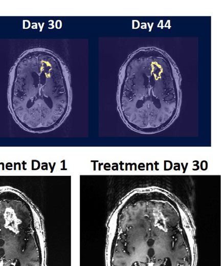
Helekar et al.                                                                                                           Oncomagnetic Treatment of Recurrent Glioblastoma

Foundation, the Gary and Marlee Swarz Foundation, the                                      Cykowski, MD, Department of Pathology and Genomic
Methodist Hospital Foundation, and the Veralan Foundation.                                 Medicine, who provided pathologic description and images.
The John S. Dunn Foundation also supports the Distinguished                                We thank Blessy S. John and Alvin Saldon for aiding in
Professorship of MS.                                                                       device construction.

ACKNOWLEDGMENTS                                                                            SUPPLEMENTARY MATERIAL
The authors thank the patient for graciously volunteering to be a                          The Supplementary Material for this article can be found online at:
research subject in this study and the rest of his family for                              https://www.frontiersin.org/articles/10.3389/fonc.2021.708017/
supporting him. We appreciate the assistance of Dr. Matthew                                full#supplementary-material

                                                                                                 Cancer Center Experience. Neurology (2019) 92:e2483–91. doi: 10.1212/
REFERENCES                                                                                       WNL.0000000000007529
 1. Stupp R, Mason WP, van den Bent MJ, Weller M, Fisher B, Taphoorn MJ,                   14.   Wen PY, Chang SM, Van den Bent MJ, Vogelbaum MA, Macdonald DR, Lee
    et al. Radiotherapy Plus Concomitant and Adjuvant Temozolomide for                           EQ. Response Assessment in Neuro-Oncology Clinical Trials. J Clin Oncol
    Glioblastoma. N Engl J Med (2005) 352:987–96. doi: 10.1056/NEJMoa043330                      (2017) 35:2439–49. doi: 10.1200/JCO.2017.72.7511
 2. Henriksson R, Asklund T, Poulsen HS. Impact of Therapy on Quality of Life,             15.   Robins HI, Nguyen HN, Field A, Howard S, Salamat S, Deming DA.
    Neurocognitive Function and Their Correlates in Glioblastoma Multiforme:                     Molecular Evolution of a Glioblastoma Controlled With Tumor Treating
    A Review. J Neurooncol (2011) 104:639–46. doi: 10.1007/s11060-011-0565-x                     Fields and Concomitant Temozolomide. Front Oncol (2018) 8:451. doi:
 3. Stupp R, Wong ET, Kanner AA, Steinberg D, Engelhard H, Heidecke V, et al.                    10.3389/fonc.2018.00451
    NovoTTF-100A Versus Physician’s Choice Chemotherapy in Recurrent                       16.   Kreisl TN, Zhang W, Odia Y, Shih JH, Butman JA, Hammoud D, et al. A Phase II
    Glioblastoma: A Randomised Phase III Trial of a Novel Treatment                              Trial of Single-Agent Bevacizumab in Patients With Recurrent Anaplastic
    Modality. Eur J Cancer (2012) 48:2192–202. doi: 10.1016/j.ejca.2012.04.011                   Glioma. Neuro Oncol (2011) 13:1143–50. doi: 10.1093/neuonc/nor091
 4. Stupp R, Taillibert S, Kanner A, Read W, Steinberg D, Lhermitte B, et al. Effect       17.   Friedman HS, Prados MD, Wen PY, Mikkelsen T, Schiff D, Abrey LE, et al.
    of Tumor-Treating Fields Plus Maintenance Temozolomide vs Maintenance                        Bevacizumab Alone and in Combination With Irinotecan in Recurrent
    Temozolomide Alone on Survival in Patients With Glioblastoma: A                              Glioblastoma. J Clin Oncol (2009) 27:4733–40. doi: 10.1200/JCO.2008.19.8721
    Randomized Clinical Trial. JAMA (2017) 318:2306–16. doi: 10.1001/                      18.   Daniels D, Guez D, Last D, Hoffmann C, Nass D, Talianski A, et al. Early
    jama.2017.18718                                                                              Biomarkers From Conventional and Delayed-Contrast Mri to Predict the
 5. Tuszynski JA, Wenger C, Friesen DE, Preto J. An Overview of Sub-Cellular                     Response to Bevacizumab in Recurrent High-Grade Gliomas. AJNR Am J
    Mechanisms Involved in the Action of TTFields. Int J Environ Res Public                      Neuroradiol (2016) 37:2003–9. doi: 10.3174/ajnr.A4866
    Health 13 (2016) 13:1–23. doi: 10.3390/ijerph13111128                                  19.   Vredenburgh JJ, Desjardins A, Herndon JE2nd, Marcello J, Reardon DA,
 6. Saliev T, Begimbetova D, Masoud AR, Matkarimov B. Biological Effects of                      Quinn JA, et al. Bevacizumab Plus Irinotecan in Recurrent Glioblastoma
    non-Ionizing Electromagnetic Fields: Two Sides of a Coin. Prog Biophys Mol                   Multiforme. J Clin Oncol (2007) 25:4722–9. doi: 10.1200/JCO.2007.12.2440
    Biol (2019) 141:25–36. doi: 10.1016/j.pbiomolbio.2018.07.009                           20.   Vredenburgh JJ, Desjardins A, Herndon JE2nd, Dowell JM, Reardon DA,
 7. Jimenez H, Blackman C, Lesser G, Debinski W, Chan M, Sharma S, et al. Use                    Quinn JA, et al. Phase II Trial of Bevacizumab and Irinotecan in Recurrent
    of non-Ionizing Electromagnetic Fields for the Treatment of Cancer. Front                    Malignant Glioma. Clin Cancer Res (2007) 13:1253–9. doi: 10.1158/1078-
    Biosci (Landmark Ed) (2018) 23:284–97. doi: 10.2741/4591                                     0432.CCR-06-2309
 8. Helekar SA, Convento S, Nguyen L, John BS, Patel A, Yau JM, et al. The                 21.   Chinot OL, Wick W, Mason W, Henriksson R, Saran F, Nishikawa R, et al.
    Strength and Spread of the Electric Field Induced by Transcranial Rotating                   Bevacizumab Plus Radiotherapy-Temozolomide for Newly Diagnosed
    Permanent Magnet Stimulation in Comparison With Conventional                                 Glioblastoma. N Engl J Med (2014) 370:709–22. doi: 10.1056/NEJMoa1308345
    Transcranial Magnetic Stimulation. J Neurosci Methods (2018) 309:153–60.               22.   Wick W, Gorlia T, Bendszus M, Taphoorn M, Sahm F, Harting I, et al.
    doi: 10.1016/j.jneumeth.2018.09.002                                                          Lomustine and Bevacizumab in Progressive Glioblastoma. N Engl J Med
 9. Helekar SA, Voss HU. Transcranial Brain Stimulation With Rapidly Spinning                    (2017) 377:1954–63. doi: 10.1056/NEJMoa1707358
    High-Field Permanent Magnets. IEEE Access (2016) 4:2520–8. doi: 10.1109/               23.   Gilbert MR, Dignam JJ, Armstrong TS, Wefel JS, Blumenthal DT, Vogelbaum
    ACCESS.2016.2568739                                                                          MA, et al. A Randomized Trial of Bevacizumab for Newly Diagnosed
10. Helekar S, Sharpe M, Pichumani K, Ijare O, Nguyen L, Baskin D. CTNI-48.                      Glioblastoma. N Engl J Med (2014) 370:699–708. doi: 10.1056/NEJMoa1308573
    Novel Treatment of End Stage Recurrent Glioblastoma Treated With a
                                                                                           Conflict of Interest: SH, MS, and DB are listed as inventors on a U.S. patent
    Noninvasive Oncomagnetic Device Using Oscillating Magnetic Fields – a
                                                                                           application filed by Houston Methodist Hospital for the device used in this report.
    New and Powerful Noninvasive Therapy. Neuro-Oncol (2020) 22:ii53–3. doi:
    10.1093/neuonc/noaa215.214                                                             The remaining author declares that the research was conducted in the absence of
11. Helekar S, Hambarde S, Baskin D, Sharpe M. EXTH-13. Potent Anticancer                  any commercial or financial relationships that could be construed as a potential
    Effects of a New Wearable Noninvasive Oncomagnetic Device: Cellular                    conflict of interest.
    Mechanisms of Action. Neuro-Oncol (2020) 22:ii89–9. doi: 10.1093/neuonc/
    noaa215.367                                                                            Copyright © 2021 Baskin, Sharpe, Nguyen and Helekar. This is an open-access
12. Hambarde S, Sharpe M, Baskin D, Helekar S. CBIO-07. Cell Death Induced by              article distributed under the terms of the Creative Commons Attribution License
    an Oscillating Magnetic Field in Patient Derived Glioblastoma Cells is                 (CC BY). The use, distribution or reproduction in other forums is permitted,
    Mediated by Reactive Oxygen Species. Neuro-Oncol (2020) 22:ii17–7. doi:                provided the original author(s) and the copyright owner(s) are credited and that
    10.1093/neuonc/noaa215.067                                                             the original publication in this journal is cited, in accordance with accepted
13. Andersen BM, Miranda C, Hatzoglou V, DeAngelis LM, Miller AM.                          academic practice. No use, distribution or reproduction is permitted which does
    Leptomeningeal Metastases in Glioma: The Memorial Sloan Kettering                      not comply with these terms.

Frontiers in Oncology | www.frontiersin.org                                            6                                            July 2021 | Volume 11 | Article 708017
You can also read