Virtual Reality Cardiac Surgical Planning Software (CorFix) for Designing Patient-Specific Vascular Grafts: Development and Pilot Usability Study
←
→
Page content transcription
If your browser does not render page correctly, please read the page content below
JMIR CARDIO                                                                                                                          Kim et al

     Original Paper

     Virtual Reality Cardiac Surgical Planning Software (CorFix) for
     Designing Patient-Specific Vascular Grafts: Development and
     Pilot Usability Study

     Byeol Kim1, PhD; Phong Nguyen2, BSc; Yue-Hin Loke3, MD; Vincent Cleveland3, MSc; Xiaolong Liu1, PhD; Paige
     Mass3, BSc; Narutoshi Hibino4, MD, PhD; Laura Olivieri3, MD; Axel Krieger1, PhD
     1
      Department of Mechanical Engineering, Johns Hopkins University, Baltimore, MD, United States
     2
      Department of Computer Science, University of Maryland, College Park, MD, United States
     3
      Division of Cardiology, Children's National Hospital, Washington, DC, United States
     4
      Department of Surgery, University of Chicago, Chicago, IL, United States

     Corresponding Author:
     Axel Krieger, PhD
     Department of Mechanical Engineering
     Johns Hopkins University
     200 Hackerman Hall
     3400 N. Charles St
     Baltimore, MD, 21218
     United States
     Phone: 1 410 516 8000
     Email: axel@jhu.edu

     Abstract
     Background: Patients with single ventricle heart defects receive 3 stages of operations culminating in the Fontan procedure.
     During the Fontan procedure, a vascular graft is sutured between the inferior vena cava and pulmonary artery to divert deoxygenated
     blood flow to the lungs via passive flow. Customizing the graft configuration can maximize the long-term benefits. However,
     planning patient-specific procedures has several challenges, including the ability for physicians to customize grafts and evaluate
     their hemodynamic performance.
     Objective: The aim of this study was to develop a virtual reality (VR) Fontan graft modeling and evaluation software for
     physicians. A user study was performed to achieve 2 additional goals: (1) to evaluate the software when used by medical doctors
     and engineers, and (2) to explore the impact of viewing hemodynamic simulation results in numerical and graphical formats.
     Methods: A total of 5 medical professionals including 4 physicians (1 fourth-year resident, 1 third-year cardiac fellow, 1 pediatric
     intensivist, and 1 pediatric cardiac surgeon) and 1 biomedical engineer voluntarily participated in the study. The study was
     pre-scripted to minimize the variability of the interactions between the experimenter and the participants. All participants were
     trained to use the VR gear and our software, CorFix. Each participant designed 1 bifurcated and 1 tube-shaped Fontan graft for
     a single patient. A hemodynamic performance evaluation was then completed, allowing the participants to further modify their
     tube-shaped design. The design time and hemodynamic performance for each graft design were recorded. At the end of the study,
     all participants were provided surveys to evaluate the usability and learnability of the software and rate the intensity of VR
     sickness.
     Results: The average times for creating 1 bifurcated and 1 tube-shaped graft after a single 10-minute training session were
     13.40 and 5.49 minutes, respectively, with 3 out 5 bifurcated and 1 out of 5 tube-shaped graft designs being in the benchmark
     range of hepatic flow distribution. Reviewing hemodynamic performance results and modifying the tube-shaped design took an
     average time of 2.92 minutes. Participants who modified their tube-shaped graft designs were able to improve the nonphysiologic
     wall shear stress (WSS) percentage by 7.02%. All tube-shaped graft designs improved the WSS percentage compared to the
     native surgical case of the patient. None of the designs met the benchmark indexed power loss.
     Conclusions: VR graft design software can quickly be taught to physicians with no engineering background or VR experience.
     Improving the CorFix system could improve performance of the users in customizing and optimizing grafts for patients. With
     graphical visualization, physicians were able to improve WSS percentage of a tube-shaped graft, lowering the chance of thrombosis.

     https://cardio.jmir.org/2022/1/e35488                                                            JMIR Cardio 2022 | vol. 6 | iss. 1 | e35488 | p. 1
                                                                                                            (page number not for citation purposes)
XSL• FO
RenderX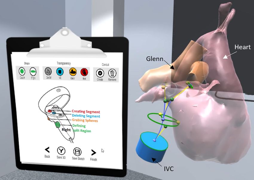
JMIR CARDIO                                                                                                                             Kim et al

     Bifurcated graft designs showed potential strength in better flow split to the lungs, reducing the risk for pulmonary arteriovenous
     malformations.

     (JMIR Cardio 2022;6(1):e35488) doi: 10.2196/35488

     KEYWORDS
     virtual reality; congenital heart disease; surgical planning; usability study; heart; surgery

                                                                           is a tablet-based heart-specific surgical planning software [12].
     Introduction                                                          It provides hole filling, stenosis repair, and Fontan graft design
     Congenital heart disease is the most common birth defect found        features. In SURGEM, the diameter, center line, and
     in nearly 1% of births worldwide [1]. Those patients who are          anastomosis region are defined, supporting the design of a
     diagnosed with single ventricle heart defect (SVHD), a rare type      cylindrical Fontan graft. The designed grafts using SURGEM
     of congenital heart disease, experience mixed circulation of          may not match the size of the native IVC because they are
     oxygenated and deoxygenated blood flows. Patients with SVHD           limited to cylindrical designs [16]. Furthermore, since anatomies
     receive 3 stages of life-saving surgery—Norwood, Glenn, and           are complex and volumetric, lack of depth perception may
     Fontan—to direct the deoxygenated blood flow to the lungs             challenge the design process. Unconstrained clay modeling
     without going through the heart. Stage I, or the Norwood              involves molding physical clay onto a 3D-printed model of the
     procedure, reconstructs the aortic arch, connecting it to the right   total cavopulmonary connection (TCPC) anatomy [13]. This
     ventricle, and a systemic-to-pulmonary artery shunt is placed         method does not require significant training to operate.
     [2]. At stage II, the Glenn procedure, a superior cavopulmonary       However, relying on 3D-printed TCPC anatomy and clay makes
     anastomosis is created by connecting the superior vena cava           it difficult for precise control, and small form changes can have
     (SVC) to the right pulmonary artery (PA) [3,4]. Stage III, the        dramatic consequences. Additionally, detailed viewing and
     Fontan procedure, involves suturing a vascular graft between          reporting, design saving, and future edits are not straightforward
     the inferior vena cava (IVC) to the PA to allow passive flow of       with these techniques.
     venous blood to the lungs for oxygenation. When post-Fontan           The ability to produce graft designs alone is not sufficient to
     surgery circulation does not provide ideal hemodynamics,              optimize Fontan procedures. Without accounting for the flow
     patients may have increased risk of elevated PA pressure,             inside each graft design, a patient may experience increased
     anatomic abnormalities of the PAs, atrial-ventricular valve           risk of medical complications. Multiple studies have emphasized
     regurgitation, and poor ventricular function [4].                     the importance of a low indexed power loss (iPL) [17,18], a
     Advances in medical imaging scanning and 3D-printing                  balanced hepatic flow distribution (HFD) [19,20], and a low
     techniques have been showing great potential for customizing          nonphysiologic wall shear stress percentage (%WSS) [13]. High
     Fontan grafts. One of the customization approaches is known           iPL is correlated with a greater chance of exercise intolerance
     as tissue-engineered vascular grafts (TEVGs), which uses              [21], an unbalanced HFD is associated with pulmonary
     biocompatible material to facilitate the growth of neotissue,         arteriovenous malformations [22], and low %WSS regions are
     including collagen, vascular muscle, and endothelial cells [5,6].     associated with a higher chance of clot formation [23].
     One of the prominent strengths of the growth of neotissue is          Evaluating these hemodynamic performances can be done using
     the patency [7], allowing an implanted graft to grow over time        physical models or computational fluid dynamic (CFD)
     along with patients [8]. It is also believed to be more               simulations. The physical setup entails 3D printing a modeled
     thrombo-resistant and less infectious than are comparable             graft and running blood-mimicking fluid through it. Advanced
     synthetic grafts [9]. These characteristics could support             imaging techniques, such as 4D flow magnetic
     long-term benefits for Fontan procedures. TEVGs involve               resonance imaging, and optical imaging methods, such as
     3D-printing techniques, such as casting, electrospinning, and         particle image velocimetry [24], are used to measure the flow
     modular construction, that can fabricate any shape of a TEVG          velocity field for computing WSS and HFD. iPL can be
     scaffold [10]. Since synthetic grafts are conventionally limited      measured by pressure sensors at the boundaries on the printed
     to specific designs (ie, cylindrical tube-shaped and bifurcated),     grafts [25,26]. These approaches, however, require each design
     being able to fabricate a scaffold allows for more patient-specific   modification to be printed and tested. Thus, the physical setup
     operations.                                                           for measuring hemodynamic performances is labor and time
                                                                           intensive. Physical testing is also limited by spatial resolution,
     3D-printed scaffolds can be modeled using various approaches          signal noise, and segmentation errors. As a mathematical method
     including computer-aided design (CAD) software [11], graft            for calculating fluid flow, CFD can reduce or even overcome
     modeling software (such as SURGEM [12]), and unconstrained            these limitations [27,28]. It can visualize multiple flow
     clay modeling [13]. CAD software is the most widely used tool         properties inside any shape of grafts on a computer without the
     for parameterizing a graft design [11,14,15]. Despite its             need to purchase any devices or print the actual models. The
     popularity, CAD’s complex parametric design process requires          accuracy of CFD simulations is widely recognized and has been
     extensive training and practice, which can be a significant           validated by multiple in vivo and in vitro studies [25,29-31].
     challenge for physicians. SURGEM and unconstrained clay               However, there needs to be further development in tools that
     modeling are 2 great alternatives which enable physicians to          bridge 3D modeling and CFD. Most available tools for
     perform the modeling tasks more easily and quickly. SURGEM            performing these tasks are complicated and require hours of
     https://cardio.jmir.org/2022/1/e35488                                                               JMIR Cardio 2022 | vol. 6 | iss. 1 | e35488 | p. 2
                                                                                                               (page number not for citation purposes)
XSL• FO
RenderX
JMIR CARDIO                                                                                                                                  Kim et al

     training. In our previous study, we developed our first prototype        formats. Our study included usability testing and design
     of virtual reality (VR) vascular graft design software, CorFix           performance evaluations where we compared CorFix designs
     [32]. The first prototype of CorFix integrated diagnosis,                created by 4 medical doctors and 1 biomedical engineer for an
     tube-shaped graft design, free-form graft design, and 3D export          actual surgical case.
     features. The diagnosis feature consisted of rotation, zoom in
     and out, anatomy clipping, annotation, and screenshot. The               Methods
     free-form graft design included pushing and pulling methods
     for manipulating a surface mesh of a designed tube-shaped graft.         Ethics Approval
     Even when the software was evaluated by engineers with                   This study was approved by the investigational review board
     extensive CAD training, CorFix outperformed CAD software                 at Children’s National Hospital (reference number:
     in time and graft design quality. In this study, we developed a          Pro00009721).
     significantly improved second version of CorFix, modifying
     the VR interface and adding bifurcated graft design, design              Medical Image Selection and Acquisition
     export and import, and CFD visualization features. Although              One anonymized post-Fontan procedure imaging data set was
     engineers were proven capable of completing the graft design             acquired via magnetic resonance imaging. The data set was
     task, we focused here on enabling and evaluating the ability for         exported as a DICOM file and then manually segmented into
     physicians to manage the design task. By evolving the CorFix             two 3D models using Mimics software (Materialise): a (1) TCPC
     software, we expect to remove the uncertainty around the                 model and (2) a heart model without the TCPC anatomy (Figure
     evaluation of surgical feasibility and preference. We also               1a). CFD simulation was performed on the TCPC anatomy to
     anticipate reducing communication and discussion times for               evaluate its hemodynamic performance. For the experiment,
     patient-specific surgical planning by avoiding the back-and-forth        the sutured vascular graft was virtually removed from the TCPC
     communication between multiple parties. There are 2 objectives           anatomy, resulting in 2 separate anatomies including the IVC
     to this study: (1) to evaluate the use of the software by medical        and Glenn (ie, PA and SVC). The native IVC surface was
     doctors and engineers and (2) to explore the impact of viewing           extruded 10 mm inferiorly using SolidWorks software (Dassault
     hemodynamic simulation results in numerical and graphical                Systèmes) to show the direction of the native IVC (Figure 1b).
     Figure 1. Patient’s Fontan anatomy. (a) The 3D models of the anonymized patient anatomy: heart (dark gray) and total cavopulmonary connection
     (light gray). (b) Patient anatomy with Fontan IVC to Glenn conduit removed and 10-mm inferior extrusion on the IVC. LPA: left pulmonary artery;
     IVC: inferior vena cava. RPA: right pulmonary artery; SVC: superior vena cava.

                                                                              (ie, zoom, rotation, label, ruler, and clipping) and modeling (ie,
     CorFix Development                                                       cutting vessels, parametric modeling, and free-form modeling)
     The VR surgical planning software, CorFix, was developed                 tasks. This version of CorFix had a modified user interface to
     based on the Unity 3D engine. The software-running platform              accommodate clinicians untrained in VR, modeling software
     was an Alienware Aurora R8 (Dell) with an Intel Core i7-9700             (eg, CAD), or CFD. The interface was adapted to allow users
     processor, a NVIDIA GeForce RTX-2080Ti, and 16 GB of                     to intuitively design patient-specific vascular grafts in a short
     RAM. An Oculus Rift S was used for displaying CorFix in                  amount of time and integrate image analysis in the workflow.
     full-immersive VR. Touch controllers (Oculus Rift) were
     integrated into the system for interacting with the interface.
     CorFix was previously designed to perform simple diagnosis

     https://cardio.jmir.org/2022/1/e35488                                                                    JMIR Cardio 2022 | vol. 6 | iss. 1 | e35488 | p. 3
                                                                                                                    (page number not for citation purposes)
XSL• FO
RenderX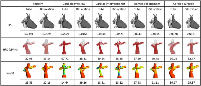
JMIR CARDIO Kim et al
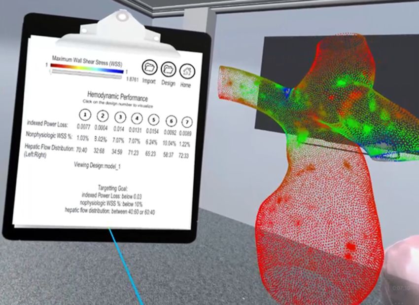
CorFix Interface for Graft Design geometry and anatomy. In the center of the clipboard, a diagram
The Corfix interface was designed to support simple memory of the Oculus controller and its functionality were visualized.
recall, allowing for a short, 1-time, 10-minute tutorial. A virtual The bottom row contained menus that are necessary when the
clipboard was used as an access point for menu and Oculus design process is completed (Figure 2). A “Save 3D” menu
controller information. The top row contained icons that support option was included for exporting the designed graft to a
the designing of a tube-shaped or bifurcated graft. Icons were 3D-formatted OBJ file. The “Save Sketch” menu was included
designed to match the color and shape of the corresponding as a newly developed feature to save the current graft design
for future edits.
Figure 2. Screenshot of a user creating a bifurcated graft using CorFix. IVC: inferior vena cava
Bifurcated Design Feature
Design Export and Import Feature
The minimum design parameters for a bifurcated graft were 2
The import process saves all information needed to reconstruct
anastomosis regions and 1 split region (Figure 3a). For defining
the graft designs using the same algorithm used to construct the
the anastomosis location, a center blue sphere was modified.
conduits. It first saves the transform information of the heart,
Two yellow spheres were located near their respective geometry
Glenn, and the graft. The location and radii of the Bezier curve
control points for defining the radii of the ellipse. These yellow
are then stored. These data are then exported into 1 CSV
spheres were grabbed and adjusted by the Touch controller.
(comma-separated value) file in the aforementioned order. The
Subsequently, through use of a polar equation for an ellipse,
design import feature works by parsing the saved file from top
multiple radii along the ellipse were then calculated and stored.
to bottom and then reconstructing the scene in that order.
https://cardio.jmir.org/2022/1/e35488 JMIR Cardio 2022 | vol. 6 | iss. 1 | e35488 | p. 4
(page number not for citation purposes)
XSL• FO
RenderX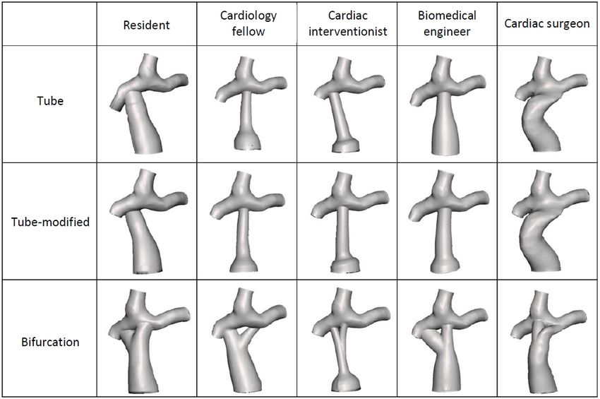
JMIR CARDIO                                                                                                                                         Kim et al

     Figure 3. Schematic of Fontan graft designs. (a) Minimum design parameters for a bifurcated graft and (b) the cubic Bezier curve and radii interpolation
     diagram. IVC: inferior vena cava.

     These calculated points were connected to the center of the                  the anastomosis region and r0 could be one of the radii from the
     ellipse to make triangular meshes, forming a surface. Two cubic              native IVC surface. f(t) is the interpolation adjustment factor at
     Bezier curves were used to define the pathways and girths of                 t. The letter t represents any location on the Bezier curve. The
     the bifurcated graft. The first Bezier curve used the center of              center points of r0 and r1 are defined as 0 and 1 for t.
     the native IVC surface and a user-defined anastomosis region.
     The second Bezier curve used the center of another anastomosis               Hemodynamic Simulation Visualization
     region and a user-specified split region of the graft. The formula           Hemodynamic simulation results were outputted in .h5 format.
     for the pathways was as follows:                                             A data import and transform script was developed using
                                                                                  MATLAB (MathWorks) since the .h5 format is not supported
            P(t) = P0 (1 + t)3+P1 (3t(1 – t)2)+P2 (3t2 (1 – t)) + P3              in Unity. The script consisted of 3 parts: data size, hemodynamic
           (t3) (1)                                                               performance summary, and raw WSS values. The data size rows
     where P0 and P3 are anchor points which represent the center                 summarized the number of graft designs that were simulated
     points of 2 different surfaces; and P1 and P2 are handles which              and the total length of the raw WSS values. The hemodynamic
                                                                                  performance summary contained information on iPL, %WSS,
     define the direction and strength of the pathways, with the
                                                                                  and HFD on each graft. The raw WSS values are composed of
     variable t ranging from 0 to 1 (Figure 3). Users were given an
                                                                                  actual WSS values on each x, y, and z coordinate of a graft.
     option to add as many anchor points as they wanted for more
                                                                                  These parts are concatenated into 1 CSV file, which is then
     precise and complex control of the pathways. Adding an
                                                                                  imported into Unity. As a default, minimum and maximum
     additional anchor point splits a single Bezier curve into 2 cubic
     Bezier curves. Two adjacent handles were automatically created               WSS are set to 0 and 1       , respectively. The maximum WSS
     at each anchor point. Connecting elliptic meshes and the native              can be changed by scrolling a slider on the clipboard towards
     IVC surface along the pathway required the 3 following steps                 the right. The maximum threshold of the slider is automatically
     of interpolation:                                                            identified by calculating the biggest WSS value from the CSV
                                                                                  file. All points with nonzero WSS values are rendered using
            ∆ = r1 – r0(2)
                                                                                  graphics processing unit acceleration and display relevant data
            f(t)       (3)                                                        regarding that point cloud. The graphics processing unit
                                                                                  acceleration approach enables real-time point cloud rendering
            rt = r0 + ∆ ∙f(t) (4)
                                                                                  that corresponds to the slider. The rendered point cloud was
     where ∆ is the difference between the radii from one center                  grabbable and rotatable for users to study in detail or to match
     point to another. For example, r1 could be one of the radii from             its orientation to their view of current designed graft (Figure
                                                                                  4).

     https://cardio.jmir.org/2022/1/e35488                                                                           JMIR Cardio 2022 | vol. 6 | iss. 1 | e35488 | p. 5
                                                                                                                           (page number not for citation purposes)
XSL• FO
RenderX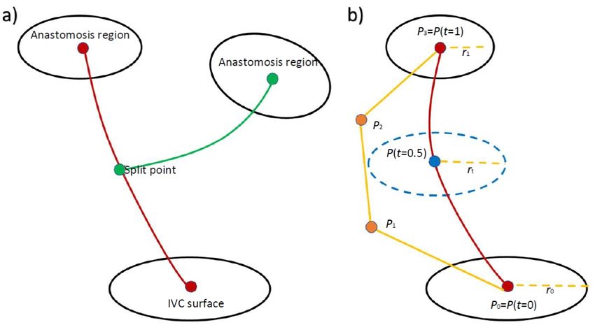
JMIR CARDIO                                                                                                                                     Kim et al

     Figure 4. Screenshot of the hemodynamic simulation results of clipboard and point cloud rendering using the percentage of nonphysiologic wall shear
     stress output values.

     CFD Simulations
     Benchmark Hemodynamic Performance Parameters
                                                                                where NA is the number of WSS values below 1 dyne/cm2 on
     The hemodynamic performance parameters included iPL,
     %WSS, and HFD. iPL is a dimensionless value of a pressure                  the graft, and        is the total number of WSS values. The
     difference between the Fontan graft and the PA. It is normalized           HFD is a ratio of the flow split to the PA from the Fontan.
     using a patient’s body surface area. High iPL values have an               Unbalanced flow split may result in higher risk of pulmonary
     increased chance of deteriorated cardiac performance and                   arteriovenous malformation [35]. HFD was calculated using a
     exercise capacity [33]. The iPL is calculated as follows:                  1-way coupling Lagrangian particle-tracking method. This
                                                                                involves releasing massless infinitesimal particles at the IVC
            iPL =                            (5)                                (NIVC). The number of particles that pass through each side of
                                                                                the PA (NLPA and NRPA) is then counted as follows:
     where BSA is the body surface area of the patient,               is the
     static pressure, ρ is the density of the blood,     is the velocity,
     Q is the flow rate, and Qs is the systemic venous flow that is
     equivalent to the sum of all inlet flow rates. The WSS is defined
     as a force created against the surface of the graft by the blood.
     A healthy physiologic range of venous WSS falls between 1
                                                                                The number of total particles varies and depends on the surface
     and 10 dyne/cm2. If WSS is below the lower threshold, there                area of the inlets. The particles are equally spaced from each
     could be an increased chance in thrombus formation on the                  other. This study set the healthy ranges of each benchmark
     surface of the graft [34]. The ratio of the areas that are below 1         parameter as below 0.03 for iPL, below 10% for %WSS, and
     dyne/cm2 has been identified as the nonphysiologic regions. Its            within the range of 40% to 60% for the HFD ratio.
     percentage against the total area was calculated for %WSS as
     follows:                                                                   CFD Simulations
                                                                                Ansys Fluent 19 (ANSYS Inc) was used to make extensions at
                                                                                inlet and outlet boundaries. The inlet, IVC, and SVC, were

     https://cardio.jmir.org/2022/1/e35488                                                                       JMIR Cardio 2022 | vol. 6 | iss. 1 | e35488 | p. 6
                                                                                                                       (page number not for citation purposes)
XSL• FO
RenderX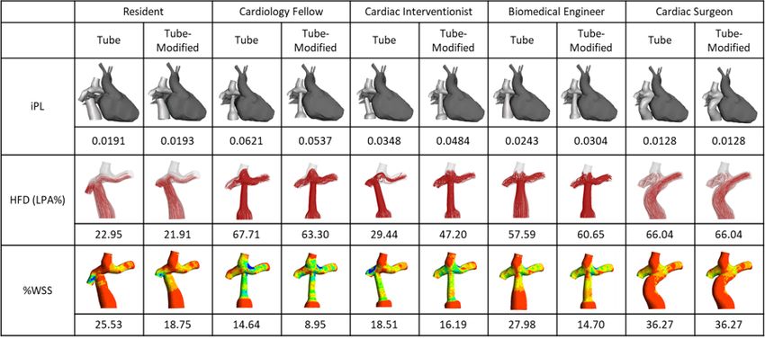
JMIR CARDIO                                                                                                                                       Kim et al

     extruded by 10 times their largest diameter. The outlets, that is           is negatively correlated with iPL. Healthy ranges of each
     the left and right PA, were extruded by 50 mm. These extensions             benchmark parameter were visually provided inside the VR
     acted as a mechanism for developing a stable blood velocity                 environment as a reference. The last tutorial was for the CorFix
     profile. The CFD simulation was performed by solving steady                 interface and took about 10 minutes. None of the participants
     3D Navier-Stokes equations with Newtonian fluid and rigid                   had prior experience with VR, requiring the CorFix tutorial to
     wall assumptions. A calculation for the Reynolds number was                 include information about the hardware (Oculus). During the
     implemented to assess the laminar flow of a patient’s anatomy.              CorFix tutorial, participants wore the gear and went through
                                                                                 the following topics with verbal feedback: importing anatomies,
     Pilot Usability Testing                                                     interacting with the anatomies, designing basic tube-shaped and
     Recruitment                                                                 bifurcated Fontan grafts, making anatomies transparent,
                                                                                 visualizing CFD results, and modifying the existing tube-shaped
     The institutional review board at the Children’s National
                                                                                 design.
     Hospital in Washington, DC, approved this study. The study
     was advertised by sending emails to the groups of residents,                After it was confirmed there were no further questions about
     fellows, cardiac specialists, and medical engineers. A total of             the VR gear or CorFix software, participants created and
     5 voluntary participants were recruited including 1 fourth-year             exported 3D models of 1 bifurcated and 1 tube-shaped Fontan
     resident, 1 third-year cardiac fellow, 1 pediatric intensivist, 1           graft (Figure 5). There was no time limit for designing the graft.
     pediatric cardiac surgeon, and 1 biomedical engineer. All                   The second part of the experiment involved evaluating
     participants gave informed consent prior to their participation.            hemodynamic performances of the patient’s anatomy along
                                                                                 with 6 other anatomies that had 1 design parameter variation.
     Experimental Process
                                                                                 The variations included the suturing region angled leftward,
     Before the experiment, all participants were queried about their            rightward, and upward; having a smaller anastomosis region;
     knowledge on the Fontan procedure and vascular grafts. Those                and offsetting the suturing region toward the left and right. None
     who did not have a strong understanding about the topics were               of these anatomies were optimal in any of the 3 benchmark
     given a short tutorial. The tutorial covered anatomy of patients            parameters. All participants made individual decisions about
     with SVHD, surgical repair for SVHD, and the shapes of Fontan               the designs to find patterns or improvements for further
     grafts in 3 PowerPoint (Microsoft) slides. All participants then            modifying a previously designed tube-shaped graft. The
     received a tutorial on the 3 benchmark parameters that would                participants were not required to modify their design. Three
     be calculated to identify the performance of their Fontan graft             hard-printed surveys were provided at the end of the design
     designs. This tutorial did not include any information about the            modification. The entire experiment was scripted to provide a
     relationships between each benchmark parameter and the graft                uniform experience.
     design parameters. The participants were informed that %WSS
     Figure 5. A participant creating a (a) tube-shaped and a (b) bifurcated Fontan graft on CorFix during the experiment.

                                                                                 usability of the system. To identify the level of sickness when
     Surveys                                                                     using VR gear, the Simulator Sickness Questionnaire (SSQ)
     All participants filled out a digital demographic survey prior to           was provided.
     the experiment, including questions about their position, level
     of VR experience, knowledge and experience on the Fontan
     procedure, and the level of training on fluid dynamics. Three
     hard-printed surveys were provided at the end of the study. The
     System Usability Scale (SUS) and the Usefulness, Satisfaction,
     and Ease of Use Questionnaire (USE) were used to measure the
     https://cardio.jmir.org/2022/1/e35488                                                                         JMIR Cardio 2022 | vol. 6 | iss. 1 | e35488 | p. 7
                                                                                                                         (page number not for citation purposes)
XSL• FO
RenderX
JMIR CARDIO                                                                                                                         Kim et al

                                                                        An average of 2.92 minutes was spent modifying the tube shape
     Results                                                            after it was created. This time includes reviewing the native
     Design Times                                                       patient model and the 6 design variations. The summary of
                                                                        design times and actual designs are provided in Table 1 and
     Participants spent an average of 5.49 minutes creating 1           Figure 6.
     tube-shaped graft and 13.40 minutes creating 1 bifurcated graft.

     Table 1. Summary of graft design and modification times.
                                                    Tube-shaped                   Modified tube-shaped                       Bifurcation
      Time (min), mean (SD)                         5.49 (2.35)                   2.92 (1.67)                                13.40 (3.48)
      Minimum time (min)                            2.50                          2.49                                       9.45
      Maximum time (min)                            8.10                          5.07                                       16.57

     Figure 6. Summary figure of the Fontan graft designs.

                                                                        Fontan anatomy under nonphysiologically optimal WSS,
     Hemodynamic Performance                                            unbalanced HFD with 72.72% of hepatic flow going to the left
     Native Fontan Patient                                              PA, and an iPL of 0.0086, indicating minimal flow change
                                                                        within the anatomy (Figure 7).
     The patient Fontan data set without modifications showed
     suboptimal hemodynamic performance. with 55.36% of the

     https://cardio.jmir.org/2022/1/e35488                                                           JMIR Cardio 2022 | vol. 6 | iss. 1 | e35488 | p. 8
                                                                                                           (page number not for citation purposes)
XSL• FO
RenderX
JMIR CARDIO                                                                                                                                   Kim et al

     Figure 7. Hemodynamic performance of the provided Fontan data set without any modifications. %WSS: percentage of nonphysiologic wall shear
     stress; HFD: hepatic flow distribution; iPL: indexed power loss; LPA: left pulmonary artery.

                                                                              were able to create designs that were much lower in %WSS
     Tube-Shaped and Bifurcated Grafts                                        compared to that of the surgical case. However, none of the
     Each participant produced 1 tube-shaped and 1 bifurcated Fontan          designs were under the safe range of 10% or below. The
     graft. CFD simulations were performed on each of the graft               bifurcated Fontan graft generally showed an optimal range of
     designs. The detailed hemodynamic results are provided in                HFD, between 40% and 60%. All graft designs had higher iPL
     Figure 8. Regardless of the shape of the graft, all participants         values than did the native Fontan surgical case.
     Figure 8. Summary of computational fluid dynamics simulations on the participants’ Fontan graft designs. %WSS: percentage of nonphysiologic wall
     shear stress; HFD: hepatic flow distribution; iPL: indexed power loss; LPA: left pulmonary artery.

                                                                              hemodynamic benchmark parameter. After evaluating the design
     Tube-Shaped Graft Modification                                           variations, the participants were given the freedom to modify
     All participants were asked to review 7 Fontan graft design              their tube-shaped graft design to attempt to optimize the
     variations based on the native Fontan surgical case. None of 7           hemodynamic parameters. All those who modified their
     design variations were considered optimal for the patient. The           tube-shaped Fontan graft were able to reduce %WSS with an
     variations were created to assist the participants in identifying        average improvement of 7.02%, ranging from 2.32% (cardiac
     important design parameters that contribute to each                      interventionalist) to 13.28% (biomedical engineer; Figure 9).

     https://cardio.jmir.org/2022/1/e35488                                                                     JMIR Cardio 2022 | vol. 6 | iss. 1 | e35488 | p. 9
                                                                                                                     (page number not for citation purposes)
XSL• FO
RenderX
JMIR CARDIO Kim et al
Figure 9. Summary table of computational fluid dynamic values after participants were presented with a set of prompt design variations; the %WSS
values improved. %WSS: percentage of nonphysiologic wall shear stress; HFD: hepatic flow distribution; iPL: indexed power loss; LPA: left pulmonary
artery.
of use (mean 4.47, SD 1.38), (3) ease of learning (mean 5.10,
Surveys SD 1.13), and (4) satisfaction (mean 4.60, SD 1.64).
CorFix scored an average of 57 on the SUS questionnaire with
a minimum score of 42.5 and a maximum of 67.5. The average The SSQ (Table 3) showed that using VR for designing,
SUS value suggests that the usability of our prototype was reviewing, and modifying Fontan grafts in less than 30 minutes
marginal. The average total score of USE (Table 2) for all could still cause a high level of nausea, oculomotor, and
participants was 4.38 out of a maximum of 7. This indicates disorientation problems: on average, participants gave 11.45,
that CorFix provides a good degree of usefulness, satisfaction, 24.26, and 16.70 for each parameter of SSQ, respectively. The
and ease of use. The results for the 4 dimensions associated SD was 10.45, 26.48, and 30.18, respectively. The high SD of
with USE were (1) usefulness (mean 3.75, SD 1.03), (2) ease disorientation is due to 3 out of 5 participants reporting no
disorientation problems.
Table 2. Summary table for the Usefulness, Satisfaction, and Ease of Use Questionnaire.
Usefulness Ease of use Ease of learning Satisfaction Overall
Score, mean (SD) 3.75 (1.03) 4.47 (1.38) 5.10 (1.13) 4.60 (1.64) 4.38 (1.10)
Maximum score 2.63 2.36 3.25 1.71 2.57
Minimum score 5.38 5.64 6.00 5.71 5.53
Table 3. Summary table for the Simulator Sickness Questionnaire survey.
Nausea Oculomotor Disorientation
Score, mean (SD) 11.45 (10.45) 24.26 (26.48) 16.70 (30.18)
Maximum score 0 0 0
Minimum score 28.62 68.22 69.60
Even with design modification, less than 20 minutes could be
Discussion spent to plan a Fontan procedure for each patient. Given the
All participants were able to successfully design patient-specific busy workload of surgeons and the urgent nature of patient care,
conduits using the VR software with limited training. Although being able to evaluate and customize a surgery for a patient in
none of the participants had VR experience and CorFix was less than 20 minutes seems advantageous for the current surgical
rated with marginal acceptable usability, designing tube-shaped workflow. The study results mirror other recent studies for other
and bifurcated grafts took less than 6 and 14 minutes, surgical procedures demonstrating that VR is feasible and
respectively. We used the time spent on a task as a surrogate potentially useful but that satisfaction is limited by the technical
for task difficulty and assessment of user adoption since there limitations of the devices and the experience of disorientation
is sound literature indicating that among adult learners, time [38-40].
spent on a task is commensurate with task difficulty [36,37].
https://cardio.jmir.org/2022/1/e35488 JMIR Cardio 2022 | vol. 6 | iss. 1 | e35488 | p. 10
(page number not for citation purposes)
XSL• FO
RenderXJMIR CARDIO Kim et al
All participants expressed that if a real-time hemodynamic were presented only in numerical format. We hypothesize that
analysis of their designs were available, they would be able to with supplementary graphical visualization, users may be able
better pinpoint the flaws of their designs. We, therefore, plan to improve iPL and HFD more easily.
on further developing the CorFix software to add real-time
With the development of graft modeling and evaluation software
simulation and visualization features. Our system has
like CorFix, physicians may be able to easily customize Fontan
implemented button and pointer color changes and tactile
grafts and find an optimal graft configuration for long-term
feedback (ie, vibration) to bolster the interactivity inside the
benefits. We plan to further develop CorFix by adding real-time
virtual scene. However, many participants struggled with depth
CFD simulation and automatic graft optimization features for
perception and interactivity. Grabbing design control points or
bolstering the graft design and evaluation process. Our next
even clicking buttons on the virtual menu were frequently
study will incorporate many of these changes and focus on
observed. Developing a feature or a device that could better
recruiting more cardiac surgeons and testing against a larger
support tactile feedback may enhance the usability and the innate
number of patient surgical cases.
learnability of the software.
This study had a small sample for recruitment due to the limited
The bifurcated graft designs were more successful in improving
number of doctors and their time availability despite 3 months
the hepatic flow distribution to a healthy range compared to the
of advertising and 2 additional months during the data collection
tube-shaped graft designs. During the experiment, all
period. We were able to include individuals with various levels
participants were asked to review 7 different tube-shaped Fontan
of medical experience, which provides a broad spectrum of
grafts, which were derived from the actual surgical case although
users and supports important preliminary insights. Our future
none of these design variations were surgically optimal. We
study will involve greater participation and a larger number of
hypothesized that the participants would be able to find patterns
patient cases to supplement the current results.
between design parameters and hemodynamic performance.
Our study showed that when participants decided to modify This paper reports the design of a VR software for
their designs after reviewing other cases, they were able to patient-specific designs of vascular grafts that demonstrated
design a more optimal graft by lowering %WSS. On average, feasibility and initial usability in a pilot usability study. All
%WSS was reduced by 7.02%. A biomedical engineer with a participants were able to create patient-specific graft designs
strong fluid dynamics education background showed the with minimal training, needing on average only 5.49 minutes
maximum %WSS reduction of 13.28%. Considering how lower to design 1 tube-shaped graft and 13.40 minutes to design 1
%WSS is related to a lower risk of thrombosis for Fontan grafts, bifurcated graft. Participants rated the design software with a
this design could provide a significant long-term improvement good degree of usefulness, satisfaction, and ease of use. Further
for the patient. We therefore infer that showing problematic design improvements are needed to visualize hemodynamics
regions in color, like a contour map, may help doctors without during the design process, and a larger study is required to fully
an engineering background to sufficiently identify low %WSS. compare the VR design to current state-of-the-art surgical
iPL and HFD improvements were not consistent throughout the procedures.
participants. Unlike %WSS, these hemodynamic parameters
Acknowledgments
This paper was supported by the National Institutes of Health (award #R01HL143468 and #R21HD090671).
Conflicts of Interest
BK, XL, and AK are founders of and hold shares of stock options in CorFix Medical, Inc. The results of the study discussed in
this publication could affect the value of CorFix Medical Inc. This arrangement has been reviewed and approved by the Johns
Hopkins University in accordance with its conflict of interest policies.
References
1. Reller MD, Strickland MJ, Riehle-Colarusso T, Mahle WT, Correa A. Prevalence of congenital heart defects in metropolitan
Atlanta, 1998-2005. J Pediatr 2008 Dec;153(6):807-813 [FREE Full text] [doi: 10.1016/j.jpeds.2008.05.059] [Medline:
18657826]
2. Primeaux J, Salavitabar A, Lu JC, Grifka RG, Figueroa CA. Characterization of post-operative hemodynamics following
the norwood procedure using population data and multi-scale modeling. Front Physiol 2021;12:603040 [FREE Full text]
[doi: 10.3389/fphys.2021.603040] [Medline: 34054563]
3. Salik I, Mehta B, Ambati S. Bidirectional Glenn procedure or hemi-Fontan. In: StatPearls Internet. Treasure Island, FL:
StatPearls Publishing; 2022. URL: https://www.ncbi.nlm.nih.gov/books/NBK563299/
4. Fredenburg TB, Johnson TR, Cohen MD. The Fontan procedure: anatomy, complications, and manifestations of failure.
Radiographics 2011 Mar;31(2):453-463. [doi: 10.1148/rg.312105027] [Medline: 21415190]
5. Yeung E, Inoue T, Matsushita H, Opfermann J, Mass P, Aslan S, et al. In vivo implantation of 3-dimensional printed
customized branched tissue engineered vascular graft in a porcine model. J Thorac Cardiovasc Surg 2020
May;159(5):1971-1981.e1 [FREE Full text] [doi: 10.1016/j.jtcvs.2019.09.138] [Medline: 31864694]
https://cardio.jmir.org/2022/1/e35488 JMIR Cardio 2022 | vol. 6 | iss. 1 | e35488 | p. 11
(page number not for citation purposes)
XSL• FO
RenderXJMIR CARDIO Kim et al
6. Ravi S, Chaikof EL. Biomaterials for vascular tissue engineering. Regen Med 2010 Jan;5(1):107-120 [FREE Full text]
[doi: 10.2217/rme.09.77] [Medline: 20017698]
7. Skovrind I, Harvald E, Juul Belling H, Jørgensen CD, Lindholt J, Andersen D. Concise review: patency of small-diameter
tissue-engineered vascular grafts: a meta-analysis of preclinical trials. Stem Cells Transl Med 2019 Jul;8(7):671-680 [FREE
Full text] [doi: 10.1002/sctm.18-0287] [Medline: 30920771]
8. Matsuzaki Y, John K, Shoji T, Shinoka T. The Evolution of Tissue Engineered Vascular Graft Technologies: From Preclinical
Trials to Advancing Patient Care. Appl Sci (Basel) 2019 Apr;9(7):1274 [FREE Full text] [doi: 10.3390/app9071274]
[Medline: 31890320]
9. Gupta P, Mandal BB. Tissue‐engineered vascular grafts: emerging trends and technologies. Adv Funct Materials 2021
Jun 12;31(33):2100027. [doi: 10.1002/adfm.202100027]
10. Wang P, Sun Y, Shi X, Shen H, Ning H, Liu H. 3D printing of tissue engineering scaffolds: a focus on vascular regeneration.
Biodes Manuf 2021;4(2):344-378 [FREE Full text] [doi: 10.1007/s42242-020-00109-0] [Medline: 33425460]
11. Kim B, Loke Y, Stevenson F, Siallagan D, Mass P, Opfermann JD, et al. Virtual cardiac surgical planning through
hemodynamics simulation and design optimization of Fontan grafts. In: Lecture Notes in Computer Science. 2019 Presented
at: Medical Image Computing and Computer Assisted Intervention; Oct 13-17; Shenzhen, China. [doi:
https://doi.org/10.1007/978-3-030-32254-0_23]
12. Luffel M, Sati M, Rossignac J, Yoganathan AP, Haggerty CM, Restrepo M, et al. SURGEM: A solid modeling tool for
planning and optimizing pediatric heart surgeries. Computer-Aided Design 2016 Jan;70:3-12. [doi: 10.1016/j.cad.2015.06.018]
13. Loke Y, Kim B, Mass P, Opfermann JD, Hibino N, Krieger A, et al. Role of surgeon intuition and computer-aided design
in Fontan optimization: A computational fluid dynamics simulation study. J Thorac Cardiovasc Surg 2020
Jul;160(1):203-212.e2 [FREE Full text] [doi: 10.1016/j.jtcvs.2019.12.068] [Medline: 32057454]
14. Aslan S, Liu X, Wu Q, Mass P, Loke Y, Hibino N, et al. Virtual planning and simulation of coarctation repair in hypoplastic
aortic arches: is fixing the coarctation alone enough? 2022 Presented at: International Joint Conference on Biomedical
Engineering Systems and Technologies; Feb 9-11; Online. [doi: 10.5220/0010842600003123]
15. Liu X, Aslan S, Hess R, Mass P, Olivieri L, Loke Y, et al. Automatic shape optimization of patient-specific tissue engineered
vascular grafts for aortic coarctation. 2020 Presented at: International Conference of the IEEE Engineering in Medicine
Biology Society (EMBC); July 20-24; Montreal, Canada. [doi: 10.1109/embc44109.2020.9176371]
16. Restrepo M, Luffel M, Sebring J, Kanter K, Del Nido P, Veneziani A, et al. Surgical planning of the total cavopulmonary
connection: robustness analysis. Ann Biomed Eng 2015 Jun 15;43(6):1321-1334 [FREE Full text] [doi:
10.1007/s10439-014-1149-7] [Medline: 25316591]
17. Whitehead KK, Pekkan K, Kitajima HD, Paridon SM, Yoganathan AP, Fogel MA. Nonlinear power loss during exercise
in single-ventricle patients after the Fontan: insights from computational fluid dynamics. Circulation 2007 Sep 11;116(11
Suppl):I165-I171. [doi: 10.1161/CIRCULATIONAHA.106.680827] [Medline: 17846299]
18. Haggerty CM, Restrepo M, Tang E, de Zélicourt DA, Sundareswaran KS, Mirabella L, et al. Fontan hemodynamics from
100 patient-specific cardiac magnetic resonance studies: a computational fluid dynamics analysis. J Thorac Cardiovasc
Surg 2014 Oct;148(4):1481-1489 [FREE Full text] [doi: 10.1016/j.jtcvs.2013.11.060] [Medline: 24507891]
19. McElhinney DB, Marx GR, Marshall AC, Mayer JE, Del Nido PJ. Cavopulmonary pathway modification in patients with
heterotaxy and newly diagnosed or persistent pulmonary arteriovenous malformations after a modified Fontan operation.
J Thorac Cardiovasc Surg 2011 Jun;141(6):1362-70.e1 [FREE Full text] [doi: 10.1016/j.jtcvs.2010.08.088] [Medline:
21146835]
20. Yang W, Vignon-Clementel IE, Troianowski G, Reddy VM, Feinstein JA, Marsden AL. Hepatic blood flow distribution
and performance in conventional and novel Y-graft Fontan geometries: a case series computational fluid dynamics study.
J Thorac Cardiovasc Surg 2012 May;143(5):1086-1097 [FREE Full text] [doi: 10.1016/j.jtcvs.2011.06.042] [Medline:
21962841]
21. Khiabani RH, Whitehead KK, Han D, Restrepo M, Tang E, Bethel J, et al. Exercise capacity in single-ventricle patients
after Fontan correlates with haemodynamic energy loss in TCPC. Heart 2015 Jan;101(2):139-143. [doi:
10.1136/heartjnl-2014-306337] [Medline: 25184826]
22. Dasi LP, Whitehead K, Pekkan K, de Zelicourt D, Sundareswaran K, Kanter K, et al. Pulmonary hepatic flow distribution
in total cavopulmonary connections: extracardiac versus intracardiac. J Thorac Cardiovasc Surg 2011 Jan;141(1):207-214
[FREE Full text] [doi: 10.1016/j.jtcvs.2010.06.009] [Medline: 20621314]
23. Buck A, Groszek J, Colvin D, Keller S, Kensinger C, Forbes R, et al. Combined In Silico and In Vitro Approach Predicts
Low Wall Shear Stress Regions in a Hemofilter that Correlate with Thrombus Formation In Vivo. ASAIO J
2018;64(2):211-217 [FREE Full text] [doi: 10.1097/MAT.0000000000000649] [Medline: 28857774]
24. Grant I. Particle image velocimetry: A review. Proceedings of the Institution of Mechanical Engineers, Part C: Journal of
Mechanical Engineering Science 2016 Aug 10;211(1):55-76. [doi: 10.1243/0954406971521665]
25. Liu X, Aslan S, Kim B, Warburton L, Jackson D, Muhuri A, et al. Computational Fontan Analysis: Preserving Accuracy
While Expediting Workflow. World J Pediatr Congenit Heart Surg 2022 May;13(3):293-301. [doi:
10.1177/21501351211073619] [Medline: 35446218]
https://cardio.jmir.org/2022/1/e35488 JMIR Cardio 2022 | vol. 6 | iss. 1 | e35488 | p. 12
(page number not for citation purposes)
XSL• FO
RenderXJMIR CARDIO Kim et al
26. Siallagan D, Loke Y, Olivieri L, Opfermann J, Ong CS, de Zélicourt D, et al. Virtual surgical planning, flow simulation,
and 3-dimensional electrospinning of patient-specific grafts to optimize Fontan hemodynamics. J Thorac Cardiovasc Surg
2018 Apr;155(4):1734-1742 [FREE Full text] [doi: 10.1016/j.jtcvs.2017.11.068] [Medline: 29361303]
27. Liu X, Kim B, Loke Y, Mass P, Olivieri L, Hibino N, et al. Semi-Automatic Planning and Three-Dimensional Electrospinning
of Patient-Specific Grafts for Fontan Surgery. IEEE Trans. Biomed. Eng 2022 Jan;69(1):186-198. [doi:
10.1109/tbme.2021.3091113]
28. Liu X, Hibino N, Loke Y, Kim B, Mass P, Fuge M, et al. Surgical Planning and Optimization of Patient-Specific Fontan
Grafts with Uncertain Post-Operative Boundary Conditions and Anastomosis Displacement. IEEE Trans. Biomed. Eng
2022:1-1. [doi: 10.1109/tbme.2022.3170922]
29. Medero R, Hoffman C, Roldán-Alzate A. Comparison of 4D Flow MRI and Particle Image Velocimetry Using an In Vitro
Carotid Bifurcation Model. Ann Biomed Eng 2018 Dec;46(12):2112-2122 [FREE Full text] [doi:
10.1007/s10439-018-02109-9] [Medline: 30112708]
30. Roldán-Alzate A, García-Rodríguez S, Anagnostopoulos PV, Srinivasan S, Wieben O, François CJ. Hemodynamic study
of TCPC using in vivo and in vitro 4D Flow MRI and numerical simulation. J Biomech 2015 May 01;48(7):1325-1330
[FREE Full text] [doi: 10.1016/j.jbiomech.2015.03.009] [Medline: 25841292]
31. Biglino G, Cosentino D, Steeden JA, De Nova L, Castelli M, Ntsinjana H, et al. Using 4D cardiovascular magnetic resonance
imaging to validate computational fluid dynamics: a case study. Front Pediatr 2015;3:107 [FREE Full text] [doi:
10.3389/fped.2015.00107] [Medline: 26697416]
32. Kim B, Nguyen PD, Nar P, Liu X, Loke Y, Mass P, et al. CorFix: Virtual reality cardiac surgical planning system for
designing patient specific vascular grafts. 2020 Presented at: The 26th ACM Symposium on Virtual Reality Software and
Technology; Nov 1-4; Ottawa, Canada.
33. Goldberg DJ, Avitabile CM, McBride MG, Paridon SM. Exercise capacity in the Fontan circulation. Cardiol Young 2014
Jan 09;23(6):824-830. [doi: 10.1017/s1047951113001649]
34. Sakariassen KS, Orning L, Turitto VT. The impact of blood shear rate on arterial thrombus formation. Future Sci OA 2015
Nov;1(4):FSO30 [FREE Full text] [doi: 10.4155/fso.15.28] [Medline: 28031903]
35. Nakamura Y, Yagihara T, Kagisaki K, Hagino I, Kobayashi J. Pulmonary arteriovenous malformations after a Fontan
operation in the left isomerism and absent inferior vena cava. Eur J Cardiothorac Surg 2009 Jul;36(1):69-76; discussion
76. [doi: 10.1016/j.ejcts.2009.02.046] [Medline: 19369088]
36. Thomas K, König CJ. Knowledge of Previous Tasks: Task Similarity Influences Bias in Task Duration Predictions. Front
Psychol 2018;9:760 [FREE Full text] [doi: 10.3389/fpsyg.2018.00760] [Medline: 29881362]
37. Eversheim U, Bock O. Evidence for processing stages in skill acquisition: a dual-task study. Learn Mem 2001;8(4):183-189
[FREE Full text] [doi: 10.1101/lm.39301] [Medline: 11533221]
38. Desselle MR, Brown RA, James AR, Midwinter MJ, Powell SK, Woodruff MA. Augmented and Virtual Reality in Surgery.
Comput. Sci. Eng 2020 May 1;22(3):18-26. [doi: 10.1109/mcse.2020.2972822]
39. Ayoub A, Pulijala Y. BMC Oral Health 2019 Nov 08;19(1):238 [FREE Full text] [doi: 10.1186/s12903-019-0937-8]
[Medline: 31703708]
40. Yoo JS, Patel DS, Hrynewycz NM, Brundage TS, Singh K. The utility of virtual reality and augmented reality in spine
surgery. Ann Transl Med 2019 Sep;7(Suppl 5):S171 [FREE Full text] [doi: 10.21037/atm.2019.06.38] [Medline: 31624737]
Abbreviations
%WSS: percentage of nonphysiologic wall shear stress
CFD: computational fluid dynamics
CSV: comma-separated value
HFD: hepatic flow distribution
iPL: indexed power loss
IVC: inferior vena cava
SSQ: Simulator Sickness Questionnaire
SUS: System Usability Scale
SVC: superior vena cava
TCPC: total cavopulmonary connection
USE: Usefulness, Satisfaction, and Ease of Use Questionnaire
VR: virtual reality
WSS: wall shear stress
https://cardio.jmir.org/2022/1/e35488 JMIR Cardio 2022 | vol. 6 | iss. 1 | e35488 | p. 13
(page number not for citation purposes)
XSL• FO
RenderXJMIR CARDIO Kim et al
Edited by G Eysenbach; submitted 07.12.21; peer-reviewed by H Mehdizadeh, SS Amritphale; comments to author 05.02.22; revised
version received 05.05.22; accepted 17.05.22; published 17.06.22
Please cite as:
Kim B, Nguyen P, Loke YH, Cleveland V, Liu X, Mass P, Hibino N, Olivieri L, Krieger A
Virtual Reality Cardiac Surgical Planning Software (CorFix) for Designing Patient-Specific Vascular Grafts: Development and Pilot
Usability Study
JMIR Cardio 2022;6(1):e35488
URL: https://cardio.jmir.org/2022/1/e35488
doi: 10.2196/35488
PMID:
©Byeol Kim, Phong Nguyen, Yue-Hin Loke, Vincent Cleveland, Xiaolong Liu, Paige Mass, Narutoshi Hibino, Laura Olivieri,
Axel Krieger. Originally published in JMIR Cardio (https://cardio.jmir.org), 17.06.2022. This is an open-access article distributed
under the terms of the Creative Commons Attribution License (https://creativecommons.org/licenses/by/4.0/), which permits
unrestricted use, distribution, and reproduction in any medium, provided the original work, first published in JMIR Cardio, is
properly cited. The complete bibliographic information, a link to the original publication on https://cardio.jmir.org, as well as
this copyright and license information must be included.
https://cardio.jmir.org/2022/1/e35488 JMIR Cardio 2022 | vol. 6 | iss. 1 | e35488 | p. 14
(page number not for citation purposes)
XSL• FO
RenderXYou can also read