White Paper on Ophthalmic Imaging for Choroidal Nevus Identification and Transformation into Melanoma
←
→
Page content transcription
If your browser does not render page correctly, please read the page content below
Article

            White Paper on Ophthalmic Imaging for Choroidal Nevus
            Identification and Transformation into Melanoma
            Carol L. Shields1 , Sara E. Lally1 , Lauren A. Dalvin2 , Mandeep S. Sagoo3 ,
            Marco Pellegrini4 , Swathi Kaliki5 , Ahmet Kaan Gündüz6 , Minoru Furuta7 ,
            Prithvi Mruthyunjaya8 , Adrian T. Fung9 , Jay S. Duker10 , Sara M. Selig11 , Antonio Yaghy1 ,
            Sandor R. Ferenczy1 , Malvina B. Eydelman12 , and Mark S. Blumenkranz13
            1
              Ocular Oncology Service, Wills Eye Hospital, Thomas Jefferson University, Philadelphia, PA, USA
            2
              Department of Ophthalmology, Mayo Clinic, Rochester, MN, USA
            3
              Ocular Oncology Service, Moorfields Eye Hospital and NIHR Biomedical Research Centre for Ophthalmology at Moorfields Eye Hospital
            and University College London Institute of Ophthalmology, London, UK
            4
              Eye Clinic, Department of Biomedical and Clinical Science “Luigi Sacco”, Luigi Sacco Hospital, University of Milan, Milan, Italy
            5
              The Operation Eyesight Universal Institute for Eye Cancer, LV Prasad Eye Institute, Hyderabad, India
            6
              Department of Ophthalmology, Ankara University School of Medicine, Ankara, Turkey
            7
              Department of Ophthalmology, Fukushima Medical University, Fukushima, Japan and Department of Ophthalmology, Yachiyo Medical
            Center, Tokyo Women’s Medical University, Tokyo, Japan
            8
              Ocular Oncology Service, Department of Ophthalmology, Byers Eye Institute, Stanford University, Palo Alto, CA, USA
            9
              Westmead and Central Clinical Schools, Discipline of Clinical Ophthalmology and Eye Health, University of Sydney, Australia and
            Department of Ophthalmology, Faculty of Medicine, Health and Human Sciences, Macquarie University, Sydney, Australia
            10
               New England Eye Center, Tufts Medical Center, Tufts University School of Medicine, Boston, MA, USA
            11
               Melanoma Research Foundation, Washington, DC, USA
            12
               Office of Ophthalmic, Anesthesia, Respiratory, Ear, Nose and Throat (ENT), and Dental Devices, Food & Drug Administration (FDA),
            Washington, DC
            13
               Department of Ophthalmology, Ophthalmology Innovation Program, Byers Eye Institute, Stanford University, Palo Alto, CA, USA

            Correspondence: Carol L. Shields,            Purpose: To discuss the evolution of noninvasive diagnostic methods in the identifica-
            Ocular Oncology Service, Suite 1440,         tion of choroidal nevus and determination of risk factors for malignant transformation as
            Wills Eye Hospital, 840 Walnut Street,       well as introduce the novel role that artificial intelligence (AI) can play in the diagnostic
            Philadelphia, PA 19107, USA.                 process.
            e-mail: carolshields@gmail.com
                                                         Methods: White paper.
            Received: October 5, 2020
            Accepted: January 3, 2021                    Results: Longstanding diagnostic methods to stratify benign choroidal nevus from
            Published: February 17, 2021                 choroidal melanoma and to further determine the risk for nevus transformation into
                                                         melanoma have been dependent on recognition of key clinical features by ophthalmic
            Keywords: artificial intelligence;           examination. These risk factors have been derived from multiple large cohort research
            multimodal imaging; choroid; nevus;          studies over the past several decades and have garnered widespread use throughout the
            melanoma                                     world. More recent publications have applied ocular diagnostic testing (fundus photog-
            Citation: Shields CL, Lally SE, Dalvin       raphy, ultrasound examination, autofluorescence, and optical coherence tomography)
            LA, Sagoo MS, Pellegrini M, Kaliki S,        to identify risk factors for the malignant transformation of choroidal nevus based on
            Gündüz AK, Furuta M, Mruthyunjaya            multimodal imaging features. The widespread usage of ophthalmic imaging systems
            P, Fung AT, Duker JS, Selig SM, Yaghy        to identify and follow choroidal nevus, in conjunction with the characterization of
            A, Ferenczy SR, Eydelman MB,                 malignant transformation risk factors via diagnostic imaging, presents a novel path to
            Blumenkranz MS. White paper on               apply AI.
            ophthalmic imaging for choroidal
            nevus identification and
                                                         Conclusions: AI applied to existing ophthalmic imaging systems could be used for both
            transformation into melanoma.
                                                         identification of choroidal nevus and as a tool to aid in earlier detection of transforma-
            Trans Vis Sci Tech. 2021;10(2):24,
                                                         tion to malignant melanoma.
            https://doi.org/10.1167/tvst.10.2.24         Translational Relevance: Advances in AI models applied to ophthalmic imaging
                                                         systems have the potential to improve patient care, because earlier detection and treat-
                                                         ment of melanoma has been proven to improve long-term clinical outcomes.

            Copyright 2021 The Authors
            tvst.arvojournals.org | ISSN: 2164-2591                                                                                                 1
Downloaded from tvst.arvojournals.org on 09/23/2021
                     This work is licensed under a Creative Commons Attribution-NonCommercial-NoDerivatives 4.0 International License.
White Paper Nevus                                                  TVST | February 2021 | Vol. 10 | No. 2 | Article 24 | 2

                                                                        Hollowness, absence of Halo and absence of Drusen,
             Introduction                                               creating a new mnemonic “To Find Small Ocular
                                                                        Melanoma - Using Helpful Hints Daily” (TFSOM -
                Choroidal nevus is the most common primary              UHHD). A combination of any three factors posed a
            intraocular tumor.1 Based on several studies, the           mean risk of >50% for transformation.12
            overall prevalence of choroidal nevus in the adult              Singh et al.13 calculated the rate for transforma-
            population ranges from 5% to 25%.2–7 Choroidal              tion of choroidal nevus into melanoma in the United
            nevus is often found incidentally on routine ophthalmic     States population at 1 in 8845, but with the assump-
            funduscopic examination. This lesion typically occurs       tion that all choroidal melanoma arises from a preex-
            as a well-circumscribed pigmented or nonpigmented           isting nevus, which might not be the case. Kivelä and
            lesion, residing deep to the retina within the choroid      Eskelin14 calculated that the lifetime risk for transfor-
            and measuring 2 mm, the presence of overlying subreti-         The word melanoma causes one to think of skin
            nal Fluid, presence of Symptoms, presence of overly-        (cutaneous) melanoma. Approximately 90% to 95%
            ing Orange pigment, and tumor location near the             of all melanomas are found in the skin. Cutaneous
            optic disc Margin, which can be remembered by               melanoma is well-publicized and is increasing in
            the mnemonic “To Find Small Ocular Melanoma”                frequency, mostly owing to overexposure to solar
            (TFSOM) (Fig. 2).10 In 2000, various combinations           radiation. Cutaneous melanoma also has the potential
            of high-risk features were evaluated, with the great-       to lead to metastasis and death. Cutaneous melanoma
            est risk for transformation into melanoma at 63%,           generally arises on sun-exposed areas of the body,
            found with the combination of three features, includ-       with ultraviolet radiation exposure being the main risk
            ing a thickness of >2 mm, the presence of subretinal        factor for development.16–19 Lesser known, the eye
            fluid, and a tumor margin near the optic disc.11 A later    (uvea [iris, ciliary body, and choroid]) is the second
            follow-up study of >2500 choroidal nevi continued to        most frequent location of malignant melanoma in
            refine the high risk clinical factors for transformation    the body, representing about 5% of all melanomas.
            into melanoma including greater Thickness, subretinal       Although cutaneous and uveal melanoma are two
            Fluid, Symptoms, Orange pigment, Margin near disc,          distinct diseases and one does not lead to the other,
            and additionally with three new features: Ultrasound        both occur most often in patients with fair skin

Downloaded from tvst.arvojournals.org on 09/23/2021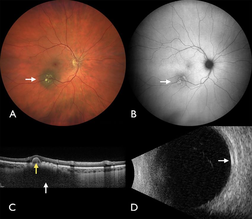
White Paper Nevus TVST | February 2021 | Vol. 10 | No. 2 | Article 24 | 3
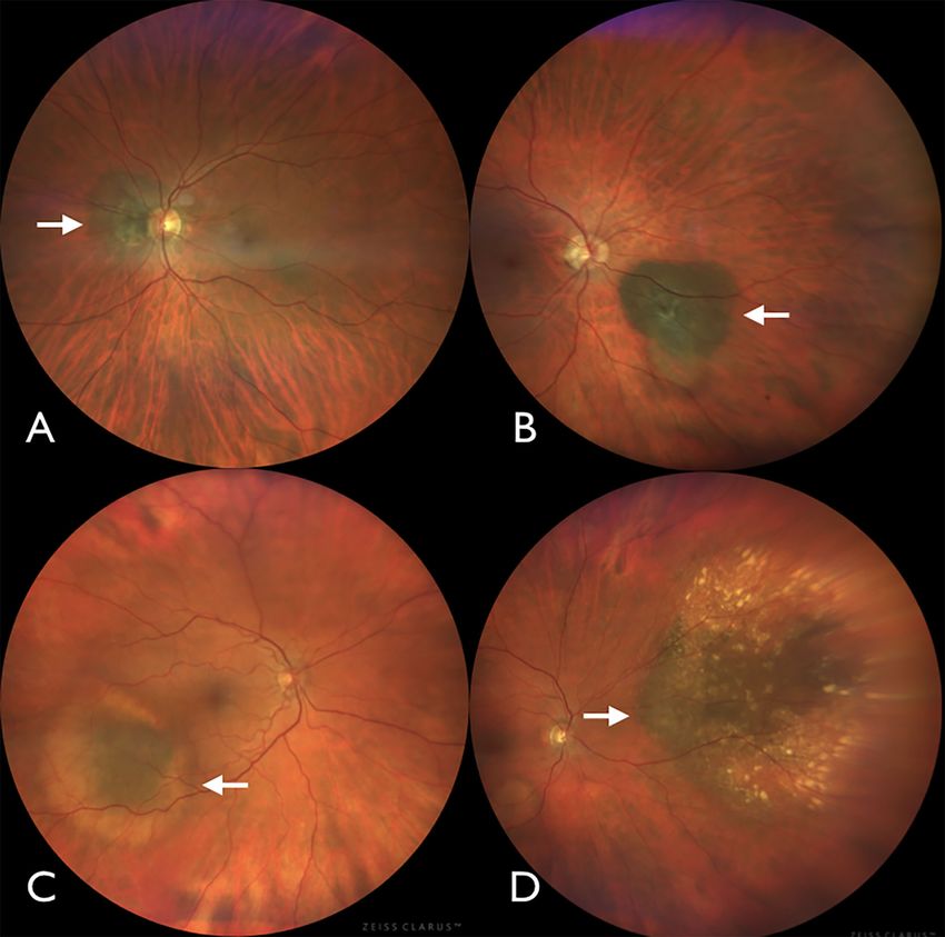
Figure 1. Spectrum of benign choroidal nevus. (A) Choroidal nevus (arrow) near the optic disc with overlying RPE atrophy. (B) Choroidal
nevus (arrow) nasal to the optic disc with overlying and dependent RPE atrophy. (C) Choroidal nevus (arrow) inferotemporal to the macula
with surrounding yellow halo. (D) Large choroidal nevus (arrow) nasal to the optic disc with overlying yellow drusen.
complexion and blue or green eyes. Uveal melanoma improve the early detection of cutaneous melanoma.19
has not clearly been found to be related to sunlight Further advances with the use of instruments like
exposure, but similar to cutaneous melanoma, uveal dermoscopy, a handheld device that allows magni-
melanoma can arise from a preexisting nevus or de fied, high-resolution visualization of the skin, has also
novo.16 For both diseases, early detection is key for improved detection.20 Automated computer-assisted
survival. differentiation has provided 95% sensitivity and 70%
In 2004, Lindholm et al.17 documented that early specificity.20
cutaneous melanoma (4 mm. choroid. This tract is a specialized vascularized tissue
Major advances in the dermatology (skin) litera- that manifests pigmented melanocytes, the source of
ture have helped in early detection of cutaneous nevus and melanoma. Uveal melanoma, similar to
melanoma. The mnemonic ABCDE (Asymmetry, cutaneous melanoma, can metastasize to distant parts
Border irregularity, Color, Diameter >6 mm, Evolv- of the body and lead to death.21 Current treatments
ing size, shape, or color) simplified the key features for uveal melanoma include enucleation (removal of
of early cutaneous melanoma and helped physicians the eye), different forms of radiotherapy, local resec-
and nonphysicians diagnose suspicious lesions before tion of the tumor, thermal laser, and novel therapies,
they grew.18 Skin self-examination has helped to still in development, using nanoparticles.22
Downloaded from tvst.arvojournals.org on 09/23/2021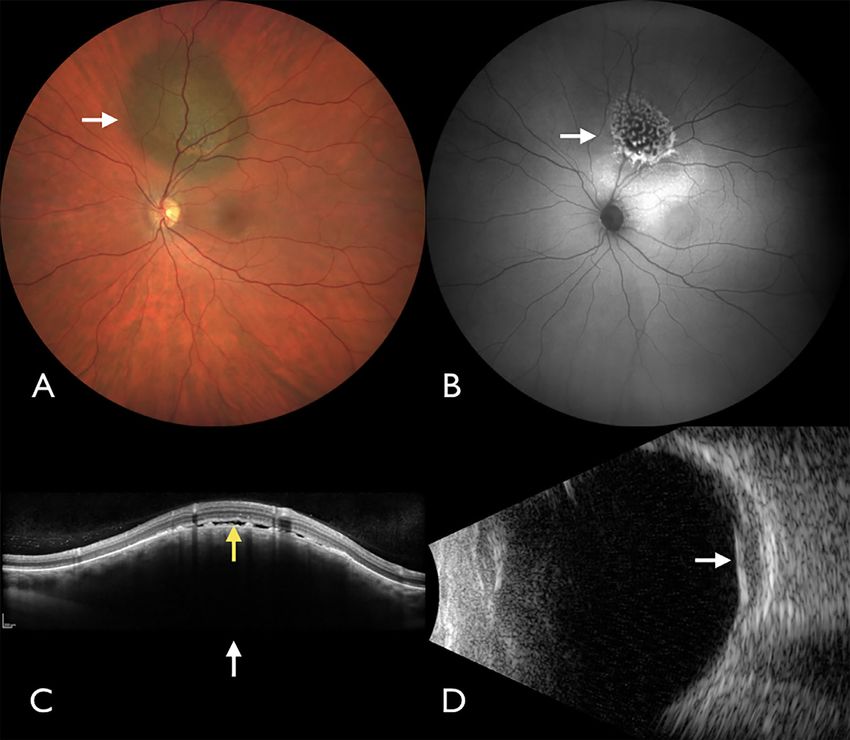
White Paper Nevus                                                            TVST | February 2021 | Vol. 10 | No. 2 | Article 24 | 4

            Figure 2. Multimodal imaging of benign choroidal nevus. (A) Choroidal nevus (arrow) with overlying yellow drusen. (B) Autofluorescence
            demonstrating the ring-shaped, minimally bright signal (arrow) of drusen. (C) OCT showing dark nevus with low signal (white arrow) and
            overlying pigment epithelial detachment (yellow arrow). (D) Ultrasound examination showing flat nevus (arrow) with
White Paper Nevus TVST | February 2021 | Vol. 10 | No. 2 | Article 24 | 5
Figure 3. Spectrum of malignant choroidal melanoma. (A) Small choroidal melanoma (arrow) with overlying orange pigment. (B) Elongated
choroidal melanoma (arrow) with overlying orange pigment on the inferior aspect. (C) Dome-shaped choroidal melanoma (arrow) with
central elevated nodule. (D) Large choroidal melanoma (arrow) with overlying subretinal fluid.
ocular blood flow is also possible by intravenous injec- retina, its use in the differential diagnosis between
tion of contrast agents, fluorescein angiography, and choroidal nevus and melanoma is currently limited.23
indocyanine green angiography. Fluorescein angiogra- Several imaging modalities, including ultrasound
phy can detect modifications in the internal and exter- examination, autofluorescence, and OCT, were
nal blood-retinal barriers, which may help in differ- combined to evaluate a series of 3806 choroid nevi.24,25
entiating large choroidal nevi from small choroidal Using multimodal ophthalmic imaging, objective risk
melanomas. In contrast, indocyanine green angiogra- factors for nevus transformation into melanoma
phy is mainly used to study intratumoral vasculariza- have been identified including Thickness >2 mm
tion and can be beneficial in the differentiation between (ultrasound examination), subretinal Fluid (OCT),
small amelanotic choroidal tumors, including choroidal Symptoms (Snellen visual acuity loss to ≤20/50),
nevi, choroidal melanoma, choroidal hemangiomas, Orange pigment (autofluorescence), Melanoma acous-
and small isolated metastatic tumors. Noninvasive flow tic hollowness (ultrasound examination), and tumor
detection is possible with OCT angiography, using only DIaMeter >5 mm (fundus photography) (Figs. 2 and
light stimulated OCT images and applying a decorre- 4).24,25 Based on these recent findings, the mnemonic
lation algorithm to detect flow changes and with ultra- for choroidal nevus transformation to melanoma
sound examination using Doppler color flow mapping. was updated to “To Find Small Ocular Melanoma
Although OCT angiography can detect modifications Doing IMaging” to reflect the importance of integrat-
of the superficial and deep capillary plexus of the ing objective imaging modalities into the clinical
Downloaded from tvst.arvojournals.org on 09/23/2021White Paper Nevus TVST | February 2021 | Vol. 10 | No. 2 | Article 24 | 6
Figure 4. Multimodal imaging of malignant choroidal melanoma. (A) Choroidal melanoma (arrow) with overlying orange pigment. (B)
Autofluorescence demonstrating the intense bright signal (arrow) of orange pigment. (C) OCT showing dark melanoma with low signal
(white arrow) and overlying subretinal fluid (yellow arrow). (D) Ultrasound examination showing elevated melanoma (arrow) with acoustic
hollowness, thickness >2 mm, and shallow subretinal fluid.
assessment of these tumors. As with clinical risk AI is an interdisciplinary field of research developed
factors, the mean 5-year rate for nevus growth to out of the postulation by McCarthy et al. in the invita-
melanoma increases as more multimodal imaging tion to the Dartmouth Artificial Intelligence Confer-
features are positive, because any two factors promotes ence in 1956 that “every aspect of learning or any other
a mean 22% risk, three factors show 34% risk, and four feature of intelligence can in principle be so precisely
factors promote >50% risk for transformation.24,25 described that a machine can be made to simulate
it.”28
Machine learning (ML) is a conceptual approach
Potential for Artificial Intelligence (AI) to computational analysis that developed out of the
field of AI. ML is an AI technique that can be used
Applications to design and train software algorithms to learn from
and act on.29 ML uses computer algorithms to gener-
The long-standing use of clinically identified alize from datasets and continue to perform accurately
risk factors in differentiating choroidal nevus from on future data sets without further programming. The
melanoma10,12,26 and the more recent extensive use algorithms used can differ in approach, based on the
of ophthalmic diagnostic imaging to both identify data sets and the application. They can be super-
and monitor choroidal nevus and melanoma22,24–27 vised, where the algorithm is trained on human-labeled
provide substantial data to begin to identify potential data, or unsupervised, where the algorithm is trained
applications of AI. on unlabeled data and will search for patterns by
Downloaded from tvst.arvojournals.org on 09/23/2021White Paper Nevus TVST | February 2021 | Vol. 10 | No. 2 | Article 24 | 7
itself,30 as well as numerous other methods of learning Of note, true color fundus photographs are often
and model creation. Supervised learning and models chosen as initial training data for the ML models.
tend to be excellent classifiers of disease type (e.g., With the advent of wide-field digital true color fundus
nevus vs. no nevus) and disease stage (e.g., low-risk cameras, a single photograph is enough in most cases
vs. high-risk nevus).30 For instance, a supervised ML to capture a choroidal nevus with its associated clini-
model can be trained with a dataset consisting of cal features in contrast to the other ophthalmic imaging
labeled fundus photographs of choroidal nevi and modalities such as OCT and OCT angiography where
photographs of the normal fundus. The trained model, often a series of cuts are needed to capture the entire
through supervised learning, may eventually be able lesion. This allows for easier labeling of the data as
to identify a nevus in a fundus photograph, with a well as faster training of the ML model. In addition,
sufficiently high level of accuracy, that is sensitivity training of future ML models can be expanded to
and specificity. For the model to achieve high levels include pseudocolor fundus photographs as this carries
of accuracy, a large dataset of fundus photographs the potential of providing AI assistance where clini-
of choroidal nevi is required so that the model can cal expertise might be needed the most, such as in
identify any choroidal nevus regardless of shape, size optometry offices where pseudocolor fundus photo-
(small vs. large), location (macular vs. extramacular), graph cameras are often used for patient screening.
degree of pigmentation (pigmented vs. nonpigmented), Recently, the US Food and Drug Administra-
as well as nevus associated with a variety of clinical tion (FDA) has cleared or approved several digital
features (subretinal fluid, orange pigment, overlying health technologies. In April 2018, the FDA authorized
drusen, overlying RPE atrophy, RPE hyperplasia, or the first stand-alone diagnostic system, the Diabetic
RPE fibrosis). Another supervised ML model can be Retinopathy Detection Device (IDx-DR), designed to
trained with a separate dataset of fundus photographs automatically detect the presence of more-than-mild
of choroidal nevi labeled as low risk or high risk diabetic retinopathy.31
based on high-risk clinical features identified on There are also FDA-cleared systems for use in breast
fundus examination (presence of overlying subretinal cancer detection including QuantX,32 for computer-
fluid, presence of overlying orange pigment, diameter aided imaging diagnosis detection granted in July 2017
of >5 mm). and MammoScreen,33 a radiologic computer-assisted
Deep learning is a highly specialized artificial neural software for lesions suspicious for cancer, cleared by
network subtype of ML that consists of layers of FDA in March 2020. Recently, the FDA granted the
algorithms forming complex neural networks inspired “breakthrough” designation to 3Derm Spot34 for the
by the ones constituting the human brain.30 This evaluation of skin images to autonomously detect
allows deep learning algorithms to self-iterate with the cutaneous melanoma, squamous cell carcinoma and
goal of improved performance and accuracy the more basal cell carcinoma. A full list of devices authorized
data they explore, a major advantage over classic ML for marketing including 510(k) “premarket notifica-
algorithms that eventually reach a plateau.30 Because tion” and “de novo” are available in the FDA search-
of convolutional layers and the overall architectural able medical device database.35 The FDA’s Center for
structure of deep learning systems, this technology Devices and Radiological Health the embraces devel-
allows models to not only be excellent classifiers of opment of digital health technologies which, include
disease but also excellent feature extractors.30 AI, ML, deep learning, and other types of software
In theory, an unsupervised deep learning model can systems, known as Software as a Medical Device.
be trained with unlabeled photographs of the normal Implementing the Center for Devices and Radiologi-
fundus as well as photographs of various choroidal cal Health’s latest policies for digital health technolo-
nevi with a range of risk factors for malignant trans- gies will facilitate innovation in Software as a Medical
formation. A deep learning algorithm’s process of Device for diagnosis of choroidal nevus and prediction
progressive, layered data extraction allows the model of risk for malignant transformation.
to infer qualities from the input fundus photographs Despite the potential of AI models in the diagnosis
and cluster them based on computational features and of choroidal nevus and prediction of risk for malignant
then further identify patterns within those features and, transformation, there remains a number of challenges
perhaps, learn not only to identify a nevus, but also to that should be considered, including the homogeneity
classify it based on risk of malignant transformation. of the training dataset, the potential of deep learn-
In this regard, deep learning algorithms hold the poten- ing models to be black boxes, and the use of two-
tial to find novel or unexpected computational features dimensional images that lack stereoscopic qualities.36
that might predict nevus at risk for malignant transfor- Using fundus photographs taken with a specific digital
mation. fundus camera with a preset width of field, image
Downloaded from tvst.arvojournals.org on 09/23/2021White Paper Nevus TVST | February 2021 | Vol. 10 | No. 2 | Article 24 | 8
magnification, and resolution, and from patients of None; P. Mruthyunjaya, None; A.T. Fung, None;
similar race could add bias to the results, preclud- J.S. Duker, None; S.M. Selig, None; A. Yaghy, None;
ing their generalizability and the use of the model S.R. Ferenczy, None; M.B. Eydelman, None; M.S.
in different clinical settings.30 A technical challenge Blumenkranz, None
of reliance solely on fundus photographs arises from
their two-dimensional nature. Without stereo fundus From the Collaborative Community on Ophthalmic
photographs or focus differential data, the aspect of Imaging (CCOI).
thickness is greatly diminished. This is an important
imaging feature, which was found to be a signifi-
cant predictor for malignant transformation through-
out all previous studies by Shields et al.10–12,24,25
However, AI might detect other features that could References
serve as a surrogate for thickness. Finally, consider-
ing the longstanding clinical diagnostic process, the 1. Shields JA, Shields CL. Choroidal nevus. In:
potential that AI and deep learning can base risk Intraocular Tumors. An Atlas and Textbook. 3rd
of malignant transformation on nonspecific compu- edition. Philadelphia, PA: Lippincott Wolters
tational features36 might raise questions among clini- Kluwers; 2016;69–80.
cians that will necessitate resolution before full accep- 2. Qui M, Shields CL. Choroidal nevus in the United
tance of the models. Although challenges will need to States adult population: racial disparities and
be addressed, the potential benefits of AI and ML is associated factors in the National Health and
substantial, especially with continued development of Nutrition Examination Survey. Ophthalmology.
neural networks, more refined models and higher confi- 2015;122:2071–2083.
dence in the results.37 3. Sumich P, Mitchell P, Wang JJ. Choroidal nevi in a
white population: the Blue Mountains Eye Study.
Arch Ophthalmol. 1998;116:645–650.
Conclusions 4. Jonas JB, You QS, Xu L, et al. Choroidal
nevi in adult Chinese. Ophthalmology. 2008;115:
The concept of AI-based screening of ophthalmic 1102.
imaging for both the presence of choroidal nevus and 5. Ng CH, Wang JJ, Mitchell P, et al. Prevalence and
the factors that suggest potential transformation into characteristics of choroidal nevi in an Asian vs.
melanoma would greatly enhance the detection and white population. Arch Ophthalmol. 2009;127:314–
early treatment of melanoma within the eye. Guided 319.
by the understanding that, based on melanoma size at 6. Greenstein MB, Myers CE, Meuer SM, et al.
the time of treatment, smaller melanoma (earlier detec- Prevalence and characteristics of choroidal nevi:
tion) portends a better life prognosis, this early detec- the multiethnic study of atherosclerosis. Ophthal-
tion has the potential to save lives.14,38,39 mology. 2011;118:2468–2473.
7. Nangia V, Jones JB, Agarwal S, et al. Choroidal
nevi in adult Indians: the Central India Eye and
Acknowledgments Medical Study. Br J Ophthalmol. 2012;96:1443–
1444.
Support provided by the Eye Tumor Research 8. Shields CL, Furuta M, Mashayekhi A, et al. Clini-
Foundation, Philadelphia, PA (CLS, SEL, SRF). The cal spectrum of choroidal nevi based on age at pre-
funders had no role in the design and conduct of the sentation in 3422 consecutive eyes. Ophthalmology.
study, in the collection, analysis, and interpretation of 2008;115:546–552.
the data, or in the preparation, review or approval of 9. Mashayekhi A, Siu S, Shields CL, Shields JA.
the manuscript. Carol Shields, MD, has had full access Slow enlargement of choroidal nevi: a long-term
to all the data in the study and takes responsibility follow-up study. Ophthalmology. 2011;118:382–
for the integrity of the data and the accuracy of the 388.
data analysis. No conflicting relationship exists for any 10. Shields CL, Shields JA, Kiratli H, et al. Risk
author. factors for growth and metastasis of small
choroidal melanocytic lesions. Ophthalmology.
Disclosure: C.L. Shields, None; S.E. Lally, None; 1995;102:1351–1361.
L.A. Dalvin, None; M.S. Sagoo, None; M. Pellegrini, 11. Shields CL, Cater J, Shields JA, et al. Combination
None; S. Kaliki, None; A.K. Gündüz, None; M. Furuta, of clinical factors predictive of growth of small
Downloaded from tvst.arvojournals.org on 09/23/2021White Paper Nevus TVST | February 2021 | Vol. 10 | No. 2 | Article 24 | 9
choroidal melanocytic tumors. Arch Ophthalmol. 24. Shields CL, Dalvin LA, Ancona-Lezama D, et al.
2000;118:360–364. Choroidal nevus imaging features in 3,806 cases
12. Shields CL, Furuta M, Berman EL, et al. and risk factors for transformation into melanoma
Choroidal nevus transformation into melanoma: in 2,355 cases: the 2020 Taylor R. Smith and Victor
analysis of 2514 consecutive cases. Arch Ophthal- T. Curtin Lecture. Retina. 2019;39:1840–1851.
mol. 2009;127:981–987. 25. Shields CL, Dalvin LA, Yu MD, et al. Choroidal
13. Singh AD, Kalyani P, Topham A. Estimat- nevus transformation into melanoma per millime-
ing the risk of malignant transformation of a ter increment in thickness using multimodal imag-
choroidal nevus. Ophthalmology. 2005;112:1784– ing in 2355 cases: the 2019 Wendell L. Hughes Lec-
1789. ture. Retina. 2019;39:1852–1860.
14. Kivelä T, Eskelin S. Transformation of nevus 26. Augsburger JJ, Schroeder RP, Territo C, et al.
to melanoma. Ophthalmology. 2006;113:887– Clinical parameters predictive of enlargement of
888.e1. melanocytic choroidal lesions. Br J Ophthalmol.
15. Shields CL, Furuta M, Thangappan A, et al. 1989;73:911–917.
Metastasis of uveal melanoma millimeter-by- 27. Dalvin LA, Shields CL, Ancona-Lezama DA,
millimeter in 8033 consecutive eyes. Arch Ophthal- et al. Combination of multimodal imaging fea-
mol. 2009;127:989–998. tures predictive of choroidal nevus transformation
16. Pampena R, Kyrgidis A, Lallas A, et al. A meta- into melanoma. Br J Ophthalmol. 2019;103:1441–
analysis of nevus-associated melanoma: prevalence 1447.
and practical implications. J Am Acad Dermatol. 28. McCarthy J, Minsky M, Rochester N, Shan-
2017;77:938–945. non CE. A proposal for the Dartmouth Summer
17. Lindholm C, Andersson R, Dufmats M, et al. Research Project on Artificial Intelligence, August
Invasive cutaneous malignant melanoma in Swe- 31, 1955. AI Magazine. 2006;27:12–14.
den, 1990-1999. A prospective, population-based 29. US Food and Drug Administration. Artificial
study of survival and prognostic factors. Cancer. intelligence and machine learning in software as a
2004;101:2067–2078. medical device. Available at: https://www.fda.gov/
18. Rigel DS, Friedman RJ, Kopf AW, Polsky D. medical-devices/software-medical-device-samd/
ABCDE–an evolving concept in the early detec- artificial-intelligence-and-machine-learning-
tion of melanoma. Arch Dermatol. 2005;141:1032– software-medical-device. Accessed May 20, 2020.
1034. 30. Schmidt-Erfurth U, Sadeghipour A, Gerendas BS,
19. Weinstock MA. Progress and prospects on et al. Artificial intelligence in retina. Prog Retin Eye
melanoma: the way forward for early detec- Res. 2018;67:1–29.
tion and reduced mortality. Clin Cancer Res. 31. US Food and Drug Administration. Device classi-
2006;12:2297s–1300s. fication under Section 513(f)(2)(De Novo). Avail-
20. Elbaum M, Kopf AW, Rabinovitz HS, et al. able at: https://www.accessdata.fda.gov/scripts/
Automatic differentiation of melanoma from cdrh/cfdocs/cfpmn/denovo.cfm?ID=DEN180001.
melanocytic nevi with multispectral digital der- [IDx-DR] Accessed May 20, 2020.
moscopy: a feasibility study. J Am Acad Dermatol. 32. US Food and Drug Administration. Evalu-
2001;44:207–218. ation of automatic class III designation for
21. Diener-West M, Hawkins BS, Markowitz JA, QuantX. Decision summary. Available at: https:
Schachat AP. A review of mortality from choroidal //www.accessdata.fda.gov/cdrh_docs/reviews/
melanoma. II. A meta-analysis of 5-year mortal- DEN170022.pdf. [QuantX] Accessed May 20,
ity rates following enucleation, 1966 through 1988. 2020.
Arch Ophthalmol. 1992;110:245–250. 33. US Food and Drug Administration. Radiolog-
22. Shields CL, Lim LAS, Dalvin LA, Shields ical computer assisted detection/diagnosis soft-
JA. Small choroidal melanoma: detection with ware for lesions suspicious for cancer. Avail-
multimodal imaging and management with able at: https://fda.report/PMN/K192854. [Mam-
plaque radiotherapy or AU-011 nanoparticle moScreen] Accessed May 20, 2020.
therapy. Curr Opin Ophthalmol. 2019;30:206– 34. Whooley, S. 3Derm lands FDA break-
214. through device designations for autonomous
23. Welch RJ, Newman JH, Honig SE, et al. Choroidal skin cancer AI. Available at: https://www.
amelanotic tumours: clinical differentiation of massdevice.com/3derm-lands-fda-breakthrough-
benign from malignant lesions in 5586 cases. Br J device-designations-for-autonomous-skin-cancer-
Ophthalmol. 2020;104:194–201. ai/. [3Derm Spot] Accessed May 20, 2020.
Downloaded from tvst.arvojournals.org on 09/23/2021White Paper Nevus TVST | February 2021 | Vol. 10 | No. 2 | Article 24 | 10
35. US Food and Drug Administration. Medical 38. Shields CL, Say EAT, Hasanreisoglu M, et al.
devices databases. Available at: https://www.fda. Cytogenetic abnormalities in uveal melanoma
gov/medical-devices/device-advice-comprehensive based on tumor features and size in 1059 patients:
-regulatory-assistance/medical-device-databases. the 2016 W. Richard Green Lecture. Ophthalmol-
Accessed May 20, 2020. ogy. 2017;124:609–618.
36. Poplin R, Varadarajan AV, Blumer K, et al. Pre- 39. Shields CL, Pefkianaki M, Mashayekhi A, et al.
diction of cardiovascular risk factors from retinal Cytogenetic results of choroidal nevus growth into
fundus photographs via deep learning. Nat Biomed melanoma in 55 consecutive cases. Saudi J Oph-
Eng. 2018;2:158–164. thalmol. 2018;32:28–32.
37. Ting DSW, Pasquale LR, Peng L, et al. Artificial
intelligence and deep learning in ophthalmology.
Br J Ophthalmol. 2019;103:167–175.
Downloaded from tvst.arvojournals.org on 09/23/2021You can also read


