WHO classification 2021 - CUHK
←
→
Page content transcription
If your browser does not render page correctly, please read the page content below
4/29/2021

      WHO classification 2021
                    H.K. Ng
       Chinese University of Hong Kong
Full ppt at http://www.acp.cuhk.edu.hk/hkng/

                         Sorry this is going to be a boring talk

                         45 minutes are not enough for the CNS classification as
                         We have many more entities than other systems
                         So there is only enough time to read through the entire
                         Classificatiion
                         Parts of the classification which do not have
                         many changes will not be dealt with.

                                                                                          1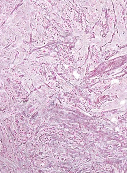
4/29/2021

                     Just too many entities

                     WHO also includes a section on hereditary brain tumor syndromes

                     Some entities I have not seen myself

                     Sorry cannot illustrate everything

       WHO Classification 2021

      Imminently in print and latest June
Look out for summary in Neuro‐oncology soon
                Louis DN et al.

                                                                                              2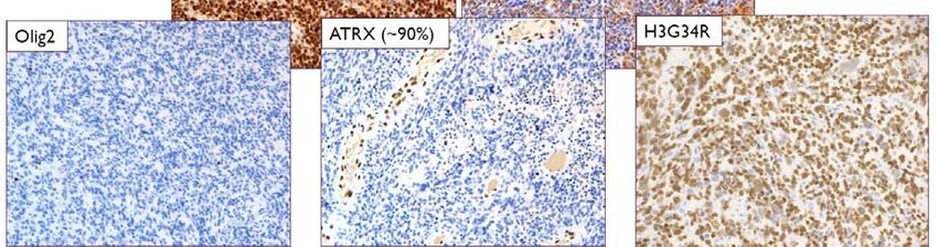
4/29/2021

    Disclaimer : Just don’t shoot the messenger

WHO 2021 – led by IARC
        Ten neuropathologists
        ‐ Brat, Dan
        ‐ Ellison, David
        ‐ Figarella‐Branger, Dominique
        ‐ Hawkins, Cynthia
        ‐ Louis, David
        ‐ Perry, Arie
        ‐ Ng, H K
        ‐ Von Deimling, Andreas
        ‐ Reifenberger, Guido
        ‐ Wesseling, Pieter

        Two clinicians
        ‐ Pfister, Stefan
        ‐ Soffietti, Riccardo

                                                         3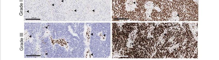
4/29/2021

Some general changes

• Pediatric and adult gliomas separate
• Grading changed to Arabic numerals, e.g. 1, 2,3, 4 and not I, II, III,IV
• Grading is WITHIN each tumor group
• Methylomes a desirable criteria in
• Many of the new and rare lesions diagnosable by methylation profiles
• Appreciate that some tumors are Not Elsewhere Classified (NEC)
• Integrated diagnosis as per Haarlem (2014) recommendation

                                                                                    4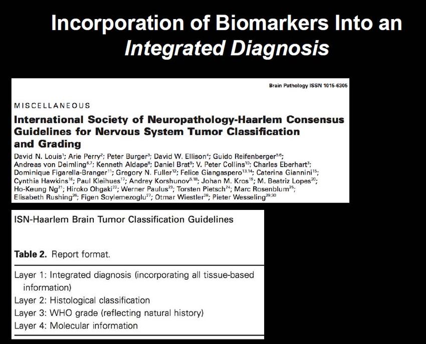
4/29/2021

Whole genome methylation profiling

                                 Binary
                                 IDAT
                                 files
                                 (1 for red,
                                  1 for green)

                                                        5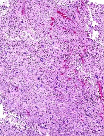
4/29/2021

Typical report from DKFZ Molecular Classifier

                                                Methylation profiling listed as
                                                DESIRABLE criteria in WHO 2021
                                                for many entities

                                                “Histological diagnosis without
                                                Histology”

   Capper D et al. Nature 2018

                                                                                         6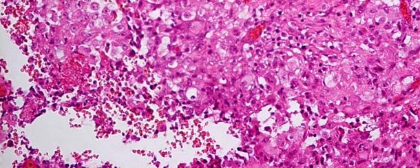
4/29/2021

  The methylomes
  A new kind of
  “histology”

 Wong, Ng
 Modern Pathology 2021

                                                                IDH glioma, subclass 1p/19q codeleted oligodendroglioma                                GBM_RTK I
                                           A_IDH_HG             IDH glioma, subclass astrocytoma                          GBM_MES

               A_IDH                                            IDH glioma, subclass high grade astrocytoma
                                                                glioblastoma, IDH wildtype, subclass RTK I
 O_IDH
                                                                glioblastoma, IDH wildtype, subclass RTK II
                                                                glioblastoma, IDH wildtype, subclass mesenchymal

                                                                IDH mutant glioblastomas (our cohort, n=86)                                      GBM_RTK II

Figure 1. Unsupervised clustering of reference cohort samples and 85 IDH mutant glioblastomas using t-SNE dimensionality reduction. The reference cohort of the DKFZ
CNS tumor classifier includes 82 tumour and 9 non-tumour classes and they are shown as circles of different colors. The 85 primary IDH mutant glioblastomas of our
cohort clustered mainly to the (I) IDH mutant high grade astrocytomas; (2) glioblastoma, IDH wildtype, subclass RTK II and (3) subclass mesenchymal (green triangles).
Mutations of IDH in our samples were tested and confirmed by independent PCR and sanger sequencing.

              Methylomes also give you the complete cytogenetic picture of copy number variation of genes

                                                                                                               Wong, Ng. Modern Pathology 2021

                                                                                                                                                                                7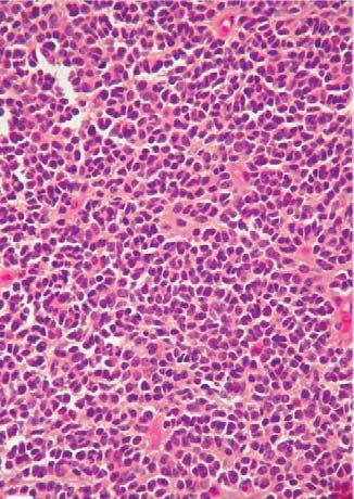
4/29/2021

Other information obtainable from
methylomes

  • MGMT
  • 1p19q status
  • G‐CIMP status (have to work
   Out yourself)

      Pediatric gliomas
       2.1.2:      Pediatric-type diffuse low-grade gliomas
                              2.1.2.1: Diffuse astrocytoma, MYB- or MYBL1-altered
                              2.1.2.2: Angiocentric glioma
                              2.1.2.3: Diffuse low-grade glioma, MAPK-altered
                              2.1.2.4: Polymorphous low-grade neuroepithelial tumour of the young

       2.1.3:      Pediatric-type diffuse high-grade gliomas
                               2.1.3.1: Diffuse midline glioma, H3 K27M-mutant
                               2.1.3.2: Diffuse hemispheric glioma, H3 G34-mutant
                               2.1.3.3: Diffuse paediatric-type high-grade glioma, H3-wildtype and IDH-wildtype
                               2.1.3.4: Diffuse midline glioma, EGFR-mutant
                               2.1.3.5: Infant-type hemispheric glioma, H3-wildtype
       2.1.4:      Circumscribed astrocytic gliomas
                               2.1.4.1: Pilocytic astrocytoma
                               2.1.4.2: High-grade astrocytoma with piloid features
                               2.1.4.3: Pleomorphic xanthoastrocytoma
                               2.1.4.6: Subependymal giant cell astrocytoma
                               2.1.4.7: Chordoid glioma
                               2.1.4.8: Astroblastoma-MN1
       2.1.5:      Glioneuronal and neuronal tumours
                    2.1.5.1: Ganglioglioma
                    2.1.5.2: Desmoplastic infantile astrocytoma / ganglioglioma
                    2.1.5.3: Dysembryoplastic neuroepithelial tumour
                    2.1.5.4: Diffuse glioneuronal tumor with oligodendroglioma-like features and nuclear clusters
                    2.1.5.5: Papillary glioneuronal tumour
                    2.1.3.13 Diffuse leptomeningeal glioneuronal tumor
                       Many others………………………………

                                                                                                                           8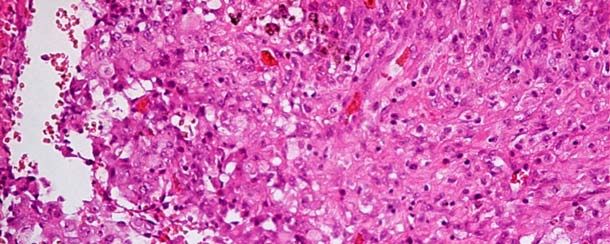
4/29/2021

          Grades 1 and 2 gliomas in children have similar prognosis

               Bandopadhayay P Pediatric Blood & Cancer 2014

                 N=4,400

Grades 3 and 4 gliomas in children have similar prognosis in children

                                                                               9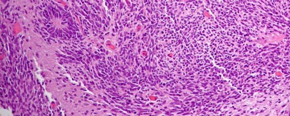
4/29/2021
H3K27M
• Midline H3K27M mutant gliomas (DIPG)
• Pitfalls : may occur outside midline; usually poor prognosis
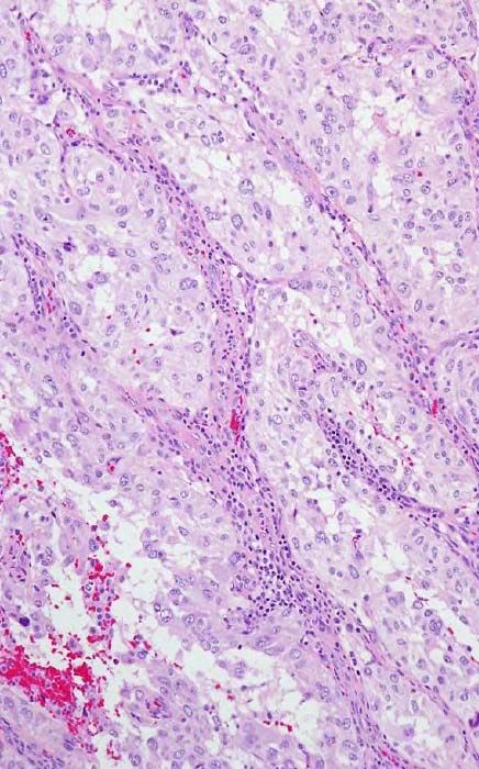
6 years old female
Thalamic GBM
K27M-H3.3 mutations
(AAG → ATG, lysine → methionine)
104/29/2021
“DIPG” – H3K27M mutant mid‐line glioma
Cynthia Hawkins, AANP
114/29/2021
Cynthia Hawkins, AANP
Pediatric high grade gliomas IDHwt, H3wt are still poorly characterized
Cynthia Hawkins, AANP web
124/29/2021
Diffuse midline glioma, EGFR mutant
134/29/2021
Infantile gliomas are a separate group in WHO 2021
2019
Infant high grade gliomas comprise multiple subgroups
Characterised by novel target fusions and better survivals
Clarke M, Mackay A…….Ng HK……Jones C
Cancer Discovery 2020
Note : NTRK inhibitors in clinical trials
Polymorphous low grade neurepithelial tumor of the young
From WHO 2021
About 50% BRAF mutated or have other MAPK aberrations,
usually FGFR2 or FGFR3
WHO 2021
CD34
144/29/2021
BRAF
8/M
? A form of PLNTY or pediatric low grade
Glioma with MAPK activation
Yang, Ng. Brain Pathology 2020
Angiocentric glioma – MYB‐QKI fusion
A Grade 1 pediatric diffuse glioma characterized by chronic intractable seizure
WHO 2021
154/29/2021
Diffuse astrocytoma, MYB or MYBL1 altered
Is a grade 1 pediatric diffusely infiltrative glioma
Previously called isomorphic glioma
BRAF V600E is a poor prognosticator for p‐LGG
Lassaletta…Ng....Tarbori
Journal of Clinical Oncology
2017
164/29/2021
Risk stratification of pLGG into low, intermediate and high groups
Low risk; n=86 Low risk
(BRAF fusion or MYB amplification
Progression free survival
Intermediate risk
p-=0.0002 (BRAFV600E or
without H3F3A/TERT/BRAF/MYB alterations)
High risk
p4/29/2021
WHO2021 defines DNET as a cortical, circumscribed
Glioneuronal tumor with alterations of FGFR1
184/29/2021
Glioneuronal tumor
Desmoplastic infantile
Astrocytoma / Ganglioglioma
Myxoid glioneuronal tumor
A tumor at septum pellucidum or periventricular
region with characteristic dinucleotide putation
of PDGFRA. Slow growing. Grade 1.
194/29/2021
Diffuse leptomeningeal glioma
Ng, Poon. Pathology 1999
May exhibit 1p loss or BRAF fusion
204/29/2021
Papillary glioneuronal tumor is defined by PRKCA‐fusions
WHO 2021
2.1.4: Circumscribed astrocytic gliomas
2.1.4.1: Pilocytic astrocytoma
2.1.4.2: High-grade astrocytoma with piloid features
2.1.4.3: Pleomorphic xanthoastrocytoma
2.1.4.6: Subependymal giant cell astrocytoma
2.1.4.7: Chordoid glioma
2.1.4.8: Astroblastoma-MN1
214/29/2021
Astroblastoma is defined now by MN1‐alterations
WHO 2021
Prototype circumscribed glioma : pilocytic astrocytoma
pilocytic
224/29/2021
BRAF Gene Rearrangement (Fusion)
Two normal signals (orange) plus a smaller third signal near one of the large signals
PXA is characterized by
BRAF mutation and CDKN2A deletion
BRAF V600E
234/29/2021
Adult diffuse gliomas
244/29/2021
Should the diagnosis of
glioblastoma just be
HISTOLOGICAL ?
Endothelial proliferation
necrosis
254/29/2021
TERT promoter mutations
Life history of a glioblastoma
TERT is a common end point
From Wesseling and Verhaak
264/29/2021
Li, Ng.
NOA 2019
CDKN2A/B homozygous deletion (FISH or methylation) : major prognosticator in IDH mutant astrocytomas
Criteria for molecular Astrocytoma Grade IV
Also Shirahata, von Deimling, ANP 2019; Yang R, Ng HK. Brain Pathology 2020
CIMPACT‐NOW Brat et al. 2020
Adult diffuse gliomas
• Astrocytoma, IDH mutant
(Grades 2‐4, “IDH mut glioblastoma” discarded;
homozygous deletion of CDKN2A/B as Grade 4 criteria for cases not
fulfilling histology criteria)
• Oligodendroglioma, IDH mutant and 1p19q codeleted
• Glioblastoma, IDH wild type
(EGFR, TERT, 7+/10‐ for cases not fulfilling histology criteria)
274/29/2021
But that does not mean :
• You need to do CDKN2A/B to diagnose an obvious glioblastoma which
is IDH mutant (Wong, Ng. Modern Pathology 2021)
• You need to do EGFR or TERT or 7+/10‐ for an obvious glioblastoma
which is IDHwt
• These are criteria for molecular glioblastoma or Grade 4 for the Grade
2‐3 lesions which do not fulfil the histological criteria
Giant cell glioblastoma is a variant enriched for p53 mutatipn (Shi, Ng. Brain Pathology 2019)
284/29/2021
Epitheloid glioblastoma typically has BRAF mutation
WHO 2021 BRAF
Ependymoma
• Supratentorial ependymoma
• Supratentorial ependymoma ZFTA (RELA) fusion‐positive
• Supratentorial ependymoma YAP1 fusion‐positive
• Posterior fossa ependymoma
• Posterior fossa ependymoma Group PFA
• Posterior fossa ependymoma Group PFB
• Spinal ependymoma
• Spinal ependymoma, MYCN‐amplified
• Myxopapillary ependymoma
• Subependymoma
294/29/2021
Posterior fossa ependymoma is the commonest
clinical scenario for ependymomas
PFA younger age and majority
Witt & Pfister Cancer Cell 2011
304/29/2021
314/29/2021
Most supratentorial ependymomas are ZFTA (RELA) fusion positive RELA
and clinically aggressive
M/3 L frontal lobe tumor
LICAM
Or p65/RELA
RELA‐positive ependymoma
M/15, ST. T and QMH, 2007‐2019
324/29/2021
From :
Ng in Russell and Rubinstein
2006
Spinal cord ependymomas are mostly low grade
Spinal ependymoma
• Without MYCN : good prognosis
• With MYCN : poor prognosis
334/29/2021
Medulloblastoma
`
Medulloblastoma
Medulloblastoma, molecularly defined
• Medulloblastoma, Wnt activated
• Medulloblastoma, SHH activated and p53 wt
• Medulloblastoma, SHH activated and p53 mt
• Medulloblastoma, non‐Wnt, non‐SHH
• Medulloblastoma, histologically defined
344/29/2021
McLendon, Ng.
Hematology /Oncology
Clinics 2021
Wnt MB ‐ Beta‐catenin
354/29/2021
Filamin
SHH medulloblastoma
Yap1
M/5. Anaplastic medulloblastoma with drop metastasis and MYC amplification
364/29/2021
Molecular grouping of medulloblastoma
• IHC (cannot distinguish Group 3 and Group 4)
• Nanostring transcriptomes
• Methylation profiling
• In addition
• FISH for example for MYC or MYCN or other cytogenetics required by
your clinicians if not doing methylation profiling
374/29/2021
Other CNS embryonal tumors
• Atypical teratoid rhabdoid tumor (AT/RT)
• Cribriform neuroepithelial tumor
• Embryonal tumor with multi‐layered rosettes (ETMR)
• CNS neuroblastoma FOXR2 activated
• CNS tumor with BCOR internal tandem duplication
SMARCB1 (INI1) loss
• Lost in ATRT
• But also in poorly differentiated chordomas
• CRINET
384/29/2021
Figure 2a. AT/RT
M/6 months, cerebellar tumor.
This area represents the
area of the tumor with rhabdoid‐
like cells
AT/RT, negative INI1‐staining
Heterozygous mutation
NM_003073.4:c.367C>T
NP_003064:p.Q123*
Germline mutation of SMARCB1
exon 4
394/29/2021
CRINET is a ventricular tumor characterized by histology and SMARCB1 mutation and INI1‐loss
Behavior is still not very certain at this stage
WHO 2021
ETMR
(embryonal tumor with multi‐
Layered rosettes)
F/1, frontal lobe tumor.
404/29/2021
ETMR – Lin28A positive and C19MC amplifictaion
All types of meningiomas
as a single tumor entity with
Different grades
414/29/2021
Mesenchymal non‐meningeal tumors ‐
selected
• Solitary fibrous tumor
• Rhabdomyosarcoma
• Intracranial mesenchymal tumor, FET‐CREB fusion positive
• CIC‐rearranged sarcoma
• Primary intracranial sarcoma, DICER‐mutant
• Ewing sarcoma
Pineal tumors
• Pineocytoma
• Pineal parenchymal tumor of intermediate differentiation
• Pineoblastoma
• Papillary tumor of the pineal gland
• Desmoplastic myxoid tumor of the pineal region, SMARCB1 mutant
424/29/2021
Tumors of the sellar region
• Adamantinomatous craniopharyngioma
• Papillary craniopharyngioma
• Pituicytoma, granular cell tumor and spindle cell oncocytoma
• Pituitary adenoma of PitNET
• Pituitary blastoma
F Stephen Vogel
Peter Burger Robin O
Barnard
S K Yee Foundation
43You can also read