Yersinia enterocolitica: Prevalence, virulence, and antimicrobial - Veterinary World
←
→
Page content transcription
If your browser does not render page correctly, please read the page content below
Veterinary World, EISSN: 2231-0916                                                                            RESEARCH ARTICLE
Available at www.veterinaryworld.org/Vol.12/July-2019/23.pdf                                                        Open Access

         Yersinia enterocolitica: Prevalence, virulence, and antimicrobial
               resistance from retail and processed meat in Egypt
                                                Gamal Younis, Mona Mady and Amal Awad

 Department of Bacteriology, Mycology and Immunology, Faculty of Veterinary Medicine, Mansoura University, Mansoura,
                                                  35516, Egypt.
                         Corresponding author: Amal Awad, e-mail: amalabdo@mans.edu.eg
                 Co-authors: GY: gamalyounis_2006@yahoo.com, MM: mona.angel221@yahoo.com
                   Received: 09-03-2019, Accepted: 12-06-2019, Published online: 21-07-2019

doi: 10.14202/vetworld.2019.1078-1084 How to cite this article: Younis G, Mady M, Awad A (2019) Yersinia
enterocolitica: Prevalence, virulence, and antimicrobial resistance from retail and processed meat in Egypt, Veterinary
World, 12(7): 1078-1084.

                                                                   Abstract
Background and Aim: The objectives of this study were to investigate the prevalence of Yersinia enterocolitica in retail
chicken meat, ground and processed beef meat, determine their virulence-associated genes, antimicrobial susceptibility
pattern, molecular detection of extended-spectrum β-lactamases, and their capability of biofilm formation in vitro.
Materials and Methods: A total of 210 samples (120 retail chicken meat, 30 ground beef, 30 beef burger, and 30 sausage
samples) were collected from different retail chicken outlets and markets located at Mansoura city between December 2016
and April 2017. Meat samples were examined bacteriologically for the existence of Y. enterocolitica; bacterial colonies
that displayed positive biochemical properties were subjected to polymerase chain reaction targeting 16 rRNA gene.
Y. enterocolitica isolates were tested for their susceptibility to six antimicrobial agents using disk diffusion method. Uniplex
PCR was used for screening Y. enterocolitica isolates for the presence of two virulence chromosome-associated genes (ail
and yst), and β-lactamases (blaTEM and blaSHV). The capability of Y. enterocolitica to form biofilms was detected by tube
method.
Results: Thirty Y. enterocolitica isolates (14.29%) were recovered including 19 (15.83%) isolates from chicken meat,
3 (10%) from ground beef, 5 (16.67%) from beef burger, and 3 (10%) from sausage samples. Regarding ail gene, it was
detected in 6.67% (2/30), while yst gene detected in 20% (6/30) Y. enterocolitica isolates. About 80%, 70%, 63.33%, and
50% of Y. enterocolitica isolates were sensitive to ciprofloxacin, gentamicin, cefotaxime, and streptomycin, respectively,
while 83.33% of Y. enterocolitica isolates were resistant to both ampicillin and cephalothin. Interestingly, 21 (70%) isolates
had the capability of biofilms formation in vitro. Among the multidrug-resistant (MDR) strains, a significant difference
(p
Available at www.veterinaryworld.org/Vol.12/July-2019/23.pdf
detected in virulent strains [10]. Two chromosomal Materials and Methods
virulent-associated genes (ail and yst) were included Ethical approval
in our analysis. The ail gene (adhesion invasion locus) There was no need for ethical approval in this
encodes a surface factor that enhances epithelial cell study because no live animals were involved. Meat
penetration, is involved in adhesion and invasion, and samples were collected from retail outlets and various
is found only among pathogenic strains [11]. The yst supermarkets.
gene encodes enterotoxin is frequently detected in
Samples collection
diarrheagenic biotype 1A strains [12].
A total of 210 samples were examined in this
Antimicrobial treatment is not necessary for
study, including 120 retail chicken meat samples, 30
treating Y. enterocolitica in immunocompetent hosts
ground beef samples, and 60 processed meat samples
because most gastrointestinal infections are self-lim-
(30 beef burger and 30 sausage). Meat samples were
iting. However, systemic, extraintestinal, and inva-
chosen randomly from retail outlets and various super-
sive infections in immunocompromised patients with
markets in Mansoura city, Egypt, from December
increased risk for developing bacteremia or septice-
2016 to April 2017. The collected samples (approx-
mia require special attention and antibiotic therapy
imately 50 g each) were kept separately in a sterile
since the mortality rate in these cases can be as high as
plastic bag, labeled, and delivered to the laboratory for
50% [10]. Ciprofloxacin, ceftriaxone, and cefotaxime
bacteriological examinations in an ice container.
have been used successfully for complicated infection
treatments such as liver abscess, endocarditis, and Isolation and identification of Y. enterocolitica isolates
septicemia [13]. About 25 g of each sample was homogenized in
Bacterial biofilm is a community of microorgan- 225 ml of peptone, sorbitol, and bile salts broth (PSB;
isms surrounded by a self-produced matrix consisting Difco, Heidelberg, Germany) and homogenized in
of an extracellular polymeric substance of polysaccha- a Stomacher for 2 min at high speed. Homogenized
rides, lipids, proteins, and nucleic acids [14]. Biofilm samples were incubated at 25°C for 72 h. Then, a
formation protects bacterial cells from antibacterial loopful of the enriched samples was streaked into
agents, phagocytes, bacteriophages, and antibodies. Cefsulodin-Irgasan-Novobiocin agar medium plates
In food industry, biofilms increase the resistance of (Oxoid) and incubated for 24 h at 30°C under aerobic
bacteria to many physical and chemical factors which conditions. Colonies with typical red bull’s eye-like
represent a food safety concern. Accordingly, the colonies (small, with a red center and translucent rim)
process of biofilm formation represents a method of were purified on the surface of tryptic soy agar plates
protection for microbial growth allows bacterial cells (Oxoid) and then stored at 4°C for further examina-
to survive and grow in several adverse environments. tion. Preliminary identification of Y. enterocolitica
In addition, the formation of biofilms may result in isolates depended on the colony morphological crite-
chronic infection due to the resistant of pathogens to ria by Gram’s stain and biochemically subjected to the
both host immunity and antibiotic treatment [15]. following biochemical tests: catalase, oxidase, urease
Therefore, this study was designed (1) to investi- production, Simmons citrate, triple sugar iron, and
gate the prevalence rate of Y. enterocolitica in chickens sugar fermentation [16]. Furthermore, a 16S rRNA-
meat, ground and processed beef meat samples col- based polymerase chain reaction (PCR) technique was
lected from Mansoura, Egypt, with the determination used for Y. enterocolitica confirmation using the prim-
of its associated chromosomal virulence genes (ail ers and the conditions listed in Table-1 [17-20].
and yst); (2) to determine the antimicrobial suscepti- Molecular detection of β-lactam resistance genes
bility patterns, β-lactam resistance genes, and biofilm and virulence-associated genes
formation; and (3) to ascertain if there is a relationship The cell lysates from Y. enterocolitica isolates
between biofilm formation and multidrug-resistant were obtained by boiling [7] to be used as DNA tem-
(MDR) isolates. plates. Confirmed Y. enterocolitica were screened for
Table-1: Oligonucleotide primers sequences used in this study.
Target Gene Primer Sequence Annealing Amplicons size References
Y. enterocolitica 16S rRNA Y1 AATACCGCATAACGTCTTCG 60 330 bp [17]
Y2 CTTCTTCTGCGAGTAACGTC
ail ail‑F TAATGTGTACGCTGCGAG 55 351 bp [18]
ail‑R GACGTCTTACTTGCACTG
yst Pr2a AATGCTGTCTTCATTTGGAGC 55 145 bp
Pr2c ATCCCAATCACTACTGACTTC
blaTEM TEM‑F ATTCTTGAAGACGAAAGGGC 60 1150 [19]
TEM‑R ACGCTCAGTGGAACGAAAAC
blaSHV SHV‑F CACTCAAGGATGTATTGTG 50 885 [20]
SHV‑R TTAGCGTTGCCAGTGCTCG
Y. enterocolitica=Yersinia enterocolitica
Veterinary World, EISSN: 2231-0916 1079Available at www.veterinaryworld.org/Vol.12/July-2019/23.pdf the presence of two virulence chromosome-associated calculated by Chi-square test. A statistically significant genes, including adhesion invasion locus gene (ail) difference did exist if p
Available at www.veterinaryworld.org/Vol.12/July-2019/23.pdf
Y. enterocolitica that contains virulence factor cephalothin resistance [38]. MDR was detected in
only has the capability to induce disease [29]. 23.33% (7/30) of Y. enterocolitica isolates, but none
Virulence genes in Y. enterocolitica located either on of the examined isolates was resistance to all of the
chromosome or on a plasmid named pYV. The most tested antimicrobial agents. However, the isolates
significant virulence genes located on chromosome show high susceptibility to ciprofloxacin, gentami-
include ail, inv, and yst. Detection of ail gene is a cin, cefotaxime, and streptomycin. The antimicro-
key factor to distinguish pathogenic and non-patho- bial resistance pattern of the isolated Y. enterocolitica
genic Y. enterocolitica [30]. In this study, Y. entero- strains is described in Table-4. By testing the correla-
colitica isolates were examined for the presence of tion between phenotypic and genotypic antimicrobial
two chromosomal genes because plasmid pYV is susceptibility, we found a positive correlation between
easily lost depending on various factors [31]. The ail the presence of blaTEM gene with the phenotypic resis-
gene is a type of protein responsible for adhesion and tance to ampicillin (R=0.5) and cephalothin (R=0.3),
invasion [11], while yst is an enterotoxin, commonly as well as, blaSHV correlated positively with the resis-
found in diarrheagenic biotype 1A strains [12]. The tance to ampicillin (R=0.3) and cephalothin (R=0.2).
obtained results from this study reveal the presence In addition, there is positive correlation between both
of ail in 6.67% (2/30) of the tested isolates, while yst genes (blaTEM and blaSHV: 0.49).
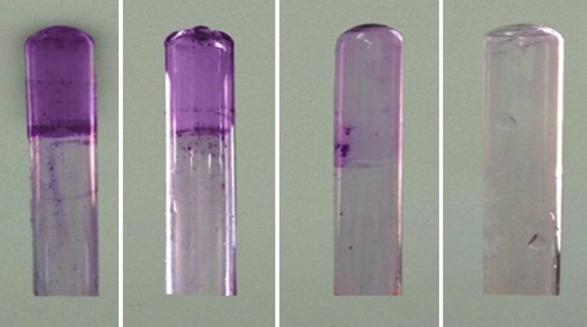
was carried by six strains (20%). The obtained results The presence of biofilms could contribute
from the present study were in accordance with Peng to Y. enterocolitica pathogenicity; in this study,
et al. [32] and lower than that described by Sacchini 21 (70%) isolates had the capability to form biofilms.
et al. [33], who could detect ail and yst from all tested Eleven isolates were strong, six isolates were moderate,
Y. enterocolitica isolates, while Bhagat and Virdi [34] four isolates were weak biofilm producer (Figure-1),
could not identify both genes in their study. The patho- while 30% (9/30) were detected as non-biofilm pro-
genicity of Y. enterocolitica associated closely with ail ducer. As previously mentioned, MDR was detected
gene, biotypes, and serotypes [35]. The diversity in in 7 strains (23.33%) of Y. enterocolitica isolates. By
the pathogenicity of Y. enterocolitica isolates may be
depended on the geographical area of isolation. By Table-3: Number and percentage of Y. enterocolitica
testing the correlation between the presence of the two antimicrobial pattern isolated from chicken and minced
meat.
virulence genes used, we found a strong positive cor-
relation between them (yst and ail: 0.56). Antimicrobial agent Code Sensitive Resist
Antimicrobial resistance among Y. enterocolitica n % n %
isolates was tested against ampicillin (AMP), ceph-
Ciprofloxacin CIP 24 80 6 20
alothin (CF), ciprofloxacin (CIP), gentamicin (GN), Gentamicin GN 21 70 9 30
cefotaxime (CTX), and streptomycin (S). Regarding Cefotaxime CTX 19 63.33 11 36.67
β-lactam resistance, 83.33% (25/30) of the examined Streptomycin S 15 50 15 50
isolates showed resistance to both ampicillin and Cephalothin CF 5 16.66 25 83.33
cephalothin. The tested strains revealed intermediate Ampicillin AMP 5 16.66 25 83.33
resistant against streptomycin (50%; 15/30) and a CF=Cephalothin, AMP=Ampicillin, S=Streptomycin,
CTX=Cefotaxime, CIP=Ciprofloxacin, GN=Gentamicin,
lower resistance was revealed by cefotaxime (36.67%;
Y.enterocolitica=Yersinia enterocolitica
11/30) followed by gentamicin (30%; 9/30) and cip-
rofloxacin (20%; 6/30) as shown in Table-3. Sixteen
Table-4: The distribution of antimicrobial resistance
resistance patterns were detected; the most common profiles among Y. enterocolitica isolates.
pattern was S, CF, AMP represented by eight strains
followed by CF and AMP displayed by five strains. Antimicrobial Number of Number
resistance profile antibiotics of isolates
MDR to ≥3 of the antimicrobial classes tested was
detected in 7 (23.33%) of Y. enterocolitica isolates; CIP, CTX, GN, CF, AMP 5 1
CTX, GN, S, CF, AMP 5 1
among them, the most common profile was CTX, S, CTX, S, CF, AMP 4 2
CF, and AMP. S, AMP, GN, CIP 4 1
Antimicrobial resistance possesses by Gram- CIP, CTX, GN, S 4 1
negative bacteria including Y. enterocolitica which GN, S, CF, AMP 4 1
represents a significant public health problem world- CTX, CF, AMP 3 2
GN, CF, AMP 3 2
wide. In this study, the antimicrobial resistance of CIP, CF, AMP 3 1
Y. enterocolitica isolates was higher in comparison S, CF, AMP 3 8
with other studies; it was noticed that Y. enteroco- GN, S, CF 3 1
litica showed higher rate of ampicillin and cephalo- CTX, AMP 2 1
thin resistance (83.33% each) which is in agreement CF, AMP 2 5
CTX, CF 2 1
with the previous studies [1,33,36,37]; the high per- CTX 1 1
centage of resistance to β-lactam antimicrobial may CIP 1 1
be attributed to the ability of Y. enterocolitica to pro- CF=Cephalothin, AMP=Ampicillin, CTX=Cefotaxime,
duce β-lactamases, which contribute to ampicillin and Y. enterocolitica=Yersinia enterocolitica
Veterinary World, EISSN: 2231-0916 1081Available at www.veterinaryworld.org/Vol.12/July-2019/23.pdf
Biofilm production
The degree of correlation (R) ranges from 0 to 1. 0 indicates the lowest correlation and 1 indicates the highest correlation. The correlation matrix was created using the R
1
0.047565
blaSHV
1
a b c d
Table-5: Correlation matrix showing the positive and negative correlation between phenotypic and genotypic features in Y. enterocolitica isolates.
Figure-1: Detection of the degree of biofilm production
using tube test. (a) Strong biofilm producer, (b) Moderate
−0.19026
0.493213
biofilm producer, (c) Weak biofilm producer, (d) Non-biofilm
blaTEM
producer.
1
testing the relationship between MDR and the capa-
bility of biofilm production, we found that there is no
0.511408
0.391077
0.252982
AMP
relation between MDR and biofilm formation capabil-
1
ity with significant differences between the MDR and
non-MDR isolates (pAvailable at www.veterinaryworld.org/Vol.12/July-2019/23.pdf
References 19. Belaaouaj, A., Lapoumeroulie, C., Caniça, M.M.,
Vedel, G.,Névot, P., Krishnamoorthy, R. and Paul, G. (1994)
1. Aghamohammad, S., Gholami, M., Dabiri, H., Nucleotide sequences of the genes coding for the TEM-like
Rahimzadeh, G., Souod, N., Goudarzi, H., Sardari, S. and β-lactamases IRT-1 and IRT-2 (formerly called TRI-1 and
Mohammadzadeh, A. (2015) Distribution and antimicrobial TRI-2). FEMS Microbiol. Lett., 120(1-2): 75-80.
resistance profile of Yersinia species isolated from chicken 20. Pitout, J.D.D., Thomson, K.S., Hanson, N.D., Ehrhardt, A.F.,
and beef meat. Int. J. Enteric Pathog., 3(4): e29009. Moland, E.S. and Sanders, C.C. (1998) β-lactamases respon-
2. Bolton, D.J., Ivory, C. and McDowell, D. (2013) A small sible for resistance to expanded-spectrum cephalosporins in
study of Yersinia enterocolitica in pigs from birth to carcass Klebsiella pneumoniae, Escherichia coli, and Proteus mira-
and characterization of porcine and human strains. Food bilis isolates recovered in South Africa. Antimicrob. Agents
Control, 33(2): 521-524. Chemother., 42(6): 1350-1355.
3. Nesbakken, T. (2015) Update on Yersinia as a foodborne 21. Clinical and Laboratory Standards Institute. (2015)
pathogen: Analysis and control. Adv. Microb. Food Saf., 2: Performance Standards for Antimicrobial Susceptibility
33-58. Testing; 25th Informational Supplement M100-S25. Clinical
4. Bonardi, S., Bruini, I. and D’Incau, M. (2016) Detection, and Laboratory Standards Institute, Wayne.
seroprevalence and antimicrobial resistance of Yersinia 22. Hassan, A., Usman, J., Kaleem, F., Omair, M., Khalid, A.
enterocolitica and Yersinia pseudotuberculosis in pig ton- and Iqbal, M. (2011) Evaluation of different detection meth-
sils in Northern Italy. Int. J. Food Microbiol., 235(October ods of biofilm formation in the clinical isolates. Braz. J.
2016): 125-32. Infect. Dis., 15(4): 305-311.
5. Tan, L.K., Ooi, P.T. and Thong, K.L. (2014) Prevalence of 23. Zadernowska, A., Chajęcka-Wierzchowska, W. and
Yersinia enterocolitica from food and pigs in selected states Łaniewska-Trokenheim, Ł. (2013) Yersinia enterocolitica:
of Malaysia. Food Control, 35(1): 94-100. A dangerous, but often ignored, foodborne pathogen. Food
6. Anju, P., Latha, C., Sunil, B. and Sethulekshmi, C. (2014) Rev. Int., 30(1): 53-70.
Detection of Salmonella and Yersinia spp. In uncooked 24. Shabana, S., Khalil, S. and Hegazy, A. (2015) Molecular
retail chicken meat in Kerala by multiplex PCR. Int. J. Curr. characterization of Yersinia enterocolitica isolated from
Microbiol. Appl. Sci., 3(6): 1028-1034. chicken meat samples. Alex. J. Vet. Sci., 46(1): 124-129.
7. Zeinali, T., Jamshidi, A., Rad, M. and Bassami, M. (2015) 25. Momtaz, H., Davood, R.M. and Safarpoor, D.F. (2013)
A comparison analysis of Listeria monocytogenes isolates Identification and characterization of Yersinia enterocolit-
recovered from chicken carcasses and human by using ica isolated from raw chicken meat based on molecular and
RAPD PCR. Int. J. Clin. Exp. Med., 8(6): 10152-10157. biological techniques. J. Appl. Poult. Res., 22(1): 137-145.
8. Ryan, K.J. and Ray, C.G., editors. (2004) Sherris Medical 26. Sirghani, K., Zeinali, T. and Jamshidi, A. (2018) Detection
Microbiology: An Introduction to Infectious Disease. 4th ed. of Yersinia enterocolitica in retail chicken meat, Mashhad,
McGraw-Hill, New York. Iran. J Pathog. 2018: Article ID 1286216..
9. Krajinović, V., Andrašević, A.T. and Baršić, B. (2007) 27. Karib, H., Boussatta, H. and Seeger, H. (1999) Yersinia
Tricuspidal valve endocarditis due to Yersinia enterocolit- enterocolitica: Verkommen in rohem fleisch und fleischpro-
ica. Infection, 35(3): 203-205. dukten in Marokko. Fleischwirtsch, 74: 1332-1333.
10. Fàbrega, A. and Vila, J. (2012) Yersinia enterocolitica: 28. Nortjé, G.L., Vorster, S.M., Greebe, R.P. and Steyn, P.L.
Pathogenesis, virulence and antimicrobial resistance. (1999) Occurrence of Bacillus cereus and Yersinia entero-
Enferm. Infecc. Microbiol. Clin., 30(1): 24-32. colitica is South African retail meats. Food Microbiol.,
11. Pierson, D.E. and Falkow, S. (1993) The ail gene of Yersinia 16(3): 213-217.
enterocolitica has a role in the ability of the organism to 29. Ye, Q., Wu, Q., Hu, H., Zhang, J. and Huang, H. (2016)
survive serum killing. Infect. Immun., 61(5): 1846-1852. Prevalence and characterization of Yersinia enterocolitica
12. Singh, I. and Virdi, J.S. (2004) Production of Yersinia sta- isolated from retail foods in China. Food Control, 61(March
ble toxin (YST) and distribution of yst genes in biotype 1A 2016): 20-27.
strains of Yersinia enterocolitica. J. Med. Microbiol., 30. Nesbakken, T., Iversen, T., Eckner, K. and Lium, B. (2006)
53(11): 1065-1068. Testing of pathogenic Yersinia enterocolitica in pig herds
13. Lupi, A., Poletti, F., Mondino, V., Canale, C., Leonardo, L., based on the natural dynamic of infection. Int. J. Food
Rognoni, A. and Nardi, F. (2013) Subacute endocarditis Microbiol., 111(2): 99-104.
caused by Yersinia enterocolitica: A case report. Eur. J. 31. Simonova, J., Vazlerova, M. and Steinhauserova, I. (2007)
Clin. Microbiol. Infect. Dis., 45(4): 329-333. Detection of pathogenic Yersinia enterocolitica sero-
14. Hall, C.W. and Mah, T.F. (2017) Molecular mechanisms of type O:3 by biochemical, serological, and PCR meth-
biofilm-based antibiotic resistance and tolerance in patho- ods. Czech J. Food Sci., 25(4): 214-220.
genic bacteria. FEMS Microbiol. Rev., 41(3): 276-301. 32. Peng, Z., Zou, M., Li, M., Liu, D., Guan, W., Hao, Q., Xu, J.,
15. Ioannidis, A., Kyratsa, A., Ioannidou, V., Bersimis, S. and Zhang, S., Jing, H., Li, Y. and Liu, X. (2018) Prevalence,
Chatzipanagiotou, S. (2014) Detection of biofilm produc- antimicrobial resistance and phylogenetic characterization
tion of Yersinia enterocolitica strains isolated from infected of Yersinia enterocolitica in retail poultry meat and swine
children and comparative antimicrobial susceptibility of feces in parts of China. Food Control, 93(Novamber 2018):
biofilm versus planktonic forms. Mol. Diagn. Ther., 18(3): 121-128.
309-314. 33. Sacchini, L., Garofolo, G., Di Serafino, G., Marotta, F.,
16. Bercovier, H. and Mollaret, H.H. (1984) Genus XIV. Ricci, L., Di Donato, G., Miracco, M.G., Perletta, F. and Di
Yersinia. In: Krieg, N.R., editor. Bergey’s Manual of Giannatale, E. (2018) The prevalence, characterization, and
Systematic Bacteriology. Vol. 1. Williams and Wilkins antimicrobial resistance of Yersinia enterocolitica in pigs
Company, Baltimore. p 498-506. from Central Italy. Vet. Ital., 54(2): 115-123.
17. Wannet, W.J., Reessink, M., Brunings, H.A. and Maas, H.M. 34. Bhagat, N. and Virdi, J.S. (2007) Distribution of viru-
(2001) Detection of pathogenic Yersinia enterocolitica by a lence-associated genes in Yersinia enterocolitica biovar 1A
rapid and sensitive duplex PCR assay. J. Clin. Microbiol., correlates with clonal groups and not the source of isolation.
39(12): 4483-4486. FEMS Microbiol. Lett., 266(2): 177-183.
18. Koua, A., Solange, K.N.E., Thomas, D.E.A., Germain, K.T., 35. Baumgartner, A., Küffer, M., Suter, D., Jemmi, T. and
Mireille, D., Sébastien, A.L. and Marcellin, D.K. (2014) Rohner, P. (2007) Antimicrobial resistance of Yersinia entero-
Characterization of Yersinia spp. Strains isolated from pigs colitica strains from human patients, pigs and retail pork in
in Abidjan, Cote d’Ivoire, West Africa. Afr. J. Microbiol. Switzerland. Int. J. Food Microbiol., 115(1): 110-114.
Res., 8(18): 1909-1915. 36. Anju, P., Latha, C., Sunil, B. and Sethulekshmi, C. (2014b)
Veterinary World, EISSN: 2231-0916 1083Available at www.veterinaryworld.org/Vol.12/July-2019/23.pdf
Antimicrobial resistance profile of Yersinia enterocolitica from chicken and fish in and around Coimbatore City, India.
and Yersinia intermedia isolates from retail pork. Int. J. Iran. J. Public Health, 43(6): 835-844.
Curr. Microbiol. Appl. Sci., 3(8): 231-234. 38. Seoane, A. and Lobo, J.M.G. (1991) Cloning of chromo-
37. Shanmugapriya, S., Senthilmurugan, T. and Thayumanavan, T. somal β-lactamase genes from Yersinia enterocolitica.
(2014) Genetic diversity among Yersinia enterocolitica isolated J. Gen. Microbiol., 137(1): 141-146.
********
Veterinary World, EISSN: 2231-0916 1084You can also read