Development of a europium nanoparticles lateral flow immunoassay for NGAL detection in urine and diagnosis of acute kidney injury
←
→
Page content transcription
If your browser does not render page correctly, please read the page content below

Yin et al. BMC Nephrology      (2022) 23:30
https://doi.org/10.1186/s12882-021-02493-w

 RESEARCH                                                                                                                                            Open Access

Development of a europium nanoparticles
lateral flow immunoassay for NGAL
detection in urine and diagnosis of acute
kidney injury
Moli Yin1, Yuanwang Nie2, Hao Liu2, Lei Liu1, Lu Tang1, Yuan Dong2, Chuanmin Hu1 and Huiyan Wang1*

  Abstract
  Background: AKI is related to severe adverse outcomes and mortality with Coronavirus Disease 2019 (COVID-19)
  patients, that early diagnosed and intervened is imperative. Neutrophil gelatinase-associated lipocalin (NGAL) is one
  of the most promising biomarkers for detection of acute kidney injury (AKI), but current detection methods are
  inadequacy, so more rapid, convenient and accuracy methods are needed to detect NGAL for early diagnosis of
  AKI. Herein, we established a rapid, reliable and accuracy lateral flow immunoassay (LFIA) based on europium
  nanoparticles (EU-NPS) for the detection of NGAL in human urine specimens.
  Methods: A double-antibody sandwich immunofluorescent assay using europium doped nanoparticles was
  employed and the NGAL monoclonal antibodies (MAbs) conjugate as labels were generated by optimizing electric
  fusion parameters. Eighty-three urine samples were used to evaluate the clinical application efficiency of this
  method.
  Results: The quantitative detection range of NGAL in AKI was 1-3000 ng/mL, and the detection sensitization was
  0.36 ng/mL. The coefficient of variation (CV) of intra-assay and inter-assay were 2.57-4.98 % and 4.11-7.83 %,
  respectively. Meanwhile, the correlation coefficient between europium nanoparticles-based lateral fluorescence
  immunoassays (EU-NPS-LFIA) and ARCHITECT analyzer was significant (R2 = 0.9829, n = 83, p < 0.01).
  Conclusions: Thus, a faster and easier operation quantitative assay of NGAL for AKI has been established, which is
  very important and meaningful to diagnose the early AKI, suggesting that the assay can provide an early warning
  of final outcome of disease.
  Keywords: neutrophil gelatinase-associated lipocalin (NGAL), monoclonal antibody, lateral flow immunoassay, acute
  kidney injury (AKI)

* Correspondence: jlmpcwhy@163.com
1
 Jilin Collaborative Innovation Center for Antibody Engineering, Jilin Medical
University, 132013 Jilin, PR China
Full list of author information is available at the end of the article

                                         © The Author(s). 2022 Open Access This article is licensed under a Creative Commons Attribution 4.0 International License,
                                         which permits use, sharing, adaptation, distribution and reproduction in any medium or format, as long as you give
                                         appropriate credit to the original author(s) and the source, provide a link to the Creative Commons licence, and indicate if
                                         changes were made. The images or other third party material in this article are included in the article's Creative Commons
                                         licence, unless indicated otherwise in a credit line to the material. If material is not included in the article's Creative Commons
                                         licence and your intended use is not permitted by statutory regulation or exceeds the permitted use, you will need to obtain
                                         permission directly from the copyright holder. To view a copy of this licence, visit http://creativecommons.org/licenses/by/4.0/.
                                         The Creative Commons Public Domain Dedication waiver (http://creativecommons.org/publicdomain/zero/1.0/) applies to the
                                         data made available in this article, unless otherwise stated in a credit line to the data.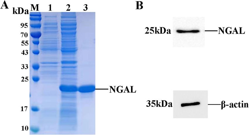
Yin et al. BMC Nephrology   (2022) 23:30                                                                          Page 2 of 9

Background                                                       Methods
Coronavirus Disease 2019 (COVID-19) has widely spread            Expression and purification of NGAL
in the worldwide scale with serious disaster[1].Acute kid-       The human NGAL gene sequence from Genbank (NP_
ney injury (AKI) has a higher rate of morbidity and mor-         005555.2) was synthesized by Beijing Institute of Gen-
tality in common complication for critical illnesses and         omics (BGI) and added restriction enzymes HindIII and
counted about 5-7 % of hospitalized patients in world[2].-       XhoI at both ends. The plasmids were digested with Hin-
Several studies have evaluated the development of AKI is         dIII and XhoI, and then cloned into the pSecTag2A vec-
more strongly related to worse outcomes and mortality            tor. The constructed plasmids were sequenced to
rates of COVID-19, described incidence of AKI that               confirm without mutation, and then transformed into
ranges widely from 0.5 to 36.6 % in COVID-19 patient-            Chinese Hamster Ovary (CHO) cells by the Lipofectami-
s[3].Early detection and precise treatments of AKI can im-       neTM2000. After 8 days, the cell supernatant was col-
plement better preventive strategies and prevent                 lected after filtering 0.45 μm filter. The expressed
deterioration of renal function and renal failure, effectively   NGAL-6×His protein was purified with the Nickel Nitri-
contain progression of the COVID-19 hospitalized pa-             lotriacetic Acid (Ni-NTA) column, and the different
tients[4]. Among them, neutrophil gelatinase-associated          fractions were collected and appraised by sodium dode-
lipocalin (NGAL) has been recognized as one of the prom-         cyl sulfate-polyacrylamide gel electrophoresis (SDS-
ising biomarkers candidate for detection of AKI. NGAL is         PAGE). The bicinchoninic acid (BCA) Protein Quantifi-
a 25 kDa glycoprotein associated with gelatinase from            cation Kit measured the concentration of protein.
neutrophil and usually exist at lower level in human tis-          The purified recombinant protein was subjected in
sues such as stomach, colon and kidney, but its expression       12 % SDS-PAGE, then adsorbed onto a polyvinylidene
is dramatical increased in serum and urine when the kid-         fluoride (PVDF) membrane. After the membrane was
ney was with ischemic or nephrotoxic injury[5].                  blocked in tris buffer with 1 % Tween-20 (TBST) solu-
   Lateral flow immunoassays (LFIA) has been regard as           tion containing 5 % skimmed milk at room temperature
desired screening assays on account of simplicity, in-situ       (RT) for 2 h, incubated with HRP-conjugated anti-6×His
analysis and easy to work[6]. The LFIA with fluorescent          tag antibody (1:5000) in the dark for 1 h at RT, visual-
microparticles have already been used for detection of           ized with Benzidine after washed with TBST and visual-
various microbial pathogens and several inflammation             ized by Bio-Rad Western blotting detection system
markers[7, 8]. Several novel nanoparticles have been             (DNR Bio-Imaging Systems Ltd., Israel).
generally applied to improve the sensitivity of LFIA, in-
cluding carbon nanoparticles, quantum dots, fluorescent          Generation and purification of MAbs
dyes, magnetic nanolabels and europium nanoparticles             In the first immunization, the female BALB/c mice (age
(EU-NPS)[9]. EU-NPS as carriers can improve 100-fold             of 6 weeks, a total of 10) were immunized with 50 µg
sensitivity contrast with colloidal gold nanoparticles la-       NGAL-6×His recombinant protein emulsified in equal
beled in LFIA[10]. EU-NPS are long fluorescence life-            dosage of complete Freund’s adjuvant, and accessional
time and also available with an average particle size of         immunized were accomplished with protein emulsified
75–100 nm range, the large Stokes shift of which is usu-         in incomplete Freund’s adjuvant. Two hypodermic injec-
ally over 200 nm is conducive to avoid the interference          tions on the back of mice and subsequent were intraper-
of scattered light caused by measuring excitation light.         itoneal injections spaced 21 days. The serum samples
EU-NPS have wide excitation band so that it is beneficial        were collected one week after the third injection, and
to increase the excitation energy, the sharp emission            the titre of antiserum was determined with indirect
peak, low background and high resolution, used in                enzyme-linked immunosorbent assay (ELISA)[12]. Three
sandwich-type immunoassays of medical diagnostics                days before fusion, mice were performed to booster
recently[11].                                                    immunization with 50 µg of NGAL-6×His diluted with
   Here, we established a new method with EU-NPS as labels       0.9 % NaCl.
of LFIA for the rapid, sensitive and early measurement of          The isolated immune mice spleen cells and the SP2/0
NGAL in urine based on two monoclonal antibodies (MAbs)          myeloma cells mixed at a ratio of 3:1 to fuse in a plat-
1G1 and 2F4 which are discoveried by our lab. The method         inum electrode LF498-3 fusion chamber (BEX Co., Ltd,
is double-antibody sandwich immunofluorescent assay using        Japan) as described literature[13]. Briefly, the mixed cell
EU-NPS and MAbs conjugate as labels. The mAb 1G1 was             was washed twice with 10 mL electrofusion buffer
conjugated with EU-NPS and the mAb 2F4 was used to cap-          (0.3 M mannitol, 0.1 mM CaCl2, 0.1 mM MgCl2, pH
ture EU-NPS-1G1-antigen complex in T-line. Our results           7.2), re-suspended at a concentration of 2 × 10⁷ cells/
showed that EU-NPS-LFIA could be used for the early              mL. The fusion was completed using an alternating
NGAL detection in urine and allow improvement in the             current voltage of 50 V at 0.8 MHz for 20 s, direct
treatment of AKI patients.                                       current pulse voltage of 450 V of 2 repetitions for 0.5 s,
Yin et al. BMC Nephrology   (2022) 23:30                                                                    Page 3 of 9

and post-fusion was 50 V at 0.8 MHz for 7 s. Finally, the    membrane was visualized by the Western blot detection
electric-treated cell suspension was moved from the fu-      system of enhanced chemiluminescence (ECL).
sion chamber into 4.5 mL of preheated RPMI 1640
(20 % fetal bovine serum) for 30 min at 37℃, then cul-       Competitive enzyme-linked immunosorbent assays
tured in 96-well plates and incubated with 5 % CO2 at        (cELISA)
37℃. After 24 h, hypoxanthine-aminopterin-thymidine          Two MAbs were tested for the ability to recognize to
(HAT) was supplemented to each well. The Cell culture        unique epitopes on NGAL by cELISA. MAb was conju-
supernatants were screened by ELISA after 9 days fu-         gated with HRP by using HRP Antibody Labeling Kit
sion, and calculated number of hybridoma clones. The         (Shanghai YSRIBIO industrial co., LTD), and the work-
BALB/c mice which injected with paraffin oil in advance      ing concentration of which was tested through direct
were inoculated with 1 × 10⁶ of NGAL hybridoma cells,        ELISA. 96-well Microtitre plates were coated with 2 µg/
the ascites were purified by Protein A column.               mL of NGAL antigenin overnight at 4 °C, then unlabeled
                                                             MAb (0.2 µg/ well, 2 µg/ well) was competitively bound
                                                             with the optimal dilution ratio of HRP labeled MAb and
Identification of MAbs                                       incubated for 1 h at 37 °C. The enzymatic reaction was
The immunoglobulin subclasses of antibodies were ana-        appeared with hydrogen peroxide by substrate 3,3’,5,5’-
lyzed using the antibody subclass identification kit. The    Tetramethylbenzidine (TMB) and stopped by 2 M sul-
indirect ELISA screened the specific MAbs by using           furic acid to all wells. The absorbance (OD450 nm) was
purified recombinant NGAL-6×His protein and PCT-             determined by Bio-Rad microplate reader (Bio-Rad La-
6×His protein. The interaction between antigen and           boratories, Inc). The blocking effects of MAbs was calcu-
antibodies were determined with BIAcore T200 system          lated by using the following equation: 100×[1-OD450 nm
(GE Healthcare, Stockholm, Sweden) in HBS-EP buffer          of (HRP-MAb + MAb)/OD450 nm of HRP-MAb]. Two
(0.005 % surfactant P20, 10 mM Hepes, pH 7.4, 3 mM           MAbs recognized different epitopes if blocking effects
EDTA, 150 mM NaCl). The NGAL antigen was                     observed more than 40 %.
adsorbed on CM5 biosensor chips reaching 400–480 re-
sponse units (RU) by an amine coupling kit. The anti-        Conjugation of EU-NPS
bodies (2F4 and 1G1) were diluted in HBS-EP buffer           The Anti-NGAL monoclonal antibody 1G1 and 2F4
were slowly passed over the chip with 50 µL/min for          were covalently conjugated to EU-NPS with standard
5 min, respectively, and subsequently HBS-EP buffer          procedure of Bangs Laboratories. Briefly, 100 µL EU-
injected over the chip to monitor the dissociation phase     NPS were added to 900 µL 0.05 M MES (pH 7.0) and
for 4 min. The sensor chips were regenerated with Gly-       dispersed by ultrasound, vibrated for 15 min at RT in
cine solution (pH 3.0) following the dissociation phase.     the presence of 0.08 M N-hydroxysulfosuccinimide
For each analyte passed over the chip, the specific re-      (NHS) and 0.05 M 1-(3-Dimethylaminopropyl)-3-ethyl-
sponses from the antigen flow channel could subtract         carbodiimide hydrochloride (EDC). The activated EU-
non-specific responses for the control flow channel. The     NPS ware washed with coupling buffer (0.05 mM
fitted saturation binding curves were plotted based on       H3BO3, 0.04 mM Na2B4O7, pH 7.5) and reacted with
concentrations of analyte for equilibrium binding re-        0.3 mg antibody for 2.5 h at RT. The europium-
sponses to calculate KD.                                     conjugated compound was incubated in 1000 µL block-
   Purified anti-NGAL MAbs was analyzed in 12 % SDS-         ing buffer (10 % BSA, 20 % tween-20, 0.05 M Tris-HCl)
PAGE under non-reducing conditions and reducing con-         for 1 h, added to 1000 µL stock solutions (10 % BSA,
ditions. Briefly, sample was mixed with 5 x non-reducing     20 % trehalose, 20 % tween-20, 0.05 M Tris-HCl ) to
buffer or 5 x protein loading buffer and loaded onto         store at 4℃.
12 % SDS-PAGE. The specificity of anti-NGAL MAbs
was determined by Western Blot. The NGAL proteins            The development of LFIA
were done to 12 % SDS-PAGE, then adsorbed onto               The LFIA strips were consisted of nitrocellulose mem-
PVDF membrane that activated by soaking in methanol          brane, conjugate pad, sample pad and absorbing pad.
for 15 s, and then subjected to the electrophoresis condi-   The glass fiber membranes were soaked in the blocking
tions in 100 V for 2 h. After blocking in Tris-HCl buffer    buffer (20 % trehalose, 10 % BSA, 20 % tween-20, 0.05 M
with 1 % Tween-20 solution (TBST) containing 5 %             Tris, 3.2 mM EDTA.Na2, pH 8.6) for 1.5 h. The concen-
skimmed milk at RT for 2 h, the membrane was incu-           tration of 1 mg/mL MAbs (2F4 or 1G1) was coated on
bated with mouse anti-NGAL MAbs as the primary anti-         the test line (TL) of nitrocellulose membrane, and goat-
bodies at 4 °C. The next day, membrane was washed            anti-mouse IgG 1 mg/mL was coated on the same NC
with TBST and incubated with anti-mouse conjugated           membrane at a distance of 4 mm to form a quality con-
HRP IgG in the dark for 1.5 h at RT. Finally, the            trol line (CL), the spray volume of dispenser instrument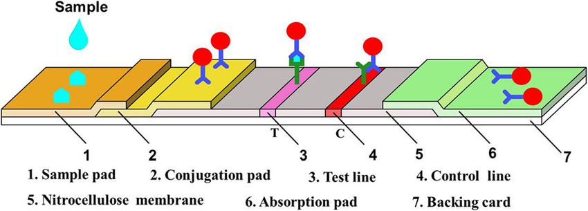
Yin et al. BMC Nephrology   (2022) 23:30                                                                    Page 4 of 9

was set at 1 µL/cm. The membrane and glass fiber mat        T line (HT) and the C line (HC) were recorded by the
were dried for 48 h at 45℃ before tested. The different     reader. Quantitative detection was completed by the
EU-NPS-1G1 and EU-NPS-2F4 conjugate particles were          HT/HC ratio to effectively eliminate strips (T and C) dif-
coated on the conjugate pad, and fluorescence signal        ference and matrsample standard matrix effects[15]. The
was measured by immunofluorescent analyzer                  standard curve was plotted against each concentration of
(Guangzhou Labsim Biotech Co., Ltd).                        NGAL and HT/HC ratio.

Urine sample collection and patients                        Statistical analysis
The total of 83 Human urine samples from AKI patients       The Passing-Bablok regression analysis and Bland-
were harvested from Affiliated Hospital of Jilin Medical    Altman plot were performed by analysis of variance
University. The ethical guidelines were strictly complied   (ANOVA) of MedCalc and SPSS 17.0 software. All data
in the experiment, was provided by the Affiliated Hos-      were showed as mean value with standard deviation
pital of Jilin Medical University (No.2018-LW029). All      (mean ± S.D. ).
subjects received oral and in written informed consent
in Chinese for the study of urine samples. All experi-      Results
ments were performed in accordance with the Declar-         Expression and purification of recombinant NGAL protein
ation of Helsinki Ethical Principles. The average age of    The pSecTag2A-NGAL recombinant plasmid was trans-
patients was 62 years ranging from 20 to 80 years who       fected into CHO cells. The NGAL-6×His protein mainly
were not infected by COVID-19 ,and AKI stage 1–3 was        expressed in cell supernatant and subsequently purified
classified according to Kidney Disease Improving Global     by Ni Sepharose. The purified recombinant NGAL pro-
Outcomes AKI criteria[14]. Urine samples were col-          tein was obtained about 95 % purity and analyzed by
lected for NGAL analysis up to 12 h before AKI was di-      SDS-PAGE with molecular approximate weight of
agnosed and at frequent intervals after operation at        23.7 kDa (Fig. 1 A). Western blot also confirmed the re-
various time points (12, 24, 48 h). For minimize poten-     combinant protein NGAL expression and purification
tial confounding factors, urine samples were analyzed       (Fig. 1B).
rapidly at clinical chemistry laboratory including urine
biochemistry (total protein concentrations < 1 g/dL,        Generation of MAbs
urea < 12 g/dL, glucose < 1 g/dL, Urine Creatinine < 1 g/   First, we compared the effects of DC voltage on cell
dL, albumin < 2.5 g/dL, pH 4.5-9.0) and carried out in      membrane perforation under different electric field in-
duplicates. Within 30 min of samples collection, they       tensities, and the pulse amplitude was 400 V, 450 V, 500
were centrifuged at 3000 rpm at 4℃ for 15 min. A mini-      and 550 V respectively, so as to optimize the electric fu-
mum of 100 µL of supernatant was dispensed into sterile     sion scheme. According to previous reports, cell concen-
containers and stored at -80℃ for further analyses, to      tration has a significant impact on fusion efficiency[16].
avoid repeated freeze-thaw cycles.                          The mixed suspension of isolated spleen cells and SP2/0
   The measurement of NGAL levels was performed             myeloma cells was transferred into the fusion chamber
using ARCHITECT urine NGAL reagent Kit (Lisna-              at different concentrations of approximately 2 × 10⁶, 2 ×
muck, Longford, Co. Longford, Ireland) utilized a non-      107, and 2 × 10⁸ cells /mL. The fusion efficiency was the
competitive, sandwich format with chemiluminescent          highest at 450 V DC, and the optimal cell concentration
signal detection. Urinary NGAL was recognized by anti-      was 2 × 10⁷ cells/mL (Fig. 2).
NGAL antibody which was covalently attached to para-
magnetic particles in microparticle reagent, and the con-   Characterization of MAbs
jugate of second anti-NGAL antibody associated with         The MAbs were purified by Protein A column. The
acridinium. Following the manufacturer’s instructions,      purified MAbs were separated by 12 % SDS-PAGE, two
the calibration assay was carried out in the range of 0-    bands with molecular weight 55 kDa and 25 kDa were
1500 ng/mL and the concentration of NGAL was                observed under reducing condition. The clear bands of
measured.                                                   170 kDa were noted under non-reducing condition,
                                                            which are the intact protein of 2F4 and 1G1(Fig. 3 A).
Clinical sample testing and analysis                        Western blot indicated that all MAbs specifically bind to
The serial concentrations of NGAL standards antigen         human NGAL protein (Fig. 3B). After cell electrofusion
(10, 50, 100, 200, 400, 800, 1500, 2000 and 3000 ng/mL)     and sub-cloning, the supernatants of hybridomas were
were prepared by using FBS to strengthen specific reac-     detected by indirect ELISA with NGAL-6×His and PCT-
tion of bioconjugate, each concentration done three rep-    6×His to ensure antibodies specific binding to the
licates. After the clinical samples were added onto the     NGAL. Two highly positive hybridomas (1G1, 2F4) with
sample pads, the results of fluorescence intensity on the   specific NGAL binding while without cross-reaction to
Yin et al. BMC Nephrology (2022) 23:30 Page 5 of 9
Fig. 1 Expression and purification of NGAL-6×His. (A) Purification of NGAL-6×His protein were appraised by SDS-PAGE, Lane M, protein marker; Lane
1, supernatant of CHO cell culture; Lane 2, supernatant of induced sample; Lane 3, purified protein. (B) Western blot analysis of NGAL-6×His expression
PCT-6×His were successfully selected for production pairing was 63 % by cELISA detection, indicating that
and purification of MAbs (Fig. 3 C). To detect the affin- they recognized different epitopes.
ity of two MAbs, the interaction between antibodies with
different concentrations and NGAL protein was ana- EU-NPS-LFIA Procedures
lyzed by BIAcore T200 system. The kinetic diagram Based on EU-NPS as labels and the sandwich-type im-
showed that the affinity of 2F4 and 1G1 were 4.5 × 10− 7 munoassay, the LFIA was established for detection of
and 6.0 × 10− 7, respectively (Fig. 3D). The isotypes of NGAL. MAb 2F4 was coated on nitrocellulose mem-
those two MAbs which were detected by commercial brane and paired with EU-NPS-1G1 based on antibodies
kits were IgG1.The blocking rate of 2F4 and 1G1-HRP pairing in lateral flow immunochromatography platform.
As schematically illustrated (Fig. 4), TL and CL of nitro-
cellulose membrane were coated with the MAbs 2F4
and goat anti-mouse IgG. EU-NPS-1G1 was labeled on
the conjugate pad. The sample containing NGAL anti-
gen migrated towards the conjugate pad to combine
with EU-NPS-1G1 and form antigen-antibody com-
plexes. Subsequently, the complexes were captured by
mAb 2F4 in T-line while migrating to form sandwich
complexes. Excess complex was combined the goat anti-
mouse IgG. Test strips were measured with a fluores-
cence detector after 15 min.
Performance evaluation
The antibody pairs (2F4- labled1G1) as detector and
capture antibodies were used to establish fluorescent
immunochromatographic, and the analytical perfor-
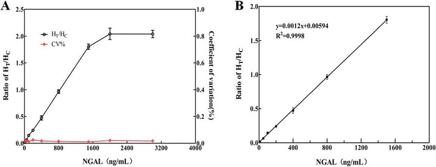
mances of EU-NPS-LFIA was evaluated by building the
standard curve. The relative fluorescence intensity
Fig. 2 Comparison of fusion efficiency using different the puncture
ratio(HT/HC) was increased with NGAL concentration.
pulse height. The fusion cells were seeded into 96-well plates at
approximately 2 × 10⁵ cells/well. A total of 480 wells was assessed EU-NPS-1G1 showed reaction to mAb 2F4 (Fig. 5 A).
for each condition. The calculated method of the fusion efficiency The high-dose hook influence wasn’t detected when
(%): the total number of colonies in 480 wells were counted,then concentration of antigen reached 3000 ng/mL. The re-
divided by the number of input B cells and multiplied by 100. The gression equation was exhibited as follows: y = 0.0012
columns represent the average fusion efficiency (%) of 3
x + 0.0059 (R2 = 0.99), where y represents the ratio of
experiments, the error bars represent the SD
HT/HC, x represents the concentrations of NGAL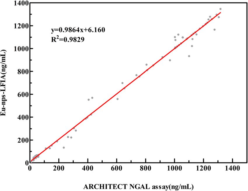
Yin et al. BMC Nephrology      (2022) 23:30                                                                                             Page 6 of 9

 Fig. 3 Characterization of MAbs. (A) The ascites were assessed by SDS-PAGE under non-reducing and reducing, Lane M, protein marker; Lane 1
 and 3, purified the ascites of 2F4; Lane 2 and 4, purified the ascites of 1G1.(B) The ascites were assessed by Western blot, Lane 1, Western blot
 analysis of mAb 2F4; Lane 2, Western blot analysis of mAb 1G1. (C) The Cross-reactivity of 2 hybridoma lines was tested by Indirect ELISA, NGAL-
 6×His and PCT-6×His were coated onto microtiter plates. The positive control was a NGAL-6×His immune serum; PBS were used as blank control.
 Red and black dots were meant 1G1 and 2F4 ELISA OD450 nm values, respectively. (D) Relative affinity of MAb 2F4 and 1G1

(Fig. 5B).The detection limit (LOD) was 0.36 ng/mL (3                      with three replicates at each spiked concentration in
times the standard deviation of the blank, n = 20) calcu-                  1 day, and inter-assay precision was calculated with
lated by the Clinical Laboratory Standards Institute                       three replicates at each spiked concentration at every
(CLSI) Guideline EP17-A2, the NGAL concentration                           three days for fifteen days continuously[18]. The
had linear relationship in the range of 1-1500 ng/                         results were shown in Table 1, the calculated intra-
mL[17].                                                                    assay coefficient of variation (CV) ranged from 2.57
                                                                           to 4.98 % (n = 10), lower than 10 %. The inter-assay
                                                                           CV ranged from 4.11 to 7.83 % (n = 15), lower than
Precision                                                                  10 % too.
Recovery experiments of the intra-assay and inter-                           Intra-assay and inter-assay precision were verified by
assay were performed to evaluate precision and re-                         two evaluator devices (Guangzhou Wondfo Biotech
producibility of immunological method. The different                       Co.,Ltd and Guangzhou Labsim Biotech Co., Ltd), and
concentrations (50, 200 and 800 ng/mL) of NGAL                             the result is eventually consistent (Supplement data).
standard substance were added to negative urine                            These results explained that the precision of the devel-
samples. The intra-assay precision was calculated                          oped LFIA was a high level, and reproducibility was an
                                                                           acceptable level.

                                                                           Specificity
                                                                           The specificity of the test strips was evaluated by adding
                                                                           endogenous substances in normal and different concen-
                                                                           trations urine samples, including creatinine, glucose and
                                                                           urea nitrogen. As shown in Table 2, the results indicated
                                                                           that all relative deviations (RD) was in the range of ±
                                                                           10 %, suggesting antigen-antibody interaction were stable
 Fig. 4 Schematic description of fluorescence immunoassay system
                                                                           and illustrating the specificity of test strips was accept-
 employed Europium-conjugated NGAL MAbs
                                                                           able toward NGAL.
Yin et al. BMC Nephrology (2022) 23:30 Page 7 of 9
Fig. 5 Standard curves for NGAL. (A) The ratio of HT/HC, by antibody pairing in LFIA, the antibody pairs 2F4-labled1G1 ratio of HT/HC rose with
increasing concentration of NGAL. (B) The standard curve of NGAL
Clinical samples tests concentration in urine is significantly associated with
In order to appraise applicative competence of the sCr concentration[22]. In recent years, various electro-
NGAL based on EU-NPS-LFIA for determination of chemical and immunological methods have been devel-
clinical samples, a total of 83 urine samples containing oped for the detection of NGAL, instance of
26 low value samples (11–70 ng/mL), 19 median value electrochemical determination, solid-phase proximity
samples (110–800 ng/mL), 38 high value samples (800– ligation assay and enzyme-free electrochemical immuno-
1740 ng/mL), were measured on the ARCHITECT urine assay[23, 24]. However, these techniques require precise
NGAL assay. The results of correlation coefficient (R2) equipment, specialized personnel and professional inter-
of the regression curve was 0.9829 (p < 0.01), indicating pretation of the results. Therefore, to develop an effect-
that the two detection methods had a significant linear ive and convenient detection assay for NGAL
relationship (where x represents concentrations of concentration in the urine is critical for this disorder
NGAL obtained by the ARCHITECT analyzer, y repre- monitoring. The low efficiency of polyethylene glycol
sents values measured by developed test strips) (Fig. 6). (PEG) fusion in conventional methods to causes difficul-
Thus, the developed EU-NPS-LFIA for determination of ties in obtaining functional antibodies, we optimized
NGAL was very accurate in clinical testing. electric fusion parameters that enabled enhancement of
fusion efficiency to prepare of viable hybridomas, and
Discussion obtained two anti-NGAL MAbs. MAbs are not only high
The EU-NPS-LFIA for detection of NGAL in human affinity, but also directed towards different epitopes,
urine and diagnosis of AKI has been developed in our used to establish high quality of diagnostic assays.
study. Current diagnosis of AKI is confirmed by the con- In recent years, many studies have been reported to
centration of serum creatinine (sCr), which is steady un- develop several NGAL rapid diagnostic immunoassays,
less at least 50 % of damaged kidney function[19]. AKI including the photoelectrochemical immunosensor and
developed in 89 % of severe patients with COVID-19, three electrochemical immunosensors, the solid-phase
and a majority of patients had predominantly oliguria proximity ligation assay and lateral flow assay[25]. In
when appears earlier than plasma Creatinine[20]. Bio- one study, immunosensor has been made by NGAL cap-
markers increased elevated levels at admission, which as- ture antibodies immobilized to screen-printed-modified
sociated with increased mortality. NGAL is one of these carbon electrode and labeled addition of secondary anti-
biomarkers, levels of which may positive correlation with body against NGA to PB-NP-decorated g-C3N4 nano-
risk for mortality in COVID-19 hospitalized patients[21]. sheets forming a sandwich on the SPCE[24]. The other
In AKI, several studies have discovered NGAL study developed antibody against NGAL was immobi-
lized on a screen printed electrode (SPCE) modified with
Table.1 Reproducibility analysis of the EU-NPS-LFIA test strip by electropolymerized aniline deposited on top of an elec-
intra-assay and inter-assay precision
trosprayed graphene/poly-aniline (G/PANI)[25]. Two re-
NGAL Intra-Assay Precision (n = 10) Inter-Assay Precision (n = 15)
(ng/
ports showed the LOD varied widely from 0.6 pg/mL to
Mean ± SD (ng/mL) CV (%) Mean ± SD (ng/mL) CV (%) 21.1 ng/mL, depending on the g-C3N4 nanosheets with
mL)
50 49.57 ± 0.003 4.98 51.28 ± 0.037 5.68 N element enhanced electrocatalytic efficiency of the
200 199.18 ± 0.009 3.84 202.78 ± 0.019 7.83
nanohybrids than graphene nanosheets, and the LOD of
the NGAL assay varies depending on immunosensor,
800 799.76 ± 0.025 2.57 801.68 ± 0.039 4.11
conjugated-complex and antibodies[26]. According toYin et al. BMC Nephrology (2022) 23:30 Page 8 of 9
Table.2 The specificity study of the EU-NPS-LFIA with different interfering endogenous substances
Interfering Substance NGAL (27.64ng/mL) NGAL (53.72ng/mL)
Value RD (%) Value RD (%)
control 0.037 ± 0.002 0.54 0.069 ± 0.003 2.35
Creatinine(10 mg/mL) 0.035 ± 0.008 -4.55 0.067 ± 0.009 -2.06
glucose(10 mg/mL) 0.036 ± 0.007 -2.55 0.071 ± 0.006 3.83
urea nitrogen(100 mg/mL) 0.036 ± 0.004 2.72 0.064 ± 0.008 -5.98
Note: RD = (Value-Standard value) / Standard value
those studies, we enlightened that sensitivity of LFIA about 7.0 ng/mL in healthy individuals[32]. The develop-
could be improved by basing on different fluorescence ments and application about the EU-NPS-LFIA of
nanoparticles mentioned in the preamble section.The NGAL are still in initial phase, this assay need to test
high sensitivity nanoparticles labeled in LFIA as fluores- NGAL of serum and urine samples with COVID- 19 pa-
cent probes has been continually researched over past tients for which improvement in the treatment in the fu-
decades, gold nanoparticles are the most widely used, ture of works.
but the application of the lateral flow tests based on
traditional labels is limitedfor there poor identification
Conclusions
and weaker the signal[27]. Currently there are antibodies In the study, we successfully got two mouse anti-NGAL
available for the development of NGAL have been ap-
MAbs (2F4, 1G1) and were labeled with EU-NPS to es-
plied to different platforms for NGAL detection, but not
tablish the lateral flow immune technique. The EU-NPS-
available in LFIA based on EU-NPS labels experimented LFIA was found to detect NGAL in a wide range of 1-
on the detection of NGAL in addition to UCP
3000 ng/mL within 15 min, the detection sensitivity
technology-based lateral flow assay [28, 29].
reached 0.36 ng/mL. These anti-NGAL MAbs could be
Fluorescence immunoassays have advantange in vari- reliability utilized in fluorescence LFIA of NGAL detec-
ous detection fields, especially in mature quantum dots,
tion in the urine samples, so that it should be applicable
that sensitivity was influenced by the MAbs and fluores-
in the AKI diagnosis.
cent materials, and could greatly improve by EU-NPS
and specific MAbs to use the fluorescent probes[30].
The EU-NPS were applied to fluorescence LFIA strips Supplementary information
The online version contains supplementary material available at https://doi.
had matured in the detection field, and enhanced several org/10.1186/s12882-021-02493-w.
advantages of LFIA for assay sensitivity, specificity and
stability[31]. Due to excellent properties of the EU-NPS Additional file 1 zip
that the limit of detection for NGAL in this method was
0.36 ng/mL, while the mean urine NGAL levels was
Abbreviations
COVID-19: Coronavirus Infection Disease 2019; AKI: Acute kidney injury;
SCr: serum creatinine; NGAL: neutrophil gelatinase-associated lipocalin;
LFIA: Lateral flow immunoassays; EU-NPS: europium nanoparticles;
ELISA: enzyme-linked immunosorbent assay; MAbs: monoclonal antibodies
Acknowledgements
Not applicable.
Authors’ contributions
HYW designed the experiments; MLY, YWN, HL and LL performed study
planning, data analysis, manuscript drafting and manuscript review.LT, YD
and CMH revised the manuscript.All authors gave intellectual input to the
manuscript and approved final version.
Funding
The authors disclosed receipt of the following financial support for the
research, authorship, and/or publication of this article:This work was
supported by the ‘Research and Development of Industrial Technology’
Program of Jilin Province, PR China (grant nos. 20180623045TC and
20170204005YY), National Natural Science Foundation of China (no.
Fig. 6 Comparison of EU-NPS-LFIA with ARCHITECT urine assay 21827812) and National Training Program of Innovation and
estimated correlation of the results for NGAL clinical test Entrepreneurship for Undergraduates (nos. 201913706017 and
202013706002).Yin et al. BMC Nephrology (2022) 23:30 Page 9 of 9
Availability of data and materials 14. Nephron KJ: KDIGO clinical practice guidelines for acute kidney injury. 2012,
The datasets used and/or analyzed during the current study are available 120(4):c179-c184.
from the corresponding author on reasonable request. 15. Huang X, Aguilar Z, Xu H, Lai W, Xiong Y: Membrane-based lateral flow
immunochromatographic strip with nanoparticles as reporters for detection:
Declarations A review. Biosensors bioelectronics 2016, 75:166–180.
16. Weeratna R, Comanita L, Davis H: CPG ODN allows lower dose of antigen
Ethics approval and consent to participate against hepatitis B surface antigen in BALB/c mice. Immunology cell biology
The study was performed on the basis of institutional ethical guidelines, 2003, 81(1):59–62.
approved by the Affiliated hospital of Jilin Medical University in China. All 17. Tong Q, Chen B, Zhang R, Zuo C: Standardization of clinical enzyme analysis
procedures were followed the principles Declaration of Helsinki, informed using frozen human serum pools with values assigned by the International
consent was obtained from all subjects. Federation of Clinical Chemistry and Laboratory Medicine reference
measurement procedures. Scandinavian journal of clinical laboratory
investigation 2018, 78:74–80.
Consent for publication 18. Huang D, Ying H, Jiang D, Liu F, Tian Y, Du C, Zhang L, Pu X: Rapid and
Not applicable. sensitive detection of interleukin-6 in serum via time-resolved lateral flow
immunoassay. Analytical biochemistry 2020, 588:113468.
Competing interest 19. Shapiro N, Trzeciak S, Hollander J, Birkhahn R, Otero R, Osborn T, Moretti E,
The authors declare that they have no conflict of interest. Nguyen H, Gunnerson K, Milzman D et al: The diagnostic accuracy of
plasma neutrophil gelatinase-associated lipocalin in the prediction of acute
Author details kidney injury in emergency department patients with suspected sepsis.
1
Jilin Collaborative Innovation Center for Antibody Engineering, Jilin Medical Annals of emergency medicine 2010, 56(1):52–59.e51.
University, 132013 Jilin, PR China. 2Academy of laboratory, Jilin Medical 20. Luther T, Bülow-Anderberg S, Larsson A, Rubertsson S, Lipcsey M, Frithiof R,
University, 132013 Jilin, PR China. Hultström M: COVID-19 patients in intensive care develop predominantly
oliguric acute kidney injury. Acta anaesthesiologica Scandinavica 2021, 65(3):
Received: 15 March 2021 Accepted: 2 August 2021 364–372.
21. Abers M, Delmonte O, Ricotta E, Fintzi J, Fink D, de Jesus A, Zarember K, Alehashemi
S, Oikonomou V, Desai J: An immune-based biomarker signature is associated with
mortality in COVID-19 patients. JCI insight 2021, 6(1).
References 22. Wagener G, Jan M, Kim M, Mori K, Barasch J, Sladen R, Lee H: Association
1. Zheng X, Zhao Y, Yang L: Acute Kidney Injury in COVID-19: The Chinese between increases in urinary neutrophil gelatinase-associated lipocalin and
Experience. Seminars in nephrology 2020, 40(5):430–442. acute renal dysfunction after adult cardiac surgery. Anesthesiology 2006,
2. Mandelbaum T, Scott D, Lee J, Mark R, Malhotra A, Waikar S, Howell M, 105(3):485–491.
Talmor D: Outcome of critically ill patients with acute kidney injury using 23. Kannan P, Tiong H, Kim D: Highly sensitive electrochemical determination of
the Acute Kidney Injury Network criteria. Critical care medicine 2011, 39(12): neutrophil gelatinase-associated lipocalin for acute kidney injury. Biosensors
2659–2664. bioelectronics 2012, 31(1):32–36.
3. Al-Hwiesh A, Mohammed A, Elnokeety M, Al-Hwiesh A, Al-Audah N, Esam S, 24. Zhang F, Zhong H, Lin Y, Chen M, Wang Q, Lin Y, Huang: A nanohybrid composed
Abdul-Rahman I: Successfully treating three patients with acute kidney of Prussian Blue and graphitic CN nanosheets as the signal-generating tag in an
injury secondary to COVID-19 by peritoneal dialysis: Case report and enzyme-free electrochemical immunoassay for the neutrophil gelatinase-associated
literature review. Peritoneal dialysis international 2020, 40(5):496–498. lipocalin. Mikrochimica acta 2018, 185(7):327.
4. Shabaka A, Rovirosa-Bigot S, Guerrero Márquez C, Alonso Riaño M, 25. Yukird J, Wongtangprasert T, Rangkupan R, Chailapakul O, Pisitkun T,
Fernández-Juárez G: Acute kidney injury and nephrotic syndrome secondary Rodthongkum N: Label-free immunosensor based on graphene/polyaniline
to COVID-19-associated focal segmental glomerulosclerosis. Nefrologia 2021. nanocomposite for neutrophil gelatinase-associated lipocalin detection.
5. Gabbard W, Milbrandt E, Kellum J: NGAL: an emerging tool for predicting Biosensors bioelectronics 2017, 87:249–255.
severity of AKI is easily detected by a clinical assay. Critical care 2010, 14(4): 26. Gong Y, Li M, Wang Y: Carbon nitride in energy conversion and storage:
318. recent advances and future prospects. ChemSusChem 2015, 8(6):931–946.
6. Zuk R, Ginsberg V, Houts T, Rabbie J, Merrick H, Ullman E, Fischer M, Sizto C, 27. Chen A, Yang S: Replacing antibodies with aptamers in lateral flow
Stiso S, Litman D: Enzyme immunochromatography–a quantitative immunoassay. Biosensors bioelectronics 2015, 71:230–242.
immunoassay requiring no instrumentation. Clinical chemistry 1985, 31(7): 28. Li H, Mu Y, Yan J, Cui D, Ou W, Wan Y, Liu S: Label-free
1144–1150. photoelectrochemical immunosensor for neutrophil gelatinase-associated
7. Juntunen E, Myyryläinen T, Salminen T, Soukka T, Pettersson K: Performance lipocalin based on the use of nanobodies. Analytical chemistry 2015, 87(3):
of fluorescent europium(III) nanoparticles and colloidal gold reporters in 2007–2015.
lateral flow bioaffinity assay. Analytical biochemistry 2012, 428(1):31–38. 29. Lei L, Zhu J, Xia G, Feng H, Zhang H, Han Y: A rapid and user-friendly assay
8. Swanson C, D’Andrea A: Lateral flow assay with near-infrared dye for to detect the Neutrophil gelatinase-associated lipocalin (NGAL) using up-
multiplex detection. Clinical chemistry 2013, 59(4):641–648. converting nanoparticles. Talanta 2017, 162:339–344.
9. Chen Y, Fu Q, Xie J, Wang H, Tang Y: Development of a high sensitivity 30. Yeo S, Bao D, Seo G, Bui C, Kim D, Anh N, Tien T, Linh N, Sohn H, Chong C
quantum dot-based fluorescent quenching lateral flow assay for the et al: Improvement of a rapid diagnostic application of monoclonal
detection of zearalenone. Analytical bioanalytical chemistry 2019, 411(10): antibodies against avian influenza H7 subtype virus using Europium
2169–2175. nanoparticles. Scientific reports 2017, 7(1):7933.
10. Zhang F, Zou M, Chen Y, Li J, Wang Y, Qi X, Xue Q: Lanthanide-labeled 31. Chen E, Xu Y, Ma B, Cui H, Sun C, Zhang M: MonascusCarboxyl-
immunochromatographic strips for the rapid detection of Pantoea stewartii Functionalized, Europium Nanoparticle-Based Fluorescent
subsp. stewartii. Biosensors bioelectronics 2014, 51:29–35. Immunochromatographic Assay for Sensitive Detection of Citrinin in
11. Nankoberanyi S, Mbogo G, LeClair N, Conrad M, Tumwebaze P, Tukwasibwe Fermented Food. Toxins 2019, 11(10).
S, Kamya M, Tappero J, Nsobya S, Rosenthal P: Validation of the ligase 32. Bolignano D, Coppolino G, Campo S, Aloisi C, Nicocia G, Frisina N, Buemi M:
detection reaction fluorescent microsphere assay for the detection of Neutrophil gelatinase-associated lipocalin in patients with autosomal-
Plasmodium falciparum resistance mediating polymorphisms in Uganda. dominant polycystic kidney disease. American journal of nephrology 2007,
Malaria journal 2014, 13:95. 27(4):373–378.
12. Xu H, Dong Y, Guo J, Jiang X, Liu J, Xu S, Wang H: Monoclonal antibody
production and the development of an indirect competitive enzyme-linked
immunosorbent assay for screening T-2 toxin in milk. Toxicon 2018, 156:1–6. Publisher’s Note
13. Kato M, Sasamori E, Chiba T, Hanyu Y: Cell activation by CpG ODN leads to Springer Nature remains neutral with regard to jurisdictional claims in
improved electrofusion in hybridoma production. Journal of immunological published maps and institutional affiliations.
methods 2011, 373:102–110.You can also read