2019 AAHA Dental Care Guidelines for Dogs and Cats
←
→
Page content transcription
If your browser does not render page correctly, please read the page content below
VETERINARY PRACTICE GUIDELINES

2019 AAHA Dental Care Guidelines for Dogs
and Cats*
Jan Bellows, DVM, DAVDC, DABVP (Canine/Feline), Mary L. Berg, BS, LATG, RVT, VTS (Dentistry), Sonnya Dennis,
DVM, DABVP (Canine/Feline), Ralph Harvey, DVM, MS, DACVAA, Heidi B. Lobprise, DVM, DAVDC, Christopher J.
Snyder, DVM, DAVDCy, Amy E.S. Stone, DVM, PhD, Andrea G. Van de Wetering, DVM, FAVD

                                                                     ABSTRACT
      The 2019 AAHA Dental Care Guidelines for Dogs and Cats outline a comprehensive approach to support companion animal
      practices in improving the oral health and often, the quality of life of their canine and feline patients. The guidelines are an update of
      the 2013 AAHA Dental Care Guidelines for Dogs and Cats. A photographically illustrated, 12-step protocol describes the essential
      steps in an oral health assessment, dental cleaning, and periodontal therapy. Recommendations are given for general anesthesia,
      pain management, facilities, and equipment necessary for safe and effective delivery of care. To promote the wellbeing of dogs and
      cats through decreasing the adverse effects and pain of periodontal disease, these guidelines emphasize the critical role of client
      education and effective, preventive oral healthcare. (J Am Anim Hosp Assoc 2019; 55:---–---. DOI 10.5326/JAAHA-MS-6933)

AFFILIATIONS                                                                  * These guidelines were supported by a generous educational grant from
                                                                              Boehringer Ingelheim Animal Health USA Inc., Hill’s® Pet Nutrition, Inc.,
From All Pets Dental, Weston, Florida (J.B.); Beyond the Crown Veterinary
                                                                              and Midmark. They were subjected to a formal peer-review process.
Education, Lawrence, Kansas (M.L.B.); Stratham-Newfields Veterinary Hos-
                                                                              These guidelines were prepared by a Task Force of experts convened by the
pital, Newfields, New Hampshire (S.D.); Department of Small Animal Clin-
                                                                              American Animal Hospital Association. This document is intended as a
ical Sciences, College of Veterinary Medicine, University of Tennessee,
                                                                              guideline only, not an AAHA standard of care. These guidelines and recom-
Knoxville, Tennessee (R.H.); Main Street Veterinary Dental Hospital, Flower
                                                                              mendations should not be construed as dictating an exclusive protocol,
Mount, Texas (H.B.L.); Department of Surgical Sciences, School of Vet-
                                                                              course of treatment, or procedure. Variations in practice may be warranted
erinary Medicine, University of Wisconsin-Madison, Madison, Wisconsin
                                                                              based on the needs of the individual patient, resources, and limitations
(C.J.S.); Department of Small Animal Clinical Sciences, College of Veter-
                                                                              unique to each individual practice setting. Evidence-based support for spe-
inary Medicine, University of Florida, Gainesville, Florida (A.E.S.S.); and
                                                                              cific recommendations has been cited whenever possible and appropriate.
Advanced Pet Dentistry, LLC, Corvallis, Oregon (A.G.VdW.).
                                                                              Other recommendations are based on practical clinical experience and a
CONTRIBUTING REVIEWERS                                                        consensus of expert opinion. Further research is needed to document some
R. Michael Peak, DVM, DAVDC (The Pet Dentist, Tampa, Florida); Jeanne R.      of these recommendations. Because each case is different, veterinarians must
Perrone, CVT, VTS (Dentistry) (VT Dental Training, Plant City, Florida);      base their decisions on the best available scientific evidence in conjunction
Kevin S. Stepaniuk, DVM, FAVD, DAVDC (Veterinary Dentistry Education          with their own knowledge and experience.
and Consulting Services, LLC, Ridgefield, Washington).                         Note: When selecting products, veterinarians have a choice among those
                                                                              formulated for humans and those developed and approved by veterinary
Correspondence: christopher.snyder@wisc.edu (C.J.S.)                          use. Manufacturers of veterinary-specific products spend resources to have
                                                                              their products reviewed and approved by the FDA for canine or feline use.
                                                                              These products are specifically designed and formulated for dogs and cats
                                                                              and have benefits for their use; they are not human generic products. AAHA
                                                                              suggests that veterinary professionals make every effort to use veterinary
                                                                              FDA-approved products and base their inventory-purchasing decisions on
                                                                              what product is most beneficial to the patient.

                                                                              y C. Snyder was the chair of the Dental Care Guidelines Task Force.

                                                                              NAD (nonanesthetic dentistry); PD (periodontal disease staging); VOHC
                                                                              (Veterinary Oral Health Council); VTS (Dentistry) (Veterinary Technician
                                                                              Specialist[s] in Dentistry)

ª 2019 by American Animal Hospital Association                                                                                   JAAHA.ORG              1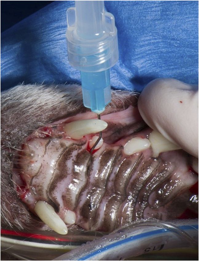
excellent, basic resource for clinicians, the 2019 guidelines published
Introduction
                                                                             here provide important new information. This includes (1) an ex-
The concept that a pet is suffering from oral pain, infection, and
                                                                             panded and updated discussion of commonly performed veterinary
inflammation that may not be apparent but is affecting their quality
                                                                             dental procedures, supported by photos that illustrate oral pathol-
of life is a reality that may not always be fully appreciated by
                                                                             ogy and therapeutic techniques; (2) criteria for periodontal disease
the veterinary profession and often not understood by the pet-
                                                                             staging; (3) the importance for addressing pain and stress in dental
owning public. Compromised dental health can affect a pet’s over-
                                                                             patients; and (4) client communication tips for explaining the im-
all health, longevity, quality of life, and interaction with its owner
                                                                             portance and rationale behind specific dental and oral procedures.
without exhibiting obvious clinical signs of disease. The purpose of
                                                                             Client education is a particularly important and often underap-
this document is to provide guidance to veterinary professionals that
                                                                             preciated aspect of veterinary dentistry. Without the pet owner’s
will enable them to recognize dental pathology and deliver appropriate
                                                                             understanding and acceptance of the veterinarian’s oral health rec-
preventive and therapeutic care to their patients, as well as to provide
                                                                             ommendations, the decision to pursue dental cleaning, oral evalu-
essential dental education to their clients.
                                                                             ation, and treatment will seem optional. Applying the AAHA Dental
     In consideration of our patients’ welfare, veterinary profes-
                                                                             Care Guidelines with an emphasis on client communication will
sionals must understand that dental patients often experience con-
                                                                             enhance your practice by providing your clients with services that
siderable fear, anxiety, stress, pain, and suffering. In order to achieve
                                                                             address a critical component of canine and feline healthcare.
optimal clinical success and client satisfaction, it is essential that the
veterinary team address these concerns with every client, beginning
                                                                             Dental Terminology
with the first interaction when scheduling an appointment.
                                                                             Although dental terminology is constantly being defined, current
     The guidelines are based on evidence-based information
                                                                             definitions applicable to veterinary dentistry are shown in Table 1.
whenever possible, although we recognize that relevant data and well-
                                                                             Readers will find it helpful to review these definitions before reading
designed veterinary dental studies have not always been conducted
                                                                             the remainder of the guidelines.
for all the topics covered in these guidelines. As a result, expert
                                                                                  Veterinary dentistry is a discipline within the scope of veterinary
opinion and the extensive clinical experience of the Task Force
                                                                             practice that involves the professional consultation, evaluation, di-
members have been used in writing the guidelines. The collective goal
                                                                             agnosis, prevention, and treatment (nonsurgical, surgical, or related
of the Task Force was to apply the highest level of evidence-based
                                                                             procedures) of conditions, diseases, and disorders of the oral cavity
information available when preparing the guidelines.                         and maxillofacial area and their adjacent and associated structures.
     The guidelines are intended primarily for general practitioners         Veterinary dental diagnoses are made and treatments performed by a
and veterinary team members without advanced dental training. The            licensed veterinarian, within the scope of his or her education,
Task Force encourages all veterinary professionals to continuously           training, and experience, in accordance with the ethics of the pro-
improve their veterinary dentistry knowledge, skills, and treatment          fession and applicable law.
capabilities and to recognize cases needing referral. It is well known            The term “dental” has lost favor as an all-purpose descriptive
that many pet owners use the internet as a default resource for pet          term because it does not adequately define a particular procedure to
healthcare information and home treatment.1 However, because of              be performed. For example, specific diagnostic and treatment ter-
the specialized nature of dental procedures, including diagnosis and         minology should be used to describe procedures such as a complete
treatment, professional veterinary care is necessary for maintaining         oral health assessment, orthodontics, periodontal surgery, and ad-
pet oral health. Therefore, veterinary dentistry represents an op-           vanced oral surgery. Using specific diagnostic and treatment ter-
portunity for a primary care practice to demonstrate a high level of         minology will help staff and clientele understand the importance
service and professional expertise to its clients and to positively          and specifics of a scheduled procedure.
impact patient comfort and wellbeing.                                             Additional information on veterinary dental nomenclature can
     The guidelines are intended to be a first-line resource in helping       be found on the American Veterinary Dental College (AVDC) website
practitioners achieve that essential goal. Readers should consider the       (avdc.org/Nomenclature/Nomen-Intro.html).
guidelines to be an extension and update of the 2013 AAHA Dental
Care Guidelines for Dogs and Cats (hereafter referred to as the 2013         Anatomy and Pathology
AAHA Dental Care Guidelines), which continue to be a relevant                A comprehensive knowledge of oral and dental anatomy and
source of medically appropriate information on veterinary den-               physiology is imperative for recognizing and treating disease in the
tistry.2 Although the 2013 AAHA Dental Care Guidelines are an                oral cavity and teeth. Veterinarians must understand the location,

2    JAAHA |     55:2 Mar/Apr 2019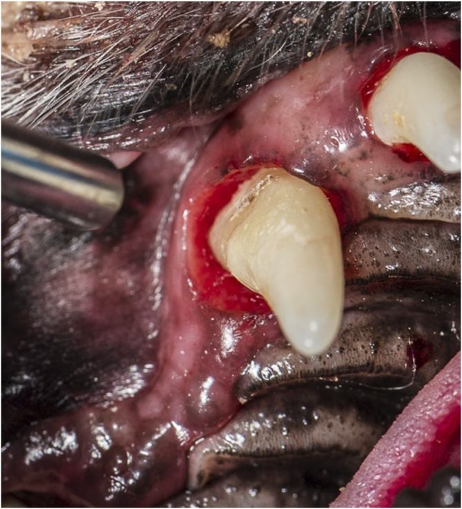
Dental Guidelines

TABLE 1
Definitions That Pertain to Dental Care Guidelines

Terminology                                                                                         Definition

Dental chart                          A written and graphical representation of the mouth, with adequate space to indicate pathology and procedures (see the “2013
                                        AAHA Dental Care Guidelines” for included items).
Dental prophylaxis                    A procedure performed on a healthy mouth that includes oral hygiene care, a complete oral examination, and techniques to prevent
                                         disease and to remove plaque and calculus above and beneath the gum line under anesthesia before periodontitis has developed.
                                         Note: The words “prophy,” “prophylaxis,” and “dental” are often misused in veterinary medicine. More descriptive terms to use
                                         for the dental procedures that are commonly performed in companion animal dentistry to prevent periodontitis are COPAT,
                                         COHAT, and an oral ATP visit.
Dentistry                             The evaluation, diagnosis, prevention, and/or treatment of abnormalities in the oral cavity, maxillofacial area, and/or associated
                                        structures. Nonsurgical, surgical, or related procedures may be included.
Endodontics                           The treatment and therapy of conditions affecting the pulp.
Exodontia (extraction)                A surgical procedure performed to remove a tooth.
Gingivitis                            Inflammation of the gingiva with or without loss of the supporting structure(s) shown with X-rays.
Home oral hygiene                     Measures taken by pet owners that are intended to control or prevent plaque and calculus accumulation.
Oral surgery                          The practical manipulation and incising of epithelium of hard and soft tissue for the purpose of improving or restoring oral health and
                                        comfort.
Orthodontics                          The evaluation and treatment of malpositioned teeth for the purposes of improving occlusion and patient comfort and enhancing the
                                        quality of life.
Periodontal disease                   A disease process beginning with gingivitis and progressing to periodontitis when left untreated.
Periodontitis                         A destructive process involving the loss of supportive structures of the teeth, including the periodontium (i.e., gingiva, periodontal
                                         ligament, cementum, and/or alveolar bone).
Periodontal surgery                   Invasive treatment necessary to re-establish or rehabilitate periodontal attachment structures. This is indicated for patients with
                                         pockets .5 mm, stage 2 and 3 furcation exposure, or inaccessible root structures.
Periodontal therapy                   Treatment of tooth-supporting structures in the presence of existing periodontal disease; includes dental cleaning as defined below
                                         and one or more of the following procedures: gingival curettage for nonsurgical removal of plaque, calculus, and debris in gingival
                                         pockets; root planing periodontal flaps; regenerative surgery; gingivectomy-gingivoplasty; and the local application of
                                         antimicrobials.
Periodontium                          The supporting structures of teeth, including (1) periodontal ligament, (2) gingiva, (3) cementum, and (4) alveolar and supporting
                                        bone.
Pocket                                A pathologic space between supporting structures and the tooth, extending apically from the normal attachment location of the
                                        gingival epithelial attachment.
Professional dental cleaning          Scaling (supragingival and subgingival plaque and calculus removal) of teeth with power or hand instrumentation, tooth polishing,
                                        and oral examination performed by a trained veterinary healthcare provider under general anesthesia.

Some definitions were derived from previously published descriptions2
COHAT, comprehensive oral health, assessment, and treatment; COPAT, comprehensive oral prevention, assessment, and treatment; oral ATP, oral assessment, treatment,
and prevention.

purpose, and function of the structures of the head and oral cavity                        is the inflammation of the tissues of the periodontium, or periodontal
                         3–5
shown in Figure 1.             Dogs and cats have two generations of teeth                 disease. The clinical terms used to describe the active process of
(diphyodont), with the roots being longer than crowns. Most of the                         periodontal disease include gingivitis and periodontitis. Gingivitis,
permanent tooth is composed of dentin, with the central portion of                         the earliest stage of periodontal disease, is described as inflammation
the tooth being the pulp chamber containing blood vessels, nerves,                         confined to the gingiva and commonly induced by bacterial plaque.
                                                                             6
lymphatics, connective tissue, and odontoblasts (Figure 1). The                            Gingivitis is reversible and preventable.8,9 Plaque-induced gingivitis
tooth supporting structures, or “periodontium,” consist of the                             can be reversed by removal of the bacteria above as well as below the
gingiva, periodontal ligament, cementum, and alveolar bone. The                            gingival margin and prevented with consistent plaque-removing
periodontal ligament attaches the tooth in the alveolus by being                           home oral hygiene efforts.10 Calculus, or bacterial plaque that has
affixed between the cementum and the alveolar bone (Figure 1).3,7                           become calcified by salivary minerals, is mostly an irritant and is
      There are many pathologic processes that affect the oral cav-                        relatively nonpathogenic.8,9
ity of dogs and cats (congenital, infectious, traumatic, neoplastic,                             The bacterial population at the tooth surface is initially com-
autoimmune, and others). The most common and significant disease                            posed of gram-positive, aerobic bacteria. The bacterial biofilm

                                                                                                                                                     JAAHA.ORG                  3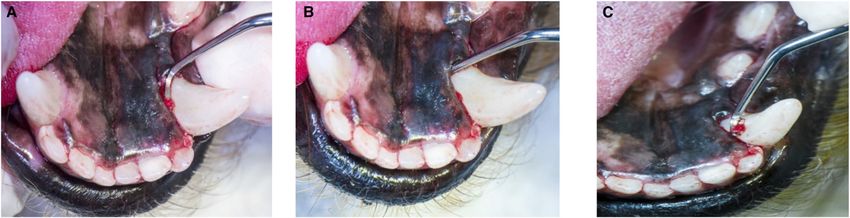
FIGURE 1        Anatomy of a tooth.
                                                                                                             ª 2019 Veterinary Information Net-
                                                                                                             work (VIN), illustration by Tamara
                                                                                                             Rees.

eventually invades the sulcus between the gingiva and the tooth,            domestic cats and fewer dogs, and is caused by odontoclastic de-
creating an environment selecting for a more destructive anaerobic,         struction of teeth. Although the etiology of these progressive lesions
                            11
gram-negative population. The bacterial byproducts directly cause           remains unproven, gingival inflammation and exposure of the pulp
tissue injury resulting in host inflammation, which directly contrib-        chamber can be the result.15 These are some of the most common
utes to loss of attachment between the tooth and periodontal struc-         pathologies encountered in veterinary general practice and are as-
tures. If left untreated, the chronic inflammatory host response can         sociated with various painful stages during the course of progres-
progress to periodontitis.9 Periodontitis is an inflammation resulting       sion. Practitioners can supplement their education and experience
in irreversible loss of the supporting tissues of the teeth, progressing    by consulting the growing body of literature and online resources on
from periodontal ligament attachment loss to the loss of alveolar bone,     the oral pathology of dogs and cats.
resulting in clinically detectable attachment loss. Although this process
can be stabilized, it is not easily reversible and can ultimately lead to   Dental Disease Prevention Strategies
tooth loss. Other factors influencing the progression and ultimate se-       It is important to communicate with pet owners the importance of
verity of periodontal disease may include breed predisposition, mal-        dental disease prevention strategies, beginning at the first visit and
                                                                12
occlusion, chewing habits, systemic health, and local irritants.            then throughout the patient’s life stages. It is particularly important
     Fractured teeth have been reported in up to 49.6% of com-              to emphasize individualized prevention strategies that should be
                  13
panion animals.        In the case of a complicated fracture (pulp          maintained on an ongoing basis. Some companion animal practices
exposure), the pulp chamber becomes contaminated by oral bacteria           use progress visits to evaluate oral health and home oral hygiene
and proceeds to infection and necrosis, resulting in periapical             efforts by pet owners. A helpful aspect of client education is for
infection.14 Tooth resorption is also common, affecting 27–72% of           veterinarians and staff to explain to clients the following three ways

4    JAAHA |     55:2 Mar/Apr 2019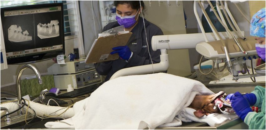
Dental Guidelines

preventive oral health products work: (1) mechanical (abrasion), (2)         treated. Ideally, periodontal therapy should then be provided
nonmechanical (chemical), and (3) a combination of mechanical                at an interval to optimally manage periodontal disease in this
and chemical modes of action. Some experts prefer oral health                preventable stage.
products that have dual action because all the teeth can benefit from            If periodontal disease with attachment loss is already present in
the combination of mechanistic activities.                                 the patient, a complete dental assessment, intraoral radiographs,
      In most patients, periodontal disease is a preventable condition.    cleaning, polishing, and any necessary treatment will help address any
Fractured teeth can often be prevented by appropriate selection of         current disease and optimally prevent further disease progression.
dental chews and toys and behavior modification for separation              Appropriate and effective home oral hygiene (see the “Client
anxiety and cage-biting.                                                   Communication and Education” section and resources at aaha.org/
                                                                           dentistry) can help maintain oral health in between dental therapy
Preventing Periodontal Disease                                             procedures. In most patients, effective periodontal prevention can
Prevention of periodontal disease begins at the first visit, either for a   help keep the oral cavity in a relatively pain-free and healthy state,
puppy or kitten, as well as for a new adult patient. Recommendations       favorably impacting the systemic health and welfare of the patient.
for young patients include the following:
·   A complete oral examination of the deciduous dentition will            Clarification of the Impact of Periodontal Health on
    assess any missing, unerupted, or slow-to-erupt teeth. The oc-         Systemic Health
    clusion should also be evaluated at this time, as well as deter-       The long-held dogma that specific oral bacteria are directly re-
    mination of abnormal jaw length and teeth that are contacting          sponsible for infection in distant organs is oversimplified and difficult
    other teeth or soft tissue. In such cases, early extraction may be     to prove.16,17 There is an association shown between periodontal
    needed.                                                                disease and systemic health parameters, and in human medicine, the
·   As permanent teeth start to erupt, it is critical to address any       presence of chronic inflammation associated with periodontitis has
    retained or persistent deciduous teeth. Immediate extraction of        been recognized to likely negatively impact overall systemic
    persistent deciduous teeth can help prevent displacement of the        health.18–25 The systemic spread of inflammatory mediators and
    erupting permanent teeth that can result in a malocclusion, or         cytokines and bacterial endotoxins from periodontal pathogens can
    that can exacerbate periodontal disease due to crowding.               impact the vascular system throughout the body and even cause
    Retained deciduous teeth without a replacement permanent               histological changes in distant organs.26–28 Management or resolu-
    tooth can remain stable, although extraction may be necessary          tion of the inflammation associated with periodontitis is likely to
    in cases of unstable dentition. Young pets with missing perma-         have greater clinical impact that just considering antibacterial ef-
    nent teeth should have intraoral dental radiographs taken to           forts.25,29,30 Although evidence demonstrating the direct correlation
    confirm that the teeth are truly not present, as unerupted teeth        between systemic disease and oral and dental infections may be
    can be problematic.                                                    difficult to prove, the positive impact on patient quality of life is
·   Home oral hygiene training can be started for clients owning           often clinically demonstrated and widely experienced.
    pets having erupted, permanent dentition. Juvenile patients
    actively exfoliating deciduous teeth may experience discomfort         Patient Assessment, Evaluation, and
    associated with home dental care efforts, and negative experi-         Documentation
    ences should be avoided.                                               History and Physical Examination
·   The owner of any puppy or kitten who will be smaller than 20–          A thorough history of patient health should always include an
    25 lbs at maturity should be informed that the level of dental         evaluation and update on systemic maladies as well as an evaluation
    care and prevention for their pet is likely to be more involved        and review of oral hygiene efforts performed by the pet owner.
    than that of a larger dog. Brachycephalic breeds also tend to have     Proactive management of oral health includes documenting any
    more dental issues due to the rotation and crowding of teeth.          efforts by the client to provide home dental care. These include tooth
·   A true dental prophylaxis (complete dental cleaning, polishing,        brushing; type of diet fed; access to “chews,” treats, and toys; in-
    and intraoral dental radiographs in the absence of obvious             formation on chewing habits; and updating any current or previous
    lesions) is recommended by 1 yr of age for cats and small- to          professional or home dental care. A thorough physical examination
    medium-breed dogs, and by 2 yr of age for larger-breed dogs.           should be performed to evaluate all body systems regardless of
    During the procedure, any hidden conditions such as unerup-            species, breed, age, health status, and temperament. Patients pre-
    ted or malformed (dysplastic) teeth can be identified and               senting for complaints separate from the oral cavity should be

                                                                                                                           JAAHA.ORG            5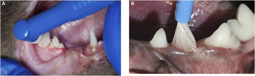
evaluated for the primary complaint. Appropriate diagnostic tests
                                                                          TABLE 2
and treatments should then be recommended. Patients with un-
derlying health conditions should be appropriately assessed so that       Items to Include in the Dental Chart or Medical Record

general anesthesia associated with dental or other procedures can be      Signalment

safely performed.                                                         Physical examination, medical, and dental history findings
                                                                          Oral examination findings

Conscious Oral Evaluation                                                 Anesthesia and surgery monitoring log and surgical findings

The conscious oral evaluation is an important first step to antici-        Any dental, oral, or other disease(s) currently present
                                                                          Abnormal probing depths (recorded for each affected tooth)
pating procedural extent and preparing and educating clients re-
                                                                          Dental chart with specific abnormalities noted, such as discoloration; worn
garding anticipated findings while under general anesthesia. In many
                                                                            areas; missing, malpositioned, supernumerary, or fractured teeth; tooth
instances, the examiner will underestimate the presence of disease          resorption; furcation exposure; and soft-tissue masses
during conscious evaluation, only to have the full extent of oral         Radiographic findings/interpretation
pathology revealed by periodontal probing and intraoral radiography.      Current and future treatment plan, addressing all abnormalities found. This
                                                                            includes information regarding initial decisions, decision-making algorithm,
     Examination of the conscious patient can be facilitated by use of
                                                                            and changes based on subsequent findings
individualized pharmacologic and nonpharmacologic protocols
                                                                          Recommendations for home dental care
designed to reduce anxiety, stress, and pain. For anxious, conscious
                                                                          Any recommendations declined by the client
patients, there should be no hesitation to recommend use of anxi-         Prognosis
olytics to facilitate an awake oral examination. For established pa-
tients, anxiety can be effectively relieved by administering trazodone
in dogs and gabapentin in cats, ideally the evening before and at least   mucous membranes, gingiva, vestibule, dorsal and ventral aspects of
2 hr before presentation if deemed safe and appropriate. For new          the tongue, tonsils, salivary glands and ducts, and assessment of the
patients who are difficult to assess, rapid-acting sedatives or anxi-      caudal oral cavity and gag reflex if it can be safely elicited. Any and
olytics such as butorphanol, acepromazine, dexmedetomidine, or            all abnormalities (including abnormal swellings or masses) should
alfaxalone are recommended. The use of anxiolytics and sedatives          be recorded in the medical record.
should not replace the need for procedure-associated analgesic                  Careful attention to a conscious oral evaluation provides the
strategies but will support the analgesic efficacy of analgesic medi-      practitioner with an opportunity to demonstrate oral pathology and
cations. Additional, nonpharmacologic techniques of compassionate         educate the client about potential treatment options. Full appreci-
restraint that can help facilitate conscious patient evaluation include   ation for the spectrum of treatment options will likely not be known
low-stress handling, use of pheromones, reduction of excess noise,        until additional information can be gathered from the radiographic
and the use of highly palatable treats as a distraction. These tech-      interpretation and additional anesthetized oral examination findings
niques reduce conflict escalation and ensure the safety of the patient,    such as pulp exposure, furcation exposure, tooth mobility, or
the client, and veterinary staff. Familiarization with techniques de-     periodontal pocketing. Pre-emptive discussion of oral findings with
scribed in the American Association of Feline Practitioners’ Feline-      the client provides additional time for the client to consider what
Friendly Handling Guidelines is recommended.31                            treatment options may be offered once anesthetized oral exam
     All physical exam findings should be recorded in the medical          findings are collected. Periodontal probing for pockets or furcation
record (Table 2). Aside from general physical exam findings, visual        exposure or dental probing to evaluate for pulp exposure or tooth
attention should be paid to the head and oral cavity, and the visual      resorption should never be performed on an awake patient. Inad-
evaluation should be performed with appropriate palpation. Specific        vertent or deliberate contact with sensitive or painful areas such as the
signs associated with oral disease include pain on palpation; hali-       exposed pulp risks hurting the pet and exposing the owner or staff to
tosis; drooling; viscous or discolored saliva; dysphagia; asymmetric      being bit. Additionally, the pet may become averse to objects being
calculus accumulation or gingivitis; resorbing teeth; discolored,         introduced into its mouth. This tends to undermine the patient’s trust
fractured, mobile, or missing teeth; extra teeth; gingival inflamma-       in human handlers and is counterproductive to coaching the client to
tion and bleeding; loss of gingiva and bone; and abnormal or painful      try various home oral hygiene tools or preventive care techniques.
temporomandibular joint range of motion. Occlusion should be
evaluated to ensure the patient has a functional, comfortable bite.32     Unconscious Oral Evaluation
The head should be evaluated and palpated including inspec-               Only after the patient has been anesthetized can a complete and
tion and retropulsion of the globes, lymph nodes, nose, lips, teeth,      thorough oral evaluation be successfully performed.33 The

6   JAAHA |      55:2 Mar/Apr 2019
Dental Guidelines
comprehensive examination includes a tooth-by-tooth visual ex- intraoral dental radiographs are essential. Although the interpretation
amination, probing, mobility assessment, radiographic examination, of full-mouth radiographs may risk overtreatment of coincidental
and oral exam charting (Figure 2). Figures 3 and 4 show AAHA findings, it has been well documented that more clinically relevant
canine and feline dental charts that can be used to record oral health pathology can only be identified radiographically.34,35
exam findings for the patient’s dental records. After collecting this As practitioners obtain the equipment necessary to take
objective information, an individualized treatment plan can be intraoral radiographs, it is essential to develop the knowledge and
discussed with the pet owner. A customized treatment plan should skills necessary to take and interpret diagnostic images. Opportu-
consider not only the extent of diagnosed pathology but also the nities to receive continuing education in these areas can be sought
practitioner’s comfort level in performing such treatments, the cli- from veterinary dental specialists (Diplomate AVDC) and Veterinary
ent’s willingness to comply with recommended anesthetized recheck Technician Specialists in Dentistry (VTS Dentistry) at national
oral exams or retreatments, and the client’s willingness and ability to veterinary conventions, the Annual Veterinary Dental Forum, in
provide supplemental home dental care. books and online courses, and at private continuing education events.
It is imperative that the practitioner recognizes that an anes- The Guidelines Task Force strongly recommends full-mouth
thetized oral examination with intraoral radiography is necessary for intraoral dental radiographs in all dental patients.
complete assessment of oral health. One study found that 28% of
grossly normal teeth in dogs actually had clinically important Considering When to Refer
findings radiographically, and a similar study in cats reported 42% of Recommending and providing optimal dental treatment recom-
grossly normal teeth demonstrated clinically important radiographic mendations for your patients sometimes includes recognizing when
findings. 34,35
Without intraoral radiography, the full extent of disease they should be referred to a specialist. This should be done when the
can easily be underestimated, leading to inappropriate treatment capabilities of the provider, expectations of the client, or anesthetic
recommendations and failure to detect painful disease conditions. management concerns exceed the comfort level of the primary care
Additionally, because of the risk of overlooking retained tooth roots veterinarian. Referral to a veterinary dental specialist or practitioner
or causing iatrogenic jaw fracture, the American Veterinary Medical with advanced dental training, expertise, or equipment is advisable if
Association’s Professional Liability Insurance Trust considers it diffi- the dental procedure requires skills and expertise beyond the level of
cult to defend recommending dental procedures without appropriate capabilities of the primary care veterinarian. Veterinary dental spe-
client counseling and without offering intraoral dental radiography.36 cialists often have experience managing high-risk dental patients.
If full-mouth intraoral dental radiographs cannot be taken, it is the Referral may be preferable if the client expresses the desire for a higher
responsibility of the healthcare team to advise the client that a com- level of care that may exceed the capabilities of the primary care vet-
plete, comprehensive examination cannot be performed. erinarian. Even though the primary care veterinarian may possess
In order to maximize patient benefits, full-mouth intraoral dental the procedural dentistry skills necessary to treat oral pathology, re-
radiographs are necessary to avoid missing inapparent pathology and to ferral to a practice with a veterinary anesthesiologist may be beneficial
establish the patient’s baseline. At a minimum, pre- and postextraction to address anesthetic risk factors and comorbidities. Additionally, such
FIGURE 2 A “four-handed” den-
tistry procedure with the practitioner
dictating oral exam findings to a
dental assistant. Photo courtesy of Jan
Bellows.
JAAHA.ORG 7FIGURE 3 AAHA canine dental record (aaha.org/dental_resources). FIGURE 4 AAHA feline dental record (aaha.org/dental_resources).
referral practices may include access to other individuals with ex- and treatment declined, as well as future planned treatment and pre-
pertise in managing patients with underlying comorbidities that vention recommendations in the medical record.
jeopardize the safety of the anesthetic event, especially involving Practitioners should be aware that transient bacteremia from the
patients with cardiac disease, chronic renal disease, diabetes, or oral cavity is commonplace and increased during oral procedures,
hyperadrenocorticism. and therefore, risk for seeding other remote surgical locations is
possible. Combining dental and other surgical procedures should be
Dental Procedures performed with caution. The risk of multiple anesthetic events
General Considerations should be weighed against the risk of complicated healing in the
Nonsurgical dental procedures must be performed by a licensed veterinarian, presence of significant periodontal disease.37
a credentialed technician, or a trained veterinary assistant under veterinarian Positioning and safety of the patient is important. The head and
supervision in accordance with applicable state or provincial practice acts. neck should be stabilized when forces are being applied in the mouth.
Oral surgery, including surgical extractions, must be performed only by The use of spring-loaded mouth gags must be avoided as it may
trained, licensed veterinarians. State-by-state regulations concerning compromise blood flow, which may cause myalgia, neuralgia,
what licensed technicians can perform are summarized at avma.org/ blindness, or trauma to the temporomandibular joint. If a mouth
Advocacy/StateAndLocal/Pages/sr-dental-procedures.aspx. prop is necessary, do not fully open the mouth or overextend the
Anesthesia allows the practitioner and assistants to carry out temporomandibular joint.38
dental procedures in a safe and effective manner, minimizing the risk of Whenever possible, practitioners and assistants should dem-
injury. Anesthesia recommendations and techniques are discussed in onstrate healthy ergonomic practices to avoid chronic injury. Ac-
the “Anesthesia, Sedation, and Analgesia Considerations” section. tivities and procedures that cause excessive reaching, bending, and
All dental procedures need to use a consistent method to record twisting should be limited. For example, instruments and equipment
pathological findings, recommended treatments, treatment performed, should be arranged where they can be easily grasped. Supplies should
8 JAAHA | 55:2 Mar/Apr 2019Dental Guidelines
be placed as close as possible to the working area and at working tooth should be probed in at least six places parallel to the
height to decrease stretching and bending. Sufficient space should roots. The probing depth should not be greater than 2–3 mm
be allowed to enable turning the whole body, using a swivel stool. in a midsized dog and 1 mm in a midsized cat. Oral exam-
ination abnormal findings should be charted.41
Essential Steps Before, During, and After the Dental 6. Perform subgingival irrigation to remove debris and polishing
Cleaning and Periodontal Therapy paste and to inspect the crown and subgingival areas. Use the
The essential steps for a professional dental cleaning and periodontal air or water syringe to inspect the visible subgingival areas for
therapy are as follows: remaining calculus requiring removal (Figure 8).
1. Perform an oral evaluation on the conscious patient before 7. After sharing examination findings, a therapy plan, related fees,
administering anesthesia. A visual assessment can suggest whether and informed consent with the pet owner, perform indicated
periodontal disease exists and its extent. periodontal therapy or extractions. Periodontal disease staging
2. Radiograph the entire mouth of the anesthetized patient using and appropriate treatment are as follows:
intraoral film or intraoral digital radiographic systems. · Stage 1 (periodontal disease staging [PD]1, gingivitis): Den-
3. Scale the teeth supra- and subgingivally using a hand scaler tal scaling, polishing, irrigation, home dental care.
(supragingivally), curette (subgingivally), or an appropriately · Stage 2 (PD2, early periodontal disease with ,25% attach-
powered ultrasonic scaler followed by a curette inserted subgingi- ment loss): PD1 care plus locally applied antimicrobials
vally to remove additional plaque and calculus (Figure 5). Do and/or subgingival scaling if pocketing exists.
not use a rotary scaler, which excessively roughens the tooth
· Stage 3 (PD3, established periodontal disease with 25–50%
enamel.39,40 Elimination of calculus is essential because it acts as attachment loss): Periodontal treatment including periodontal
a retention matrix for plaque and toxins harmful to the tooth’s surgery will only be successful if the client is committed to
support. Curettes are designed to assist in the removal of sub-
consistently administering home dental care. Extraction indi-
gingival plaque and calculus for root planing and curettage
cated if client and patient will not commit to daily home oral
(soft tissue removal in diseased periodontal pockets). Curettes
hygiene. Periodontal therapy: closed- or open-root planing 6
have a smooth, rounded heel and toe opposite the cutting
locally applied antimicrobials or advanced periodontal treatment
surface. The rounded back makes curettes less traumatic to soft
such as guided tissue regeneration.
tissues compared with sickle scalers.
Every professional teeth cleaning should include hand scaling
· Stage 4 (PD4, advanced periodontal disease with .50% at-
tachment loss): Extraction or periodontal surgery including
of the accessible root surfaces (Figures 6, 7). Aggressive curet-
osseous resective or additive procedures followed by con-
tage and scaling causing cementum removal is discouraged.
sistently performed home dental care; prognosis is consid-
Cementum covering the roots contains cell-activating proteins
that encourage reattachment. Dentin does not contain these ered guarded.
proteins. Subgingival ultrasonic treatment causes cavitation Extraction site packing, which includes bone autografts, allografts,
and disruption of the subgingival ecosystem and biofilm. The or synthetic products, may be appropriate in select extraction sites
design and safety of thin, long, ultrasonic periodontal tips de- where the remaining supporting bone is at risk for fracture during
crease the need to aggressively root-plane teeth affected by the period of extraction site healing, for example, in a dog’s
periodontal disease. mandibular first molar or canine. These products are used to
4. Crown polishing is recommended after cleaning and scaling, to facilitate bone healing when concern over bone integrity or
assist in the reduction of microabrasions on the enamel. Polish strength exists. The use of extraction site packing is contra-
the teeth using a low-speed hand piece prophy angle and indicated in the presence of osteomyelitis or infection.42–44
polishing cup running at no more than 3000 rpm. Polish Periodontal surgery is performed to remove deep debris,
with prophy paste or fine-grit pumice because medium or eliminate pockets, and to extract teeth. When pocketing or
course grit can contribute to enamel loss, microabrasions, gingival recession exceeds 50% of the root support, extrac-
and predisposition for plaque accumulation. The use of dis- tion is indicated and should be performed by trained veter-
posable prophy angles and individually packaged prophy inarians or referred for treatment by a veterinary dental
paste is strongly recommended to avoid cross contamination. specialist when the practitioner does not have the expertise,
5. Perform oral evaluation using a periodontal probe after dental equipment, or facilities to perform treatment. It is recom-
scaling and full-mouth radiographs have been obtained. Each mended that extraction sites .1 mm should be sutured with
JAAHA.ORG 9FIGURE 5 Sequence for a dental
cleaning and periodontal therapy
procedure. (A) Plaque- and calculus-
laden right maxillary fourth premo-
lar. (B) Placement of the ultrasonic
scaler tip against the crown before
activation. (C) Activation and tuning
of the ultrasonic scaler to deliver a
cooling and irrigation mist. (D) Re-
moval of plaque and calculus. (E)
Removal of plaque and calculus
from the developmental groove. (F)
Cleaned tooth. Photo courtesy of Jan
Bellows.
absorbable suture (4-0 or smaller) to keep blood clots in and food contaminated procedures, meaning that after extractions,
and debris out. systemic antibiotics are usually not indicated.45–47
8. Administer either systemic or local perioperative antibiotics Preoperative antibiotics given several days before surgery
where indicated. The use of antibiotics in veterinary den- may be administered in cases of PD4 for the purpose of
tistry must be assessed on a case-by-case basis. Therapeutic making tissues more amenable to surgical handling. Intra-
antimicrobials should be used appropriately in the surgical operative antibiotics may be indicated in patients with sys-
setting. Most dental procedures are considered to be clean- temic risk factors, such as subaortic stenosis, systemic
FIGURE 6 The photos show hand scaling of accessible root surfaces. (A) Orientation of the curette before placement in the periodontal pocket.
(B) Insertion of curette into the periodontal pocket. (C) Removal of subgingival debris. Photo courtesy of Jan Bellows.
10 JAAHA | 55:2 Mar/Apr 2019Dental Guidelines
FIGURE 8 Compressed air used to visualize the root surface and
subgingival calculus. Photo courtesy of Jan Bellows.
of use is to improve periodontal health and encourage reat-
tachment to a normal level.48 PD4 cases require more inva-
sive periodontal debridement and management; however,
locally applied antimicrobials (LAA) may also be a compo-
nent.
FIGURE 7 The subgingival curette blade is introduced atrau- 9. Apply antiplaque substances such as barrier sealants. It is impor-
matically below the gumline with the face of the instrument nearly tant for practitioners to understand the appropriate indications
parallel to the root surface. At the bottom of the sulcus, the handle is for the use of sealants. The term “sealant” in human dentistry is
adjusted, causing the down (cutting) edge of the instrument to contact a substance applied to teeth to prevent tooth decay. In veteri-
the root surface. Plaque, calculus, and debris is removed on the upward nary medicine, barrier sealants are applied to decrease the ac-
pull stroke. ª 2019 Veterinary Information Network (VIN), illustra- cumulation of plaque (Figure 10). Although the use of barrier
tion by Tamara Rees. sealants has been shown to decrease accumulation of plaque
subgingivally, it does not totally prevent accumulation of sub-
immunosuppression, and orthopedic implants placed in the gingival plaque, the occurrence of periodontal disease, the need
last 12–18 mo. Appropriate clinical judgment for each indi- for home oral hygiene, or professional dental therapy.49–51
vidualized patient is necessary. Postoperative antibiotics are The use of resin-bonded sealants is designed to treat damaged
indicated when radiographic evidence of presumed osteomy- tooth structure (e.g., fractured or abraded teeth without pulp ex-
elitis is present. Clindamycin (5.5 mg/kg per os q 12 hr) and posure) by sealing exposed dentin tubules, thus decreasing sensi-
amoxicillin-clavulanic acid (13.75 mg/kg per os q 12 hr) are tivity and risk for bacterial migration leading to pulpitis. A
both approved for use in cases of dental infections and complete examination and intraoral radiographs are necessary be-
should be prescribed for a full 7–14 day course. fore using any bonded sealant to identify nonvital teeth and other
The use of locally applied antimicrobials (LAA), also called pathology. Application of these products requires appropriate
perioceutics, may be indicated where a .5 mm cleaned training and radiographic follow-up in 6 mo to reconfirm tooth
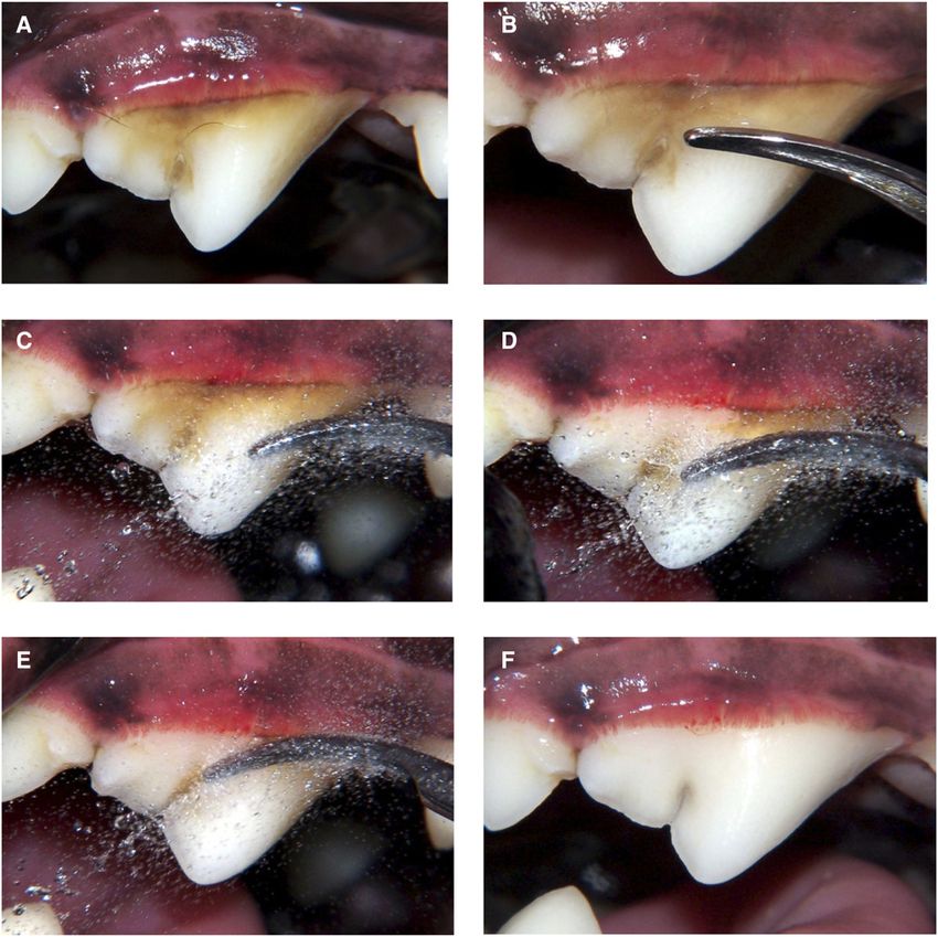
pocket exists in PD2 or PD3 cases (Figure 9). The purpose vitality. Inappropriate use may result in increased dental pain, risk
JAAHA.ORG 11need of medical or surgical procedures requiring anesthesia can be
managed to maintain a favorable balance between risk and derived
benefit. Medically important and indicated procedures should not be
absolutely discouraged based on chronologic age or most underlying
comorbidities. The most recent AAHA/AAFP Pain Management
Guidelines provide the entire veterinary care team an opportunity to
revisit the pathophysiology of pain and intervention strategies and
associated pharmacology/pharmacokinetics of treatment.
General anesthesia with endotracheal intubation, appropriate
monitoring, and physiologic support is necessary for dental proce-
dures, including dental cleaning and scaling as well as more advanced
dental care. Expert opinion and published data strongly support the
use of general anesthesia for dentistry. So-called “anesthesia-free”
dentistry has not been shown to be safer or comparable to the ca-
pacity to supra- and subgingivally clean teeth in an anesthetized
patient and is therefore unacceptable.2,61
Any dog or cat presenting for anesthesia should be considered on an
individual basis. Anesthesia for older dental patients and those with
comorbidities requires special attention. Each patient will have specific
physiologic alterations or diseases unique to that individual. Thus, the
anesthetic protocol needed for one patient typically will be quite different
from that needed for another. The use of local anesthetics as dental blocks
dramatically decreases the depth of general anesthesia needed, and thereby
FIGURE 9 Injection of perioceutic into a 5 mm cleaned, bleeding helps support blood pressure, decreases ventilatory depression, provides
periodontal pocket. Photo courtesy of Jan Bellows. analgesia, and generally increases safety. Additionally, anxiolytic adminis-
tration prior to veterinary visits has become routine to decrease stress in
for infection, and loss of tooth vitality. The use of resin-bonded some patients. The synergistic effect between anxiolytics and other drugs
sealants in cases of tooth resorption is contraindicated.52,53 necessitates consideration for decreased amount of premedication, induction
10. Biopsy all abnormal masses visualized grossly or radiograph- agents, and maintenance anesthetics necessary to achieve the desired effect
ically and submit samples for histopathologic evaluation by a and should be considered when formulating an anesthetic plan.
54 As with any patient, a thorough and complete history and
pathologist qualified in oral tissues analysis.
11. Maintain an open airway via intubation until the animal is preanesthetic examination should be completed. Any previous anes-
swallowing and is in sternal recumbency. Maintain body tem- thetic experience with the patient should be noted, and close attention
perature and continue intravenous fluid support as needed. should be paid to any anesthetic complications or abnormal responses.
Continuously monitor and record vital signs until the pa- A minimum database including laboratory evaluation and imaging will
tient is awake. Continue pain management while the pet is be individually developed. Additional diagnostics will be indicated for
55–57 some dental patients based on clinical signs, practical availability, and
in the hospital and upon discharge.
12. Provide instruction on home oral hygiene. The Veterinary client consultation. Any abnormal preanesthetic findings should be
Oral Health Council (VOHC) Accepted Products web page thoroughly evaluated and delaying the anesthesia and surgery should
(vohc.org/accepted_products.html) lists products that have be considered if necessary to address any potential problem areas
been scientifically proven to be effective in retarding accu- identified. Veterinarians must be in tune with their clients, their pa-
58,59 tient’s psychosocial issues, and the existing human–animal bond.
mulation of dental plaque and/or calculus.
Often, stressed and compromised animals do not thrive at the vet-
Anesthesia, Sedation, and Analgesia erinary practice, away from their families and homes.
Considerations Considerations should be made to make the dental stay brief
Fear of anesthesia is the most common cause of clients’ decisions to and less stressful. Outpatient techniques with prompt return of the
forego dental procedures for their pets.60 Canine and feline patients in patient to familiar settings and routines are highly desirable for all
12 JAAHA | 55:2 Mar/Apr 2019Dental Guidelines
FIGURE 10 Application of antiplaque sealant. (A) Barrier sealant gel professionally applied to a cat’s gingival sulcus; home plaque prevention
gel is then reapplied weekly by the pet owner. (B) Application of hydrophilic gingival dental sealant professionally applied to a dog’s gingival sulcus;
reapplication is recommended every 6 mo. Photo courtesy of Jan Bellows.
dental patients. A gentle approach, both in pharmacology and in the injectable general anesthetic agents need to be used with care in
application of clinical techniques, is especially important and will higher-risk patients (including geriatric animals) because of the
benefit all patients. Support of the human–animal bond is an im- typically altered hemodynamics, pharmacokinetics, and pharmaco-
portant goal, and dedicated emphasis on the reduction of fear, stress, dynamics; decreased plasma protein binding; and decreased ability
and pain is always warranted and primarily addressed through for hepatic metabolism and renal excretion in compromised animals.
management and behavioral modification. Anesthetic management Brachycephalic breeds and their associated airway conformations
represents a powerful combination of additional modalities. warrant particularly close attention during the induction and re-
covery periods to avoid hypoxia and prevent dyspnea.
General Anesthesia Inhalant general anesthetics are the anesthetics of choice in
For outpatient dental anesthesia, it is useful to select perioperative many small animal patients, especially for procedures lasting longer
medications that (1) typically provide for a rapid and complete than 10–15 min. The inhalants isoflurane and sevoflurane offer the
recovery (propofol or alfaxalone), (2) can be carefully reversed advantage for outpatient anesthesia of rapid adjustment of inhaled
(diazepam, midazolam, opioids, and dexmedetomidine), (3) can be and alveolar anesthetic dose and effect. However, inhalational in-
totally eliminated by supported ventilation (isoflurane, sevoflurane, duction of anesthesia (by either mask or chamber) is contra-
or desflurane), or (4) do not have substantial intrinsic toxicity or indicated in almost all clinical situations.64
significant adverse effects should drug effects persist (diazepam, Dose-dependent vasodilatation and hypotension preclude the use
midazolam, or butorphanol). In situations in which delayed or in- of higher doses of inhalant anesthetics in many higher-risk patients.
adequate recovery is recognized, physiologic support including judi- Dose-sparing anesthesia achieved using lower doses of synergistically
cious fluid therapy, support of body temperature, ventilatory support, acting injectable systemic agents (e.g., a fentanyl infusion) with local
and extended postanesthetic care should be provided. It is worth anesthetic techniques allows for the maintenance of partial IV anes-
noting that there is a strong consensus among veterinary anesthesi- thesia (PIVA) with comparatively low doses of inhalants. In other
ologists to reverse dexmedetomidine only when medically necessary, words, “less is more.” In more extreme cases, injectable agents (total
which allows the beneficial residual sedation to continue after the IV anesthesia [TIVA]) are best used in conjunction with intubation
completion of procedures in order to facilitate and ease recovery. If and oxygen supplementation but without inhalant anesthesia. This
62
necessary, consider using a low dose of atipamezole in cats. approach can often support markedly improved hemodynamics.
Adequate fluid replacement should be given to help prevent a Patients should be preoxygenated for 2–5 min before anesthetic
renal crisis and to help maintain a proper perioperative hemody- induction to help prevent hypoxia from developing during induction.
namic state. The rate of IV fluid administration will depend on the Every anesthetized patient should be intubated to protect and
particular patient’s needs, but will generally be in the range of 3– maintain a patent airway. The safety that often has been associated
5 mL/kg/hr.63 with inhalants, as opposed to injectable anesthetics, is partly due to
Careful planning and additional attention to drug and dosage the customary, if not obligatory, provision of supplemental oxygen as
selection is important to safely manage high-risk patients. Some the carrier gas for the volatile anesthetics. Endotracheal intubation
JAAHA.ORG 13and administration of supplementary oxygen can easily be incorpo- other analgesics, and will ease the transition to administering
rated into injectable general anesthetic techniques and substantially postoperative oral pain medications at home. Specific techniques for
adds to patient safety. If anesthesia is deep enough to allow for local anesthetic dental nerve blocks (indications, doses, and specific
placement of an endotracheal tube, then the patient is no longer able techniques) are described in detail by Niemiec et al., Beckman, and
to protect its airway from either obstruction or aspiration of regur- Gracis, and others.61,65–68 Three approaches for the maxillary nerve
gitated or foreign material. Adherence to proper techniques protects block are well described and offer choices based on anatomy and
64
our personnel and practices from waste anesthetic gases. personal preference.66 The maxillary tuberosity approach, using ei-
ther an intra- or extraoral (via the buccal pouch) access, allows for a
Sedation very short needle insertion just posterior to the caudal molar and
In select cases in which teeth cleaning, polishing, and extractions are maxillary tuberosity. Both the subzygomatic approach and the
not anticipated, heavy sedation may be appropriate and sufficient to technique of advancing the needle through the infraorbital canal
collect limited baseline information. Examples include a targeted provide access to the maxillary nerve as alternatives. Care is taken to
intraoral radiograph recheck and a more involved preliminary ex- avoid damage to the maxillary or infraorbital neurovascular bundle
amination of the oral cavity. When making the decision to use se- and inadvertent vascular or intraneural injection. Molars may not be
dation versus general anesthesia, there are three considerations: (1) adequately blocked using the infraorbital nerve block technique alone,
protecting the patient, (2) protecting personnel, and (3) protecting but anesthesia should be reliable from the third or fourth premolar
equipment. The loss of intrinsic airway protection requires us to place and the more rostral structures including the canine teeth.67
an endotracheal tube and serves as an operational distinction between The mandibular or inferior alveolar block can be performed at
sedation and anesthesia. The use of reversible agents, such as alpha- the angle of the mandible. The more successful intraoral approach
agonists, or boluses of induction agents, such as propofol combined technique is recommended.68 More rostral block at the mental fo-
with a quiet and dim environment and care to avoid stimulation, may ramen is less effective.60 Rarely, the lingual branch will be anesthetized
provide sufficient chemical restraint to meet these ends. with a mandibular nerve block, and a very few patients may bite their
Sedation-only procedures generate limitations including risking tongue during recovery. Recovery of the patient in sternal recum-
aspiration of fluids and aerosolized bacteria into the airways and bency with the tongue between the jaws may decrease this risk.
substandard ability to monitor ventilatory capacity without a proper Regardless of the local anesthetic technique or site, always as-
endotracheal tube in place. Because of the brief duration of action pirate to avoid intravascular injection of local anesthetic. Other uses
and efforts to minimize depth of sedation, challenges arise sur- of local anesthetics may contribute to the basic nerve block tech-
rounding the ability to appropriately monitor patient hemodynamics niques and include “splash blocks,” infiltration anesthesia, intra-
because time and patient handling (additional stimuli) are necessary osseus anesthesia, intraseptal injection, periodontal ligament or
to properly affix monitoring equipment. This results in difficulties intraligamentary injection, and intrapulpal injection.66
monitoring the adequacy of sedation even with well-trained and
dedicated staff. Because of the absence of reaching a surgical plane of Nonanesthetic Dentistry
anesthesia, sedation risks self-inflicted injury from the patient’s re- Nonanesthetic dentistry (NAD), also referred to as anesthesia-
flexes when attempting to probe subgingivally during an oral exam free dentistry, is a procedure in which the teeth are scaled and
and unnecessary risk for damage to equipment if bitten. Personnel polished without the benefit of general anesthesia. NAD is
health must also be considered during sedated procedures because considered not appropriate because of patient stress, injury, risk
an absence of a proper endotracheal tube while delivering inhalant of aspiration, and lack of diagnostic capabilities. Because this
gas risks human exposure to waste gas, ultrasonic scaling with in- procedure is intended to only clean the visible surface of the
appropriate irrigation results in increased bacterial aerosolization, teeth, it provides the pet owner with a false sense of benefit to
and abrupt patient response to stimuli risks bite injury. their pet’s oral health. 69,70
Veterinary dentistry relies on detailed examination by a veter-
Local Analgesia inarian with thorough knowledge of oral anatomy, physiology, and
Anyone performing oral surgical or periodontal procedures should pathology to make an accurate diagnosis. The examination includes
be familiar with dental nerve block techniques, including a thorough radiographs, requiring the animal to be motionless, as well as the use
knowledge of oral anatomy and analgesic agents and their applica- of costly equipment in the oral cavity. Periodontal probing (noxious
tion. Administration of local anesthetics will decrease the amount of stimulus) is also required to allow appropriate diagnosis and
required inhalant anesthetic, will decrease the required amount of treatment recommendations.
14 JAAHA | 55:2 Mar/Apr 2019You can also read