3-year-old male child with hypertension uncontrollable vomiting, sympathetic manifestations palor, piloerection, prolonged time of capillary ...
←
→
Page content transcription
If your browser does not render page correctly, please read the page content below
3-year-old male child with hypertension
 uncontrollable vomiting, sympathetic manifestations (palor,
 piloerection, prolonged time of capillary filling and priapism

Marcelo Garcia Leal M.D. Ribeirão Preto- São Paulo – Brazil
English
Dear Andrés,
I find that this case is very interesting for the network; a 3-year-old child, admitted in the ER of the
Hospital das Clinicas USP from Ribeiro Preto, with uncontrollable vomiting, with information from
the mother that when putting the child in bed, he started crying and yelling touching his head. In the
physical examination at admittance, he was in a poor general state, pale, piloerection, no fever,
acyanotic, anicteric, regular heart rhythm in 2 times, BP: 142x102, HR: 60 (?), respiratory auscultation
with diffuse rattles, dyspneic; respiratory rate: 40, O2 saturation: 98%, time of capillary filling greater
than 5 sec, presence of priapism, Glasgow 7 (AO: 1, RV: 2, RM: 4).
In the attachment you will find the ECG of the child.
I will send the evolution and the final diagnosis after hearing your opinions,
Warm regards,

                                               Portuguese
Caro Andrés,
Acho esse caso interessante para ir para a rede, criança de 3 anos deu entrada na unidade de
emergência do Hospital das Clínicas USP de Ribeirão Preto com vômitos incontroláveis, com
informação da mãe que ao colocar criança na cama esta começou a chorar e a gritar levando a mão a
cabeça. Ao exame físico da entrada estava em mal estado geral, descorada, piloereção, afebril,
acianótico, anictérico, ritmo cardíaco regular a 2 tempos PA: 142X102 FC:60 (?), ausculta respiratória
com estertores difusos, dispneico FR: 40, saturação de O2: 98%, tempo de enchimento capilar maior
que 5 segundos, presença de priapismo, Glasgow 7 (AO:1, RV:2, RM:4).
Segue em anexo o ECG da criança.
A evolução e o diagnóstico final mandarei após ouvir as opniões.
Um abraço,
Marcelo Garcia Leal M.D. Ribeirão Preto- São Paulo – Brazil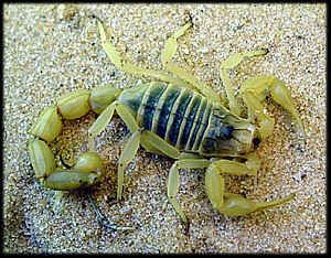
Colleagues opinions
Do you have some biological and echocardiograph data ? medications ? familial history and ECG from the
parents ? looks like lateral "Brugada like" ST elevation
I would suspect myocarditis in this child at least at first look but I'm waiting for other parameters

Philippe Maury mauryjphil@hotmail.com
Marcelo e Andres
A clinica é soberana:
Esta criança tem quadro neurologico compativel com acidente por animal peçonhento ( escorpião ou
aracnideo).
Criança de baixo peso, com menos de 6 kg, podem ter quadro severo e morte por esse tipo de acidente.
Sendo originario da região de Ribeirão Preto, esta é minha primeira hipotese.
O ECG está alterado em função do quadro neurológico.
Claudio Pinho MD,
                                                 English
Marcelo and Andrés: The Clinic is sovereign:
This child has neurological symptoms compatible with accidents caused by venomous animals
(scorpion or spider).
Underweight children, (under 6 kg ) can have severe and death from this type of accident.
Being from the region of Ribeirão Preto, this is my first hypothesis.
The ECG is abnormal consequence of neurological damage.
Claudio Pinho MD, Brazil,
Professor Cardiologia Faculdade de Medicina na PUC-Campinas Sao Paulo – Brazil,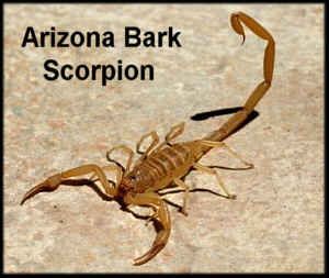
English

Dear friend Prof Andrés Ricardo Pérez Riera PhD I think that the young male boy suffers from .most
probably, a sympathetic adrenergic release . due a feocromocitoma The signs of an high lateral
myocardial injury is due to adrenaline effect ,and less noradrenalin as well the very low pressure
differences
The other possibility is a scorpion beat
my kindly regard
Samuel Sclarovsky M.D. From Israel

                                           Castellano/Spanish
Querido amigo Profesor Andrés: Pienso que este chico presenta una liberación masiva de adrenalina.
Consecuencia de un feocromocitoma. La corriente de lesión el pared lateral alta es causada por la
adrenalina, y menos por noradrenalina asi como la diferencial reducida
La otra posibilidad es la picadura de alacrán.
Samuel
Finals comments
       By Andrés Ricardo Pérez-Riera.M.D.Ph.D,

“It seems, from what I gather, to be one of those simple
cases which are so extremely difficult”

                        Sherlock Holmes.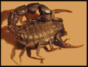
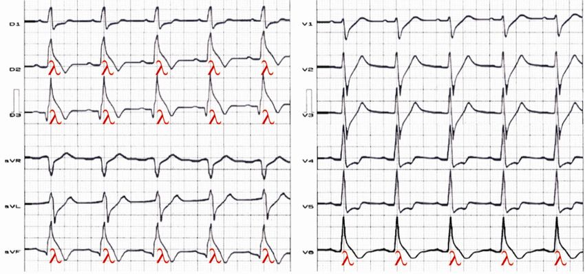
ECG diagnosis:
1. Heart rate: 167bpm (Normal maximal limit: 120 bpm at three years old of age)
2. Sinus tachycardia
3. QRS duration 78ms: Normal. (average 70 ms; maximal normal limit 80 ms at three years old.)
4. Normal QRS voltage on precordial leads There are not criteria for LVH because voltage of QRS
   Complexes In Precordial Leads in children from three to eight years old are:V1: average voltage of
   R wave 7 mm. Maximal 18 mm. Average depth of S wave 14 mm. Maximal 30 mm.V5: average voltage
   of R wave 21mm. Maximal 36 mm. Average depth of q wave 2 mm. Maximal up to 6 mm. Average
   depth of S wave 5 mm. Maximal 14 mm.V6: average voltage of R wave 14 mm. Maximal 24 mm.
   Average depth of q wave 1.5 mm. Maximal up to 4.5 mm.Average depth of S wave 1mm. Maximal 5
   mm.
5. ST segment elevation convex to the top in high lateral leads I and aVL: Subepicardial Injury current.
   “Brugada-like ECG pattern in aVL”
6. Mirror image or reciprocal changes in inferior and anteroseptal leads: Reciprocal ECG changes
   generally refer to leads with ST segment depression that face the “ischemic boundary” in patients with
   acute ST segment elevation ischemic events. During an acute ST-segment elevation myocardial
   infarction (STEMI) ST segment elevation is present in leads that face the region of transmural injury. ST
   segment depression in the setting of ST segment elevation in other leads can also be a result of an injury
   current vector resulting from transmural myocardial ischemia. Reciprocal ST-segment depression
   simulating additional subendocardial ischemia is commonly observed in STEMI. Consequently, it may
   be a marker of an independent additional zone of injury. Reciprocal electrocardiographic changes are
   considered when ST-segment elevation and broad upright T waves in leads that are mirrored perfectly by
   reciprocal ST depression and T-wave inversion in other leads. Many theories have been advanced to help
   explain reciprocal changes. Computer modeling has shown that as the ischemic zone extends from the
   endocardium to the epicardium, it creates a relatively positive area above the ischemic zone, and a
   relatively negative area at the ischemic boundaries (1).
This computer model explains why reciprocal changes may appear prior to ST segment elevation. Some
   authors have suggested that the first sign of acute inferior STEMI is a downsloping ST segment in lead I
   and aVL. Reciprocal changes can be subtle, and may present as nothing more than a flattening of the ST
   segment in the reciprocal leads. Clinical. situations where the direction of the ST injury vector is limited
   include: the presence of a previous infarction, preexisting abnormalities of the ST segment, LBBB and
   RBBB, ventricular pre-excitation, multivessel disease, abnormal site of origin of a coronary artery, and
   dominance or underdeveloped coronary arteries. When the ST segment deviation is minimal it may be
   difficult to differentiate the abnormal ST segment changes of transmural myocardial infarction from
   normal ERP. One of the following criteria is required for the diagnosis of a transmural MI: ST segment
   elevation at the J point of ≥ 1.0 mm in two or more limb leads or ≥ 2.0 mm in two or more precordial
   leads, and ST segment depression at the J point ≥ 2.0 mm in at least two of the three leads V1 to V3.
   Reciprocal changes are not the exclusive findings of ST segment elevation acute coronary syndrome.
   This phenomenon is also observed in patients with the typical type 1 ECG Brugada pattern, the
   Haïssaguerre ECG pattern (also called atypical Brugada ECG phenotype), and in overlapping Brugada /
   Haissaguerre pattern. Also seen in these patients is a lambda wave morphology (QRS complexes
   resembling the Greek letter lambda) with J point and ≥2 mm ST segment elevation in the inferior, lateral,
   or inferolateral leads with concomitant reciprocal image in the anterior wall. Recently, transient J-wave
   appearance in the inferior-lateral leads during electrical storm in a patient with BrS it has been reported.
   (see the next two slides 11 and 12)
7. QT/QTc: 278/476 Prolonged QT interval
Normal ECG paramethers in children from three to eight years old
-Heart Rate: Normal minimal limit: 80 bpm at three years old, 85 bpm at four years, 65 bpm at 6 years; after
this age the same as adults: 60 bpm. Normal maximal limit: 120 bpm at three years old, 115 bpm at 4 years;
after 6 years the same as adults: 100 bpm.

-Rhythm: sinus.

-P wave: SAP: between 0º and +90º. Average +60º. Duration: up to 100 ms. Voltage: maximal limit 2.5 mm.
P polarity: the same as the previous age group between 0º and 90º.

-PR interval: average 130ms (0.13 s). Maximal normal limit 160 ms for rates between 80 and 120 bpm.

-SAQRS In Frontal & Horizontal Planes:
 FP: average +60º. It may vary between -5º and +90º.
 HP: slightly intermediary posterior or possibly anterior. R/S ratio in V2 is lower, equal or greater than 1
respectively. Isodiphasism predominates in V2, i.e. AQRS in the HP is intermediary and pointing towards X.
 Duration of QRS complex: average 70 ms; maximal normal limit 80 ms. R/S ratio in precordial leads: The
so called “adult progression” of the R/S ratio begins in precordial leads, i.e. progressive increase of voltage of
R wave from V1 up to V5 and concomitant decrease of S up to V6.
-Voltage of QRS Complexes In Precordial Leads:
V1: average voltage of R wave 7 mm. Maximal 18 mm. Average depth of S wave 14 mm. Maximal 30 mm.
V5: average voltage of R wave 21mm. Maximal 36 mm. Average depth of q wave 2 mm. Maximal up to 6
mm. Average depth of S wave 5 mm. Maximal 14 mm.
V6: average voltage of R wave 14 mm. Maximal 24 mm. Average depth of q wave 1.5 mm. Maximal up to
4.5 mm.Average depth of S wave 1mm. Maximal 5 mm.
Reciprocal or mirror image in the anterior wall

The ECG shows persistent ST segment elevation in the inferior and apical leads, associated to concomitant
reciprocal or mirror image in the anterior wall that was not modified with the use of sublingual nitrate in
absence of hypothermia, electrolyte imbalance or ischemia.
The patient was a young symptomatic (repetitive syncope episodes) Thai man, with positive familial
background of SCD in young first degree relatives. He died 24h after performing this ECG. The ECG shows
persistent ST segment elevation in the inferior and low lateral precordial apical leads (V5-V6), associated
with concomitant reciprocal or mirror image in the anteroseptal wall that was not modified with the use of
sublingual nitrate in absence of hypothermia, electrolyte imbalance or ischemia.(2)
Typical ECG of Brugada Pattern

                    Presence of reciprocal image.

The ECG belongs to a symptomatic patient with Brugada syndrome who had three previous syncope
episodes. The ECG shows the typical ECG Brugada pattern: J point and ST segment elevation (> 2 mm),
convex to the top followed by a negative T-wave in right precordial leads. Lead aVR that faces the right
ventricular epicardium above the outflow tract shows subtle ST segment and J point elevation (red arrows).
Inferior leads show reciprocal or mirror image changes (blue arrows).
And which are the possible clinical diagnosis?
1) Pheocromocitoma? (Dr Samuel Sclarowsky Hypothesis) A pheochromocytoma is a rare,
catecholamine-secreting tumor that may precipitate life-threatening hypertension. The tumor is malignant
in 10% of cases but may be cured completely by surgical removal. Although pheochromocytoma has
classically been associated with 3 syndromes—von Hippel-Lindau (VHL) syndrome, multiple endocrine
neoplasia type 2 (MEN 2), and neurofibromatosis type 1 (NF1)—there are now 10 genes that have been
identified as sites of mutations leading to pheochromocytoma. Scholten et al concluded that
pheochromocytoma crisis should be treated with medical stabilization and elective or urgent surgery
rather than emergency surgery, based on their study of 137 patients with pheochromocytoma, including
25 who presented in crisis. Their literature review found that emergency resection of pheochromocytoma
is associated with high surgical morbidity and mortality
Signs and symptoms
Classically, pheochromocytoma manifests as spells with the following 4 characteristics:
Headaches – Palpitations – Diaphoresis - Severe hypertension( all present in this case)
Typical patterns of the spells are as follows: Frequency may vary from monthly to several times per day
Duration may vary from seconds to hours. Over time, spells tend to occur more frequently and become more
severe as the tumor grows. The following may also occur during spells: Tremor, nausea, weakness, anxiety,
sense of doom, epigastric pain. flank pain and constipation
Clinical signs associated with pheochromocytomas include the following: Hypertension: Paroxysmal in
50% of cases. postural hypotension: From volume contraction, hypertensive retinopathy, weight loss, pallor,
fever, tremor, neurofibromas, tachyarrhythmias, pulmonary edema
Diagnosis
Diagnostic tests for pheochromocytoma include the following:
Plasma metanephrine testing: 96% sensitivity, 85% specificity(3)
24-hour urinary collection for catecholamines and metanephrines: 87.5% sensitivity, 99.7% specificity(4)Test selection criteria include the following:
Use plasma metanephrine testing in patients at high risk (ie, those with predisposing genetic syndromes or a
family or personal history of pheochromocytoma)
Use 24-hour urinary collection for catecholamines and metanephrines in patients at lower risk
Imaging studies should be performed only after biochemical studies have confirmed the diagnosis of
pheochromocytoma. Studies are as follows:
Abdominal CT scanning: Has accuracy of 85-95% for detecting adrenal masses with a spatial resolution of
1 cm or greater
MRI: Preferred over CT scanning in children and pregnant or lactating women; has reported sensitivity of
up to 100% in detecting adrenal pheochromocytomas
Scintigraphy: Reserved for biochemically confirmed cases in which CT scanning or MRI does not show a
tumor.
PET scanning: A promising technique for detection and localization of pheochromocytomas.
Additional studies to rule out a familial syndrome in patients with confirmed pheochromocytoma include
the following:
1. Serum intact parathyroid hormone level and a simultaneous serum calcium level to rule out primary
hyperparathyroidism (which occurs in MEN 2A)
2. Screening for mutations in the ret proto-oncogene (which give rise to MEN 2A and 2B)(5)
3. Genetic testing for mutations causing the MEN 2A and 2B syndromes
4. Consultation with an ophthalmologist to rule out retinal angiomas (VHL disease)2 Terrestrial Venomous Animals Accidents caused by venomous animals: scorpion or spider ? Drs Claudio Pinho and Samuel hypothesis. Annually millions of scorpion stings and anaphylactic reactions to insect stings may occur worldwide, causing tens of thousands of deaths in humans each year mostly among children. Envenomation (toxic effects) is also an occupational hazard for populations involved in agriculture and forestry in these regions. Among the animals that can inflict injury on humans by the action of their venom are: 1. Invertebrates, such as Arachnida (spiders, scorpions and sun spiders) 2. Acarina (ticks and mites) 2. Chilopoda (centipedes) and 3. Hexapoda (bees, wasps, butterflies, and midges). Scorpion envenoming cause multi-system-organ failure, characterized by a massive release of counter- regulatory hormones (catecholamines, glucagon, cortisol), angiotensin-II, and changes in insulin secretion. It is a condition of fuel-energy deficits and an inability to utilize the existing metabolic substrates. Scorpion sting is a hazardous and potentially lethal condition. Venom of some variety of scorpion can cause dramatic cardiovascular and ECG changes, that have been related to heart stimulation by autonomous nervous system. González-Romero et al (7) prospectively studied 722 patients following scorpion sting. Mean age for the group was 25.5 +/- 18.3 years. 67% were less than 30 years of age. In 294 patients (40.7%) they found ECG changes. These cases were followed until those changes disappeared. First degree atrioventricular block was found in 10.2%. Intraventricular conduction disturbances in 12.8% with predominance of RBBB. In 11% the authors found arrhythmias. In 15% reversible ventricular repolarization changes. Of this no one died. This lack of mortality could be attributed to a prompt therapeutic intervention. In Ribeirâo Preto SP Brazil Cuppo et al (8) reported the clinical and laboratory data of 4 patients victims of scorpion stings by T. serrulatus, who developed heart failure and pulmonary edema, with 3 of them dying within 24 hours of the sting. Anatomopathologic study of these patients revealed diffuse areas of myocardiocytolysis in addition to pulmonary edema. The surviving child presented enzymatic, electrocardiographic and echocardiographic changes compatible with severe cardiac involvement, which were reversed within 5 days. These findings reinforce the need for continuous monitoring of patients with severe scorpion envenoming during the hours immediately following the sting.
Diagnosis & Treatment

In general, scorpions are not aggressive. They do not hunt for prey; they wait for it. Scorpions are nocturnal
creatures; they hunt during the night and hide in crevices and burrows during the day to avoid the light.
Thus, accidental human stinging occurs when scorpions are touched while in their hiding places, with most
of the stings occurring on the hands and feet.
 Scorpions use their pincers to grasp their prey; then, they arch their tail over their body to drive their stinger
into the prey to inject their venom, sometimes more than once. The scorpion can voluntarily regulate how
much venom to inject with each sting. The striated muscles in the stinger allow regulation of the amount of
venom ejected, which is usually 0.1-0.6 mg. If the entire supply of venom is used, several days must elapse
before the supply is replenished. Furthermore, scorpions with large venom sacs, such as the Parabuthus
species, can even squirt their venom.
 The venom glands are located on the tail lateral to the tip of the stinger and are composed of 2 types of tall
columnar cells. One type produces the toxins, while the other produces mucus. The potency of the venom
varies with the species, with some producing only a mild flu and others producing death within an hour.
Generally, the venom is distributed rapidly into the tissue if it is deposited into a venous structure. Venom
deposited via the intravenous route can cause symptoms only 4-7 minutes after the injection, with a peak
tissue concentration in 30 minutes and an overall toxin elimination half-life of 4.2 to 13.4 hours through the
urine. The more rapidly the venom enters the bloodstream, the higher the venom concentration in the blood
and the more rapid the onset of systemic symptoms.
Scorpion venom is a water-soluble, antigenic, heterogenous mixture, as demonstrated on electrophoresis
studies. This heterogeneity accounts for the variable patient reactions to the scorpion sting. However, the
closer the phylogenetic relationship between the scorpions, the more similar the immunological properties.
Furthermore, the various constituents of the venom may act directly or indirectly and individually or
synergistically to manifest their effects. In addition, differences in the amino acid sequence of each toxin
account for their differences in the function and immunology. Thus, any modifications of the amino acid
sequence result in modification of the function and immunology of the toxin.
The venom is composed of varying concentrations of neurotoxin, cardiotoxin, nephrotoxin, hemolytic toxin,
phosphodiesterases, phospholipases, hyaluronidases, glycosaminoglycans, histamine, serotonin, tryptophan,
and cytokine releasers. The most potent toxin is the neurotoxin, of which 2 classes exist. Both of these
classes are heat-stable, have low molecular weight, and are responsible for causing cell impairment in
nerves, muscles, and the heart by altering ion channel permeability. The long-chain polypeptide neurotoxin
causes stabilization of voltage-dependent sodium channels in the open position, leading to continuous,
prolonged, repetitive firing of the somatic, sympathetic, and parasympathetic neurons. This repetitive firing
results in autonomic and neuromuscular over-excitation symptoms, and it prevents normal nerve impulse
transmissions. Furthermore, it results in release of excessive neurotransmitters such as epinephrine,
norepinephrine, acetylcholine, glutamate, and aspartate. Meanwhile, the short polypeptide neurotoxin blocks
the potassium channels. The binding of these neurotoxins to the host is reversible, but different neurotoxins
have different affinities. The stability of the neurotoxin is due to the 4 disulfide bridges that fold the
neurotoxin into a very compact 3-dimensional structure, thus making it resistant to pH and temperature
changes. However, reagents that can break the disulfide bridges can inactivate this toxin by causing it to
unfold. Also, the antigenicity of this toxin is dependent on the length and number of exposed regions that are
sticking out of the 3-dimensional structure.
                                           Frequency Internationally
Scorpion stings occur in temperate and tropical regions, especially between the latitudes of 50°N and 50°S
of the equator. Furthermore, stings predominantly occur during the summer and evening times. In addition,
the majority of patients are stung outside their home.
 A recent 5-year surveillance study in Saudi Arabia found 6465 scorpion sting cases with a mean patient age
of 23 years, a male-to-female ratio of 1.9, and a higher incidence of stings in the months of May-October.
Furthermore, patients in rural areas tend to fare worse than patients in urban areas because of the delay in
getting medical help due to a longer travel time to medical centers. Fortunately, better public education,
improved control of the scorpion population, increased supportive therapies, and more technologically
advanced intensive care units have combined to produce a substantial decrease in mortality from these
envenomations.
Mortality & Morbidity:
The under-reporting of scorpion stings is frequent because most envenomations occur in desert and jungle
areas that do not have large medical facilities. Furthermore, reporting is not required. Most deaths occur
during the first 24 hours after the sting and are secondary to respiratory or cardiovascular failure.Children and
elderly persons are at the greatest risk for morbidity and mortality. A smaller child, a lower body weight, and
a larger ratio of venom to body weight lead to a more severe reaction. A mortality rate of 20 percent is
reported in untreated babies, 10 percent in untreated school-aged children, and 1 percent in untreated adults.
Race No racial predilection exists. Any differences in individual reactions to the scorpion sting are a
reflection of that individual's genetic composition rather than race.
Sex Females are more susceptible than males to the same amount of scorpion venom because of their lower
body weight.
Age While adults are stung more often than children, children are more likely to develop a more rapid
progression and increased severity of symptoms because of their lower body weight. Furthermore, elderly
persons are more susceptible to stings because of their decreased physiologic reserves and increased
debilitation.
Scorpion Sting - General First-aid
First aid for a scorpion sting is simple:
1. Wash the sting site with soap and water.
2. You may apply a cool compress to the sting site. Ice may or may not be recommended.
3. Numbness and tingling should pass away in time.
4. If symptoms persist or are severe, seek medical attention.
5. Keep your tetanus shots and boosters current.Wilderness First-Aid
1. Find out in advance if the wilderness area you are visiting is likely to be populated by centruroides (the
only dangerous kind of scorpion). These are found in New Mexico, Arizona, Southern Utah and Mexico.
2. Exercise caution when stepping or reaching into places where scorpions are likely to be: dark places like
wood piles, underneath rocks, inside shoes, or roaming the ground after dark.
3. Look for the signs and symptoms of a scorpion sting: burning pain, swelling or numbness at the site of
the sting.
4. Clean the sting with an antiseptic cleanser.
5. Apply an ice pack to the site of the sting.
6. Immobilize the extremity which was stung until you can establish whether the sting has produced severe
poisoning . Keep the extremity immobilized if an evacuation is necessary.
7. Administer an antihistamine such as Benadryl to reduce swelling and itching.
8. Monitor the injured person for signs and symptoms of severe poisoning: muscle spasms, convulsions,
impaired vision or speech, nausea, vomiting, difficulty breathing, impaired circulation. If any of these
symptoms are present, evacuate immediately to a hospital to receive an antivenom (antivenin).
9. Evacuate immediately if the person stung is a child or elderly person, or if you suspect the sting was from
a centruroide.
The stings from Texas scorpions produce only moderate reactions in most people because the poison has
little affect on the nervous system. However, a person who is stung by a scorpion should be watched closely
for adverse reactions. An ice pack applied to the affected area will relieve some pain. If swelling and/or pain
persists or if breathing difficulties occur, immediate medical attention is necessary. If you are stung by the
Arizona Bark Scorpion, the most dangerous of the Arizona scorpions, it is not likely to be fatal. Local
Arizona medical centers are familiar with the treatment. The Arizona Bark Scorpion is venomous. Arizona
Bark Scorpion sting symptoms are immediate pain or burning, very little swelling, sensitivity to touch, and a
numbness/tingling sensation. The Arizona Bark Scorpion sting may have additional symptoms such as
numbness or tingling of extremities or face, blurry vision, or muscle twitching. When stung by an Arizona
Bark Scorpion, children may start to exhibit hyperactivity and have roving eye movements.If you are victim of a scorpion sting, wash the area with soap and water. Apply a cool compress on the area of the scorpion sting. Ice (wrapped in a washcloth or other suitable covering) may be applied to the sting location for 10 minutes. Remove compress for 10 minutes and repeat as necessary. Call the Poison Control Center at (800) 362- 0101 or your local poison center (see your phone book). If you develop symptoms of an Arizona Bark Scorpion sting, go to the nearest emergency room. Very few people die from scorpion stings, even the sting of the Arizona bark scorpion. Scorpion stings are most dangerous to the very young and the very old. Pets are also at risk. While the dangerous scorpions are limited to the southwestern region of the United States, their stings are not necessarily deadly, however they are extremely painful. Compared with their U.S. counterparts, Mexican scorpions are another matter altogether. Some 2,000 people a year die from their stings. Any type of scorpion stings require emergency medical attention and should be approached in the same way as a snake bite. It is possible for hyper-acute (typically allergic) reactions to occur in susceptible individuals, taking the form of blurring of consciousness, unconsciousness, convulsions, a rapid drop in blood pressure, shock, and, in extreme cases, death. In general, scorpions are not aggressive. They do not hunt for prey; they wait for it. Scorpions are nocturnal creatures; they hunt during the night and hide in crevices and burrows during the day to avoid the light. Thus, accidental human stinging occurs when scorpions are touched while in their hiding places, with most of the stings occurring on the hands and feet. Scorpion stings are a major public health problem in many underdeveloped tropical countries. For every person killed by a poisonous snake, 10 are killed by a poisonous scorpion. In Mexico, 1000 deaths from scorpion stings occur per year. In the United States, only 4 deaths in 11 years have occurred as a result of scorpion stings. Furthermore, scorpions can be found outside their normal range of distribution, ie, when they accidentally crawl into luggage, boxes, containers, or shoes and are unwittingly transported home via human travelers. Scorpions are basically immune to most pesticides. If you suspect your house has scorpions, call a professional exterminator.
Clinical Patient History
For patients presenting with scorpion stings, ascertaining the following is essential:
• Time of envenomation.
• Nature of the incident.
• Description of the scorpion.
• Local and systemic symptoms.
The toxicity, variation, and duration of the symptoms depends on the following factors:
1. Scorpion species.
2. Scorpion age, size, and nutritional status.
3. Healthiness of the scorpion's stinging apparatus (telson).
4. Number of stings and quantity of venom injected.
5. Depth of the sting penetration.
6. Composition of the venom.
7. Site of envenomation: Closer proximity of the sting to the head and torso results in quicker venom
    absorption into the central circulation and a quicker onset of symptoms.
8. Age of the victim.
9. Health of the victim.
10. Weight of the victim relative to amount of venom.
11. Presence of comorbidities.
12. Treatment effectiveness.
Generally, intrathecal and intravenous routes have immediate effects, while subcutaneous and intramuscular
routes take effect several minutes to hours later.

Non-lethal scorpion species tend to produce local reactions similar to a hymenopteran sting, while lethal
scorpion species tend to produce systemic symptoms. The duration to progress to systemic symptoms ranges
from 5 minutes to 4 hours after the sting. The symptoms generally persist for 10-48 hours.
Physical Examination
The signs of the envenomation are determined by the scorpion species, venom composition, and the
victim's physiological reaction to the venom. The signs occur within a few minutes after the sting and
usually progress to a maximum severity within 5 hours. The signs last for 24-72 hours and do not have an
apparent sequence. Thus, predicting the evolution of signs over time is difficult. Furthermore, a false
recovery followed by a total relapse is common. A person who has been stung by a scorpion usually has 4
signs, with the most common being mydriasis, nystagmus, hypersalivation, dysphagia, and restlessness.
The mode of death is usually via respiratory failure secondary to anaphylaxis, bronchoconstriction,
bronchorrhea, pharyngeal secretions, and/or diaphragmatic paralysis, even though venom-induced multi-
organ failure plays a large role.
Children present with the same symptoms and signs as adults, except their symptoms are more severe and
protracted. Furthermore, they may display a restlessness that is out of proportion when compared to any
other disease. A child's symptoms have been described as inconsolable crying; uncontrollable jerking of the
extremities; and chaotic thrashing, flailing, and writhing combined with contorted facial grimaces. The
symptoms mimic a centrally mediated seizure, but the patient is awake and alert the entire time.
The grading of these scorpion envenomations depends on whether or not neurological signs predominate
and is as follows:
1. Non-neurological Predominance
2. Mild - Local signs.
3. Moderate - Ascending local signs or mild systemic signs.
4. Severe - Life-threatening systemic signs.Neurologic Predominance
Grade I - Local pain or paresthesia at the sting site (83 percent).
Grade II - Pain or paresthesia that has traveled from the sting site (9.1 percent).
Grade III - Either cranial nerve or somatic neuromuscular dysfunction (4.7 percent).
Grade IV - Both cranial nerve and somatic neuromuscular dysfunction (3 percent).
Local Signs
Neurotoxic Local Effects
1. Local evidence of a sting may be minimal or absent in as many as 50 percent of
cases of neurotoxic scorpion stings. In fact, tissue necrosis is rarely found.
2. A sharp burning pain sensation at the sting site, followed by pruritus, erythema,
local tissue swelling, and ascending hyperesthesia, may be reported. This
paresthesia feels like an electric current, persists for several weeks, and is the last
symptom to resolve before the victim recovers.
3. The tap test is administered by tapping at the sting site. A positive result is when
the paresthesia worsens with the tapping because the site is hypersensitive to touch
and temperature. In fact, wearing clothing over the area and sudden changes in
temperature exacerbate the symptoms.
Cytotoxic Local Effects
A macule or papule appears initially at the sting site, occurring within the first hour of
the sting.
The diameter of the lesion is dependent on the quantity of venom injected.
The lesion progresses to a purpuric plague that will necrose and ulcerate.
Lymphangitis results from the transfer of the venom through the lymphatic vessels.
Non-Lethal Local Effects
Pain, erythema, induration, and wheal may be present.
These are secondary to venom activation of kinins and slow-releasing substances.Neurologic signs: Most of the symptoms are due to either the release of catecholamines from the adrenal
glands (sympathetic nerves) or the release of acetylcholine from postganglionic parasympathetic neurons.
One study by Freire-Maia et al (1974) found that the adrenergic signs occur at a low venom dose, while
cholinergic signs occur at high venom dose concentrations (ie, greater than 40 mcg/100 g in Tityus
serrulatus scorpion venom). Furthermore, the adrenergic phase tended to be more dependent on the venom
dose than the cholinergic phase. However, dual manifestations of the adrenergic and cholinergic signs are
possible because of varying organ system sensitivities to these neurotransmitters.
Central Nervous System Signs
Thalamus-induced systemic paresthesia occurs in all 4 limbs.
Patients experience venom-induced cerebral thrombosis strokes.
The level of consciousness is altered, especially with restlessness, confusion, or delirium.
Patients have abnormal behavior.
Ataxia is also a sign.
Autonomic Nervous System Signs - Predominately sympathetic signs, parasympathetic signs, or a
combination of signs.
Sympathetic Signs
Hyperthermia.
Tachypnea.
Tachycardia.
Hypertension.
Arrhythmia.
Hyperkinetic pulmonary edema.
Hyperglycemia.
Diaphoresis.
Piloerection.
Restlessness and apprehension.
Hyperexcitability and convulsions.Parasympathetic Signs
• Bronchoconstriction,
• Bradycardia.
• Hypotension.
• Salivation, lacrimation, urination, diarrhea, and gastric emesis (SLUDGE).
• Rhinorrhea and bronchorrhea.
• Goose pimple skin.
• Loss of bowel and bladder control.
• Priapism.
• Dysphagia.
• Miosis.
• Generalized weakness.
Somatic Signs
• Rigid spastic muscle of the limbs and torso.
• Involuntary muscle spasm, twitching, clonus, and contractures.
• Alternating opisthotonos and opisthotonus from inactivation of sodium channels, leading to
increased sodium and calcium uptake.
• Increased tendon reflexes, especially prolongation of the relaxation phase.
• Piloerection accompanied by goose pimples.
Cranial Nerve Signs
Classic rotary eye movement may result in ptosis, nystagmus, and blurred vision.
Mydriasis is a sign.
Patients may have tongue fasciculations.
Dysphagia, dysarthria, and stridor occur secondary to pharyngeal reflex loss or muscle spasm.
Patients may present with excessive salivation and drooling.
Peripheral nervous system signs - Intense local burning pain with minimal swelling at sting site, followed by
ascending numbness and tingling, then paralysis and convulsions.Non-Neurologic Systemic Signs
Cardiovascular signs
Usually follow a pattern of a hyperdynamic phase followed by a hypodynamic phase.
Hypertension is described as follows: Secondary to catecholamine and renin stimulation. Observed as early
as within 4 minutes after the sting.
Lasts a few hours.
High enough to produce hypertensive encephalopathy.
Hypotension - Less common and occurs secondary to excess acetylcholine or catecholamine depletion.
Tachycardia is greater than 130 beats per minute, although bradycardia can be observed.
Transient apical pansystolic murmur is consistent with papillary muscle damage.
Cardiovascular collapse occurs secondary to biventricular dysfunction and profuse loss of fluids from
sweating, vomiting, diarrhea, and hypersalivation. Observed in 7-38 percent of cardiovascular cases.
Mild envenomation - Vascular effect with vasoconstriction hypertension.
Moderate envenomation - Left ventricular failure hypotension with and without an elevated pulmonary
artery wedge pressure, depending on fluid status of the patient.
Severe envenomation - Biventricular cardiogenic shock. Cardiac dysfunctions attributed to
catecholamine-induced increases in myocardial metabolism oxygen demand (leading to myocardial
ischemia-induced myocardial hypoperfusion) and to the direct effects of the toxin (leading to
myocarditis).
Respiratory Signs
Tachypnea may be present.
Pulmonary edema with hemoptysis and a normal-sized heart is observed in 7-32 percent of respiratory cases.
This is secondary to a direct toxin-induced increased pulmonary vessel permeability effect and is also
secondary to catecholamine-induced effects of hypoxia and intracellular calcium accumulation, which leads
to a decrease in left ventricular compliance with resultant ventricular dilation and diastolic dysfunction.
Respiratory failure may occur secondary to diaphragm paralysis, alveolar hypoventilation, and bronchorrhea.Allergic Signs
Urticaria, Angioedema is reported, bronchospasm and eventual anaphylaxis..
Gastrointestinal Signs
Excessive salivation, dysphagia, nausea and vomiting, gastric hyperdistention occurs secondary to vagal
stimulation, increased gastric acid output may lead to gastric ulcers, acute pancreatitis may lead to
hyperglycemia, liver glycogenolysis may occur from catecholamine stimulation and toxic Hepatitis.
Genitourinary Signs
Patients have decreased renal plasma flow, toxin-induced acute tubular necrosis renal failure may occur,
rhabdomyolysis renal failure may result from venom-induced excessive motor activity, priapism may
occur secondary to cholinergic stimulation. One small study by Bawaskar (1982) found a positive
prognostic correlation to the development of cardiac manifestations following scorpion stings.
Hematological Signs
Platelet aggregation may occur because of catecholamine stimulation, disseminated intravascular
coagulation with massive hemorrhage may result from venom-induced defibrination.
Metabolic Signs
Hyperglycemia may occur from catecholamine-induced hepatic glycogenolysis, pancreatitis, and insulin
inhibition. Increased lactic acidosis may occur from hypoxia and venom-induced increased lactase
dehydrogenase activity, electrolyte imbalance and dehydration from hypersalivation, vomiting,diaphoresis,
and diarrhea.
Pregnancy Signs - Toxin-induced uterine contraction.
Symptoms predictive of hospital admission. Priapism (odds ratio 150.59) vomiting (odds ratio 15.82)
systolic blood pressure (SBP) greater than 160 (odds ratio 13.38), temperature greater than 38°C (odds ratio
3.66) and heart rate greater than 100 beats per minute (odds ratio 3.35)
Symptomology of Specific Scorpion Species
Mesobuthus, Tityus, and Leiurus - Tend to cause severe cardiovascular symptoms
Centruroides - Tend to cause neurological symptoms
Hemiscorpius - Tend to cause tissue necrosis.Scorpion Venom & Lethal Dose Risks Scorpions are shy creatures and only sting if threatened, cornered, or disturbed (eg, being sat or stepped upon). Curious individuals are at risk because of increased interaction with the scorpion. The median lethal dose 50 (LD50) of various scorpion venoms in mg/kg of a subcutaneous injection into mice and the territorial distribution are listed below. Unfortunately, humans are much more sensitive than mice. Leiurus quinquestriatus (Middle East) - 0.25 mg/kg Androctonus crassicauda (Saudi Arabia) - 0.08-0.5 mg/kg Centruroides noxius (Mexico) - 0.26 mg/kg. Androctonus mauritanicus (North Africa) - 0.32 mg/kg. Centruroides santa maria (Central America) - 0.39 mg/kg. Tityus serrulatus (Brazil) - 0.43 mg/kg. Buthus occitanus (North Africa) - 0.9 mg/kg. Centruroides sculpturatus (Southwest United States) - 1.12 mg/kg. Mesobuthus eupeus (Iran) - 1.45 mg/kg. Generally, most lethal scorpions have an LD50 below 1.5 mg/kg. The average yield per scorpion via electrical excitation of the venom gland for a few species is listed below. Tityus species - 0.39-0.62 mg. L quinquestriatus - 0.62 mg. Buthus species - 0.38-1.5 mg. Milking the venom gland produces approximately a 4-fold increase in yield amount compared to electrical excitation.
3) Sickle cell anemia CRISIS? It is a common cause of priapism, is an inherited disorder characterized by
   abnormally shaped red blood cells. These abnormally shaped cells can block the flow of blood. Sickle
   cell anemia is the most common cause of priapism in boys.Sickle cell anemia is the most common form
   of sickle cell disease. It is is a serious disorder in which the body makes sickle-shaped red blood cells.
   “Sickle-shaped” means that the red blood cells are shaped like a crescent. Normal red blood cells are
   disc-shaped and look like doughnuts without holes in the center. They move easily through your blood
   vessels. Red blood cells contain an iron-rich protein called hemoglobin). This protein carries oxygen
   from the lungs to the rest of the body. Sickle cells contain abnormal hemoglobin called sickle
   hemoglobin or hemoglobin S. Sickle hemoglobin causes the cells to develop a sickle, or crescent, shape.
   Sickle cells are stiff and sticky. They tend to block blood flow in the blood vessels of the limbs and
   organs. Blocked blood flow can cause pain and organ damage. It can also raise the risk for infection. A
   team of researchers from the Johns Hopkins Children’s Center, Vanderbilt University and elsewhere
   have demonstrated that high blood pressure and anemia together put children with sickle cell disease at
   serious danger for symptom less or so-called “silent” strokes, although either condition alone also
   signaled high risk. Additionally, a serious complication of sickle cell disease is pulmonary hypertension.

                                     Sickle cell anemia Complications

      I      Stroke. A stroke can occur if sickle cells block blood flow to an area of your brain. Signs of
            stroke include seizures, weakness or numbness of your arms and legs, sudden speech difficulties,
            and loss of consciousness. If your baby or child has any of these signs and symptoms, seek
            medical treatment immediately. A stroke can be fatal.
      II.   Acute chest syndrome: This life-threatening complication of sickle cell anemia causes chest
            pain, fever and difficulty breathing. Acute chest syndrome can be caused by a lung infection or
            by sickle cells blocking blood vessels in your lungs. It may require emergency medical treatment
            with antibiotics and other treatments.
III. Pulmonary hypertension. People with sickle cell anemia can also develop high blood pressure
in their lungs (pulmonary hypertension). Shortness of breath and difficulty breathing are
common symptoms of this condition, which can be fatal.
IV. Organ damage. Sickle cells can block blood flow through blood vessels, immediately depriving
an organ of blood and oxygen. In sickle cell anemia, blood is also chronically low on oxygen.
Chronic deprivation of oxygen-rich blood can damage nerves and organs in your body,
including your kidneys, liver and spleen. Organ damage can be fatal.
V. Blindness. Tiny blood vessels that supply your eyes can get blocked by sickle cells. Over time,
this can damage the retina — the portion of the eye that processes visual images — and lead to
blindness.
VI. Skin ulcers. Sickle cell anemia can cause open sores, called ulcers, on your legs.
VII. Gallstones. The breakdown of red blood cells produces a substance called bilirubin. A high
level of bilirubin in your body can lead to gallstones.
VIII. Priapism. Men with sickle cell anemia may experience painful, long-lasting erections, a
condition called priapism. As occurs in other parts of the body, sickle cells can block the blood
vessels in the penis. This can damage the penis and eventually lead to impotence. This little boy
had pianism.
4. Acute Myocarditis? Hypertension is rare in Myocarditis.Definitive Diagnosis
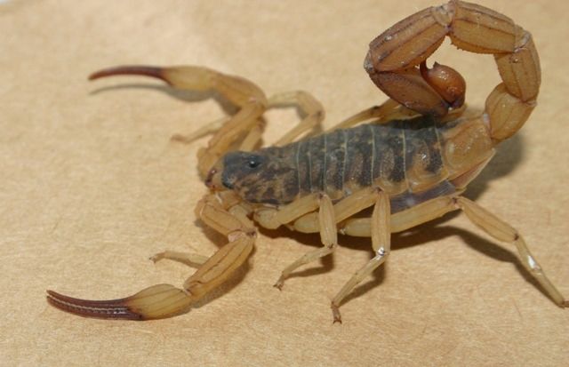
Portuguese: Caro Andrés, Ribeirão Preto é uma cidade que tem um excelente chopp, porém está
infestada de escorpiões. Parabéns para este time. Como é bela a medicina simples, apenas com o
exame clínico e eletrocardiograma. O escorpião da foto estava na cama da criança.
Evolução enzimática: CK-MB: 55- 105- 77- 44- 40 (normal:
Absolutely normal!!!!!!

Scorpion sting usually follow a pattern of a hyperdynamic phase followed by a hypodynamic phase.
Hypertension is secondary to catecholamine and renin stimulation. Observed as early as within 4
minutes after the sting.
Lasts a few hours. High enough to produce hypertensive encephalopathy.
Hypotension - Less common and occurs secondary to excess acetylcholine or catecholamine depletion.
Tachycardia is greater than 130 bpm, although bradycardia can be observed.
Eventual transient apical pansystolic murmur is consistent with papillary muscle damage.
Cardiovascular collapse occurs secondary to biventricular dysfunction and profuse loss of fluids from
sweating, vomiting, diarrhea, and hypersalivation. Observed in 7-38 percent of cardiovascular cases.
1. Mild envenomation Vascular effect with vasoconstriction hypertension.
2. Moderate envenomation - LV failure hypotension with and without an elevated pulmonary artery
wedge pressure, depending on fluid status of the patient.
3. Severe envenomation - Biventricular cardiogenic shock. Cardiac dysfunctions attributed to
- catecholamine-induced increases in myocardial metabolism oxygen demand (leading to myocardial
ischemia-induced myocardial hypoperfusion) and to the direct effects of the toxin (leading to
myocarditis).
This is the photo of the Scorpion that was in the bed of the child.
. I think that this is a Brazilian yellow scorpion T. serrulatus whose venom can cause severe symptoms
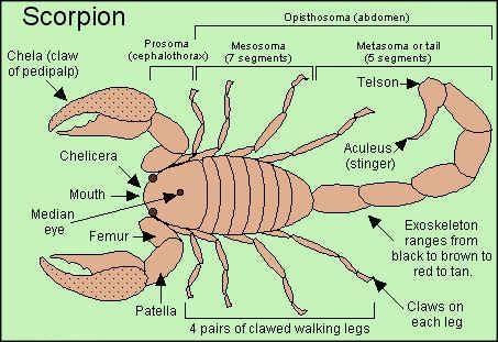
(which can include death in small children). With Mild envenomation.Scorpion Description & Habitat The scorpion is an animal closely related to spiders, mites, and ticks. Scorpions make up the order Scorpiones of the class Arachnida. They are easily recognized by their characteristic shape. Scorpions glow brightly under UV light (black light).
Brazilian Scorpions Scorpion of Other Countries And Regions Outside The United States
A) Brazilian Yellow Scorpion The Yellow Scorpion (Tityus serrulatus) is a Brazilian scorpion which is
extensively studied. Even though there are few documented deaths (death mainly comes from children under
7), the symptoms is severe and among 5 percent of the patients studied. There is systemic involvement. This
represents a very significant percentage.
B) Brazilian Scorpion (Tityus Bahiensis) Tityus is a large genus of scorpions belonging to the family
Buthidae. There are currently 185 described species distributed throughout the Caribbean and South America
and in Central America as far north as Costa Rica. The genus contains several dangerously venomous
scorpions, the best known of which is the Brazilian yellow scorpion T. serrulatus whose venom can cause
severe symptoms (which can include death in small children). Tityus serrulatus and Tityus bahiensis are
considered to be the most venomous.

A                                                       B
 The present case
A scorpion's long, slender body consists of a cephalothorax (joined head and thorax) with six pairs of
appendages and an abdomen. Scorpions have four pairs of legs and two large pincer-bearing arms
(pedipalps) in front. The first two pairs of appendages are used for catching and mashing prey and for
transferring it to the mouth; the last four pairs are legs. The abdomen is a flexible structure made up of 12
segments. All scorpions have a five-segmented tail that can be arched over the back. The tail ends in a sharp,
hollow, bulb-like poison gland or venom-injecting stinger.
 Scorpions are well equipped to defend themselves or attack prey with their pincers and stinger. Between the
last pair of legs is a comb-like structure (pectines) that is used to identify surface textures and to detect prey.
Scorpion venom can be deadly to many animals, including humans. They can be a nuisance when they
interact with humans because they will sting when disturbed.
 Scorpions have two eyes on the top of the head, and usually two to five pairs of eyes along the front corners
of the head. They do not see well, however, and must rely on the sense of touch, using their pectines and
other organs for navigation and hunting. Their bodies are flat, which allows them to hide in small cracks,
under rocks and under bark.
 The scorpion lives in warm, dry regions and in the tropics, preferring dry-and habitats. Scorpions are most
commonly found in southern Europe, Africa, the western and southwestern region of the United States, and
the tropical regions of the Western Hemisphere. There are about 1,500 species of scorpions and there may
be as many as 1,000 more species undiscovered.
 Worldwide, scorpions range in size from about 1/2 inch up to 10 inches (1.3 to 25 cm) in length (including
the tail), depending upon the species. About 70 to 75 species are found in the United States and most of
these are found in the desert region of the Southwest. These scorpions are usually about 1 to 3 inches (2.5 to
7.5 cm) in length. Scorpions may be found in many types of habitats in the United States. Although most
scorpions live in desert regions such as desert flats and sand dunes, desert and mesic mountains, they also
live in rain forests, grassy prairies and grasslands, pine forests, deciduous forests, and chaparral, and others
live only beneath the bark of palm trees. Species are most diverse in desert areas. Scorpions hide under
stones, bark, wood or other objects on the ground where they wait or search for prey.
Wherever they live, scorpions are nocturnal predators. Hunting at night, a scorpion will eat almost anything,
even other scorpions. Chief foods are small insects, spiders, centipedes, earthworms, and other scorpions. In
fact, a scorpion's favorite food is another scorpion! Most scorpions are solitary and will attempt to kill and
eat other scorpions that invade their territory. Once they capture their prey, they use the large pincers to
crush and draw it toward the mouth.

 A scorpion grabs prey with its pincers and then stings it. Some scorpions have such powerful pincers they
seldom use their stinger. First the scorpion breaks the cricket or beetle into tiny pieces. When the pile is big
enough, the scorpion spits strong digestive juices onto the pile of bug bits. The juices melt the bits into soft
sticky stew. When the stew is soft enough, the scorpion sucks the gooey pieces into its mouth. The body
juices of the prey are eaten by the scorpion.

 Some scorpions can survive a whole year with no food. Other scorpions can live for two days under water
or survive long periods of cold. Scorpions may also live for a long time. Some species may live for 20 to 25
years, but longevity of the typical scorpion is between 3 and 8 years.

 Scorpions hide during the day and become active at night. This behavior helps scorpions manage
temperature and water balance, important functions for survival in dry habitats. Many species dig burrows in
the soil. During the day, the scorpion rests in an underground burrow, emerging at night to feed on insects
and spiders, which it immobilizes and kills with the sting. The sting is also used when the scorpion is
threatened. They detect and capture prey by the sense of touch. They also have a well-developed sense of
hearing.

According to fossil records, the scorpion has been in existence for about 400 million years. Fossil remains
reveal very little change between the ancient and the present-day scorpion. Scorpions have been around for a
long time. They have changed very little in 350 million years.
Scorpions Versus Spiders

Like spiders, scorpions are arachnids. They have two main body parts and eight legs. And like spiders,
scorpions have hairs, called bristles, along their legs. These bristles feel vibrations on the ground. They tell
the scorpion, which has poor vision, when prey is near.
 Most scorpions are larger than spiders. Their body parts are larger, too. Look at the scorpion's head. Instead
of spider fangs, a scorpion has pincers that grab and tear prey. Instead of leg-like palps, a scorpion has a large
set of claws that hold and crush prey. The last few segments of the scorpion's abdomen form a long "tail.“
                                                   Scorpion Stings
 A scorpion often kills its prey with venom. But a scorpion does not use fangs to deliver its poison. It uses the
stinger at the end of its tail. First, the scorpion grabs its prey with its claws. Then it raises its tail up and over
its head to sting its prey.
 The sting of a scorpion is very powerful. It kills insects and spiders instantly. And it can be deadly to larger
animals. Still, many animals - such as lizards, snakes, owls, and mammals - feed on scorpions. Scorpions are
nocturnal animals. They hunt and feed mostly at night. During the day, scorpions hide among rocks, in cracks
on the ground, and under the bark of trees. A scorpion's sting can be painful, but most scorpions are not
dangerous to humans
                                                  Scorpion Offspring
Adult scorpions may have several broods of young. Prior to mating, the male and female grasp each other's
claws and perform a courtship dance. Following an elaborate mating process, which lasts from 24 to 36
hours, the female undergoes a gestation period ranging from 5 months to more than 1 year. A female
scorpion gives birth to a litter, or a group, of about 25 baby scorpions (the litter can range from 6 to 90
young). The young are born one at a time and alive in semi-transparent sacs (thin layer of tissue around it's
body). With its mother's help, the newborn breaks out of this thin sack. As soon as the young scorpions free
themselves from these thin wrappers, they climb onto their mother's back. Newborn scorpions are pale in
color. The mother scorpions take care of their young for a short time until they are ready to live on their own.
In the first few days of their lives, they are totally defenseless. They depend on their mothers for protection.
Young scorpions stay with their mothers a week or more, until their first molt. Already capable of stinging,
the young scorpions leave the mother after several days and begin to fend for themselves. Scorpions reach
maturity in a one to five years, depending on availability of food.
Scorpion Myths
Scorpions are often misunderstood. Many people say that scorpions are so aggressive they will sting
themselves to death, but this is not a natural behavior of scorpions in the wild. Other people say the sting of
a baby scorpion is more dangerous than the sting of an adult, but again, this is false. The venom in a
scorpion's stinger is the same all through a scorpion's life. Perhaps the biggest myth is that all scorpions are
deadly, and this is totally wrong.
Only a very few scorpions are potentially dangerous to people. Of the 1,500 known scorpion species only
25 have a sting potent enough to be considered potentially dangerous to humans. About 90 species of
scorpions have been identified in the United States. Only one lives in the United States. The most dangerous
scorpion is found in the southwest region of the U.S. and is known as the Arizona Bark Scorpion
(Centruroides sculpturatus). Arizona is home to a large number of bark scorpions and is also home of the
only scorpion antivenom program in North America. Scorpion antivenom is used to treat severe scorpion
stings. Despite the large number of people and scorpions in Arizona, only about 100 stings each year receive
scorpion antivenom therapy, and most of those are either young children or elderly adults. Most important
for severe scorpion stings is immediate access to medical treatment, whether or not antivenom is available.
In less developed countries, such as Brazil lack of transportation to medical care contributes to higher
numbers of scorpion stings resulting in illness or death. No scorpion sting-related death has been reported in
Arizona for more than 40 years.
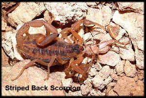
The most common species in Texas is the Striped Bark Scorpion (Centruroides vittatus). The adult
scorpion is about 2-1/2 inches long, which is typical of the size of all species found in the state. Texas has
18 species and only one species, Centruroides vittatus, occurs throughout the state. It is the only species of
scorpion found in the eastern part of Texas. The number of species found in the state increases moving west
and south.You can also read