A 3D CNN Network with BERT For Automatic COVID-19 Diagnosis From CT-Scan Images
←
→
Page content transcription
If your browser does not render page correctly, please read the page content below
A 3D CNN Network with BERT For Automatic COVID-19 Diagnosis From
                                                                       CT-Scan Images

                                                                                       Weijun Tan, Jingfeng Liu
                                                                              Shenzhen Deepcam Information Technologies
                                                                                          Shenzhen, China
                                                                                  {weijun.tan,jingfeng.liu}@deepcam.com
arXiv:2106.14403v1 [eess.IV] 28 Jun 2021

                                                                    Abstract                                and non-COVID-19 from a volume of CT-scan slice images
                                                                                                            of a patient. We use the dataset provided in the ICCV-MIA
                                              We present an automatic COVID1-19 diagnosis frame-            COVID-19 diagnosis challenge [11],. Since there is no slice
                                           work from lung CT-scan slice images. In this framework,          annotation in this dataset, 2D CNN classification network
                                           the slice images of a CT-scan volume are first proprocessed      on single slice image is not considered. Instead, 3D CNN
                                           using segmentation techniques to filter out images of closed     network is explored.
                                           lung, and to remove the useless background. Then a resam-            In this paper, we first discuss the preprocessing of CT-
                                           pling method is used to select a set of fixed number of slice    scan slice images, which is very critical for good classifica-
                                           images for training and validation. A 3D CNN network with        tion performance. The goal of preprocessing is to prepare
                                           BERT is used to classify this set of selected slice images. In   good-quality slice images per volume for training and val-
                                           this network, an embedding feature is also extracted. In         idation in the CNN classification network. In this work,
                                           cases where there are more than one set of slice images in       we propose segmentation techniques using both traditional
                                           a volume, the features of all sets are extracted and pooled      morphological transforms and a deep learning method us-
                                           into a feature vector for the whole CT-scan volume. A sim-       ing UNet [18]. Since the 3D CNN network requires fixed
                                           ple multiple-layer perceptron (MLP) network is to used to        number of images as input, we adopt and revise a resam-
                                           further classify the aggregated feature vector. The models       pling method used in [7]. In validation and testing time, we
                                           are trained and evaluated on the provided training and val-      propose a method to use all available good slice images to
                                           idation datasets. On the validation dataset, the precision is    make the final classification result.
                                           0.906 and the F1 score is 0.89.                                      A 3D network is widely used in many tasks including
                                                                                                            video understanding (recognition, detection, captioning). In
                                                                                                            videos, frame images at different time form a 3D series of
                                           1. Introduction                                                  images. A 3D CNN network is good at aggregating the tem-
                                                                                                            poral information. 3D network is also used in COVID-19
                                               The novel COVID-19 coronavirus breaking out in late          diagnosis, where the slice images at different spacing form
                                           2019 has been one of the worst disasters in the human his-       a 3D series of images. The correlation between slice images
                                           tory. As of end late June 2021, more than 142 millions in-       is just analogous to the temporal information in videos. We
                                           fections have been identified, more than 3 million lives have    are the first to use a 3D CNN network with BERT (CNN-
                                           been lost, and more than 200 countries have been drastically     BERT) for video action recognition [10] in classification of
                                           overwhelmed [1]. Therefore, it is very critical to stop the      COVID-19 from CT-scan images.
                                           spreading of the virus. So first a person has to be confirmed        Most 3D CNN networks use fixed number of images as
                                           to have COVID-19 before safety measures and treatments           input. Others can use all available images by a global pool-
                                           can be taken accordingly. One example is [19], where ther-       ing method. In our study, to avoid the out-of-memory prob-
                                           mal imaging is used to detect fever patients and face recog-     lem caused by using all slice images, we choose to use fixed
                                           nition is used to report and trace patients and their close      number of images as input to the 3D CNN-BERT network.
                                           contacts.                                                        However, since the available images may be a lot more than
                                               Among the techniques to diagnosis COVID-19, X-ray            this fixed number, if only one set of them is used, some
                                           and CT-scan images are studied extensively. In this paper,       useful information may be missed. Motivated by this in-
                                           we present an automatic diagnosis framework from chest           sight, we extract the embedding feature vector of all avail-
                                           CT-scan slice images. The goal is to classify COVID-19,          able sets of images for every CT-scan volume, then aggre-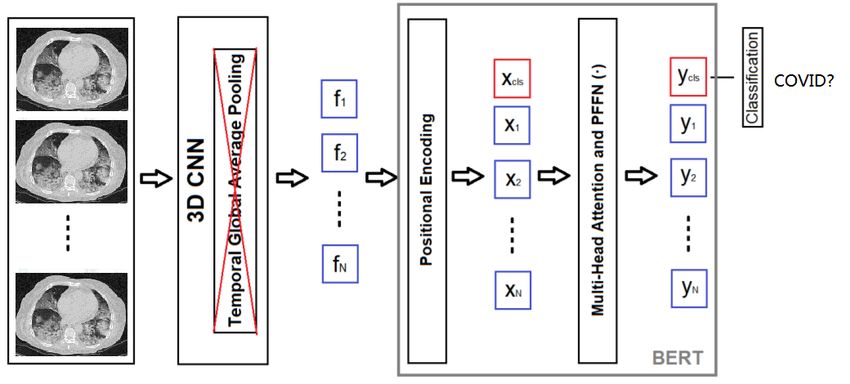
gate one global feature vector out of them. This feature         is extracted for every image, then multiple pooling methods
vector is sent to a simple MLP classifier for extra classifi-    are ensembled to generate a global feature vector before a
cation. This way, both the advanced 3D CNN network with          classification is made.
BERT and the information in all available image are used.            In the 3D CNN method, in [23], a 3D CNN network is
Evaluation results show that this pooling method and MLP         used, however, with both the slice image and a segmented
can improve the accuracy on the validation dataset by 1.5%.      lung mask as input. They cut off a portion of slice images
To our knowledge, we are the first to explore using all avail-   at the beginning and end of a CT-scan volume. In [24], the
able slice images in a 3D CNN network for classification of      authors first segment the lung mask from a slice image us-
COVID-19.                                                        ing traditional morphological transforms, then use this mask
   On the provided validation dataset, we achieve an accu-       to select good slice images and generate lung-only images
racy 0.906, and an F1 score 0.89. On the test dataset, we        (no background, bone or tissue) slice images. To make the
achieve an F1 score xxx.                                         number of images a fixed number, they use 3D cubic inter-
                                                                 polation to regenerate slice images. In [7], 3D CNN net-
2. Related Work                                                  work using a fixed number of slice images as input is used.
                                                                 Instead of using a fixed 3D CNN architecture, an autoML
   Deep learning has been used in a lot of medical imaging       method is used to search for best 3D CNN architecture in
analysis and diagnosis, e.g. in [13], [12], [14].                the network space using Mobilenet network block.
   Since the outbreak of the COVID-19 pandemic, a lot of
researches have been done to diagnose it using deep learn-       3. Data Preprocessing and Preparation
ing approaches, mostly CNNs on CT scan images or X-ray
images. For a complete review, please refer to [16] and [2].        The first goal of preprocessing is to select good slice im-
   Among the classification methods, some use 2D network         ages for training and validation. The second goal is to seg-
on slice image individually and make prediction for every        ment the lung mask of a slice image, so a masked image,
image. This is called 2D network. To make a decision for         where background, bones, vessels, tissues are all blacked
a patient, some kinds voting method is typically used [3],       out. This has been shown useful in [9], [8], [24].
[17], [8]. Others use 2D network on slice image, and gener-
                                                                 3.1. Morphological Lung Segmentation
ate embedding feature vector for every image, then all fea-
ture vectors are pooled to a single global feature vector, and      In this work, we use two segmentation techniques to seg-
a few fully-connection (FC) layers are used for classifica-      ment the lung region out of the slice image. The First tech-
tion. This is called 2D+1D network [15], [9], [4]. The third     nique is based on traditional morphological transforms. It
method is a pure 3D CNN network, where slice annotation          works well to get an bounding box of the lung (including
is not needed, and a set of or all the available slice images    bones and tissues), and a coarse mask image of the two
are used as input, and the 3D network process all these input    lungs. The bounding box can be used to remove background
images all at once in a 3D channel space [23], [24], [7].        of the slice image, and the mask can be used to tell if the
   In the 2D CNN method, some use the lung mask seg-             lungs are closed. This bounding box and the mask image
mentation, but most of them directly use the raw slice im-       are used to select slice images of a patient for training and
age. The COVID-MaskNet [20] uses a segmentation net-             validation.
work to localize the disease lesion, then use a FasterCNN-          The segmentation involves Gaussian blurring, binariza-
based approach to do the classification on the detected le-      tion, erosion and dilation, contour finding, seed filling,
sion regions. The COVID-Net Initiative [6], [5] have done        clearing border, labelling, filling holes etc. Shown in Figure
extensive studies of COVID classification on both CT scan        1 (a)-(c) is an example of this segmentation. In this exam-
images and X-ray images. They also collect and publish           ple, the raw image has infection lesions. In the lung mask,
the largest CT image dataset - so called COVIDx CT-2             many important parts are filtered out by this segmentation.
dataset. In [17], Resnet50 with FPN is used. In [3], a com-
                                                                 3.2. UNet Segmentation
bination of infection/non-infection classifier, and a COVID-
19/CAP/normal classifier is used.                                    The morphological lung segmentation may miss many
   In the 2D+1D method, in [15], a pretrained 2D Resnet          important details of the true lung mask, particularly near
network is used to extract a feature vector out of every im-     the edges, and on images with infection lesions. So it does
age, then all the features are pooled using max-pooling.         work well for segmenting the lung images for COVID-19
This feature is sent to a few FC layers to make classifica-      classification. To overcome this problem, a UNet segmen-
tion prediction. In [9], a Capsule network is used to extract    tation network [18] is trained. The datasets from Kaggle
feature vector for every image, then these feature vectors are   and CNBC [25] are used. Since the ICCV-MIA dataset [11]
pooled using max-pooling into a global feature vector and        does not have any lung mask annotations, we do not re-
a decision is made for the volume. In [4], a feature vector      train the model. We simply use the trained UNet model to
3.4. Slice Images Resampling
As we explain before, we use fixed number (32) of slice
images as the input to the 3D CNN-BERT network. How-
ever, since the number of available slice images is varying,
we need to use resampling strategy to generate the input
slice images. There are two cases down-sampling and up-
sampling. We use the resampling idea similar to [7]. On
the training dataset, random sampling is used, while on the
validation and test datasets, a symmetrical and uniform re-
sampling is used.
In the training and validation time, only one set of im-
ages is selected in every epoch. However, in the testing
time, we use a different symmetrical resampling method.
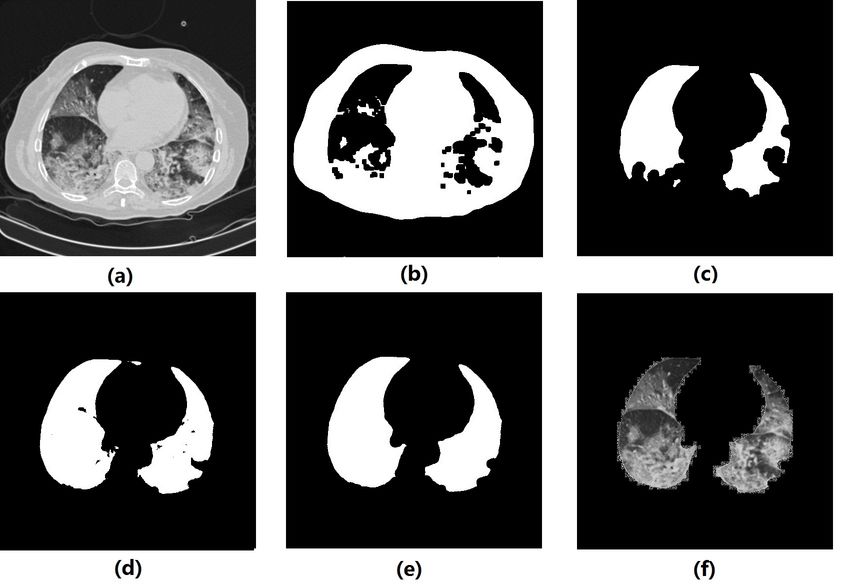
Figure 1. Example of lung segmentation: (a) raw image, (b) af- Instead of selecting one set of data, we select multiple sets
ter binarization, (c) morphological segmented mask, (d) origi- of data if there are enough available images.
nal UNet segmented mask, (e) refined UNet segmented mask, (f)
masked lung image 3.5. Input Image Composition
. The 3D CNN-BERT network [10] requires to have three-
channel input image, which is typically RGB. In CT-scan,
all slice images are in gray-scale. So we need to compose
segment the slice images in the ICCV-MIA dataset. Fur- three-channel input images.
thermore, since there are holes and noisy boundaries in the The first choice is the raw gray-scale slice image, which
segmented mask, we use further morphological transforms is denoted as an R channel, as shown in Figure 1 (a). In
to smooth the edges and fill the holes. The mask image af- [23], the authors propose to use the segmented mask as part
ter this step is used to extract the lung image for training of the input. We use it as well as one choice in our design,
and validation. Shown in Figure 1 (d)-(e) is example of the which is denoted as an M channel, as shown in Figure 1
original and refined UNet masks. Figure 1 (f) is the masked (e). In many works, the lung image masked by the mask,
lung image. i.e., the pixels inside the lung mask keep their gray-scale
Generally speaking, the UNet segmentation works better values, while all other pixels have value 0, are used as in-
than the morphological segmentation. However, it works put to the classification network. We use this lung image
poorly on the closed-lung images. That is why we still need as the third choice, which is denoted as the L channel, as
the morphological segmentation for the selection of slices shown in Figure 1 (f). So in comparison to the RGB image,
images. we have RML image, where the channels R, M, L can be
combined in many different ways. We will show different
performance of these images.
3.3. Selection of Slice Images Since we have the bounding box for every image in a CT-
scan volume, we take the largest bounding box of all images
Previous works show that some slice images, particularly as the bounding box for the volume. This bounding box can
those of closed lungs, are useless in classification the CT- be used to crop the RML image inside the bounding box.
scan volume [17], [23], [24]. Therefore, it is beneficial to
filter out these closed-lung slice images. In [23], a fixed por- 4. Classification Networks
tion of slice images at the beginning and end of a volume is
discarded. In [24], lung mask is segmented, and the images In this paper, we explore two levels of 3D classification.
whose percentages of lung mask in the whole image is less In the first level, a 3D CNN-BERT network [10] is used. In
than a threshold are filtered out. We use a method similar to the second level, feature vectors of all available set of slices
[24]. However,since some volumes have very small number images are pooled to a global feature vector for every CT-
of slice images, we make the threshold adaptive. In the first scan volume. This feature vector is sent to a simple MLP
step, the threshold 0.7 is used, i.e., slice images whose per- classifier for second level classification.
centage of lung mask is less than 0.7 of the largest percent-
4.1. 3D CNN-BERT Network
age in the whole volume are filtered out. In the next step,
if the remaining number of slice images is too few (e.g., 8), We reuse the 3D CNN-BERT network in [10], as shown
then we reduce the threshold by 0.1 until 8 images remain in Figure 2. This architecture utilizes BERT-based temporal
or the threshold reaches 0. pooling for video action recognition. In this work, we useFigure 2. The modified 3D CNN-BERT Architecture from [10] Figure 3. The MLP classification network
it to pool the correlation between CT-scan slice images. In drop out of 0.5 after first two FC layers. This MLP network
this architecture, the selected 32 slice images from the re- is depicted in Figure 3.
sampling scheme are propagated through a 3D CNN archi-
tecture without applying temporal global average pooling 5. Experiment Results
at the end of the architecture. A learned positional encod-
5.1. Dataset
ing is added to the extracted features. In order to perform
classification with BERT, additional classification embed- In the ICCV-MIA dataset [11], there are 1552 CT-scan
ding (xcls) is appended (represented as red box in Figure training volumes, 374 validation volumes, and 3455 test
2). The classification of the architecture is implemented volumes. In each volume, there are various number of slice
with the corresponding classification vector ycls which is images, ranging from 1 to more than a thousand. There are
sent the FC layer, producing the classification output. no slice annotations, so 2D CNN classification is not possi-
As the authors point out, the use of the temporal atten- ble if not using extra datasets.
tion mechanism for BERT is not only to learn the conve- Most of the sizes of the images are 512x512. However,
nient subspace where the attention mechanism works effi- there are quite a lot images whose sizes are not so. So we
ciently, but also to learn the classification embedding which first do a quick check, if the size of an image is not 512x512,
learns how to attend the temporal features of the 3D CNN it is resized to 512x512 before any other preprocessing is
architecture properly [10]. In [10] many 3D backbone net- applied.
works are studied, including the R(2+1)D [21]. On two For the Unet segmentation, we use the annotated dataset
main video action recognition benchmark datasets, this ar- we find on Kaggle and from CNBC [25]. In the CNBC
chitecture achieves state-of-the-art performance. For more annotation, three types of annotations - lung field, Ground-
detail of this architecture, including the cost function, please glass opacity, and Consolidation. We merge all three types
refer to [10]. to the lung field.
In this paper, we use the R(2+1)D backbone [21]. We No other datasets are used in training or validation of the
use 32 slice images as input. We make some modifications, 3D CNN-BERT network or the MLP network.
including using input image size 224x224, where 112x112
is used in [10], and outputting the embedding feature. 5.2. Implementation details
The 3D CNN-BERT is implemented with Pytorch frame-
4.2. MLP Classification Network
work. Input image size is set to 224x224. In order to do so,
The 3D CNN-BERT network produces a classification a few changes are made in the source code from [10]. We
result for a single set of input slice images. For CT-volumes choose to use 32 slice images as input and use the R(2+1)D
where there are more than one set of slice images, we want backbone. We use the Adam optimizer with a initial learn-
to process all available slice images in order not to miss ing rate 1E-5. A reduced learning rate on plateau with a
some useful information. Therefore, we propose to use a factor 0.1 and 5 epoch patience is used. The training runs
second level MLP classifier on the pooled embedding fea- at most 50 epochs with early stopping. The validation accu-
ture vectors out of the 3D CNN-BERT network. racy is used to select the best model.
To aggregate a global feature vector from multiple local The MLP network is also implemented with Pytorch.
feature vectors, the max-pooling and the avg-pooling are The input feature vector size is 512 or 1024. We use the
two most popular methods. We consider both of them, and Adam optimizer with a initial learning rate 1E-4 or 1D-5.
a concatenation of them. The first FC1 layer has 128 neu- A reduced learning rate on plateau with a factor 0.1 and
rons, and the second FC2 layer has 32 neurons. We consider 5 epoch patience is used. The training runs at most 300
different activation functions, including ReLU and Sigmoid epochs with early stopping. The validation accuracy is used
after each FC layer. In order to prevent overfitting, we use to select the best model.Input Augmentation Cropping? Accuracy(%) Pooling Activation Accuracy(%)
RRR MSC No 89.06 - - 91.18 (baseline)
LLL MSC No 89.84 Max ReLU 91.44
RRL MSC No 90.10 Avg ReLU 90.91
RML MSC No 91.18 Both ReLU 91.18
RML MSC Yes 87.43 Max Sigmoid 91.71
RML MSC+A No 90.91 Avg Sigmoid 91.18
RML A No 87.43 Both Sigmoid 91.98
Table 1. 3D CNN-BERT classification accuracy on validation Both Tanh 91.72
dataset for different configurations. In augmentation, two impor- Table 2. MLP classification accuracy on validation dataset.
tant approaches Affine transforms (A) and multiple scale cropping
(MSC) are studies.
Dataset 3D-CNN-BERT MLP
Validation (91.18,91.00) (92.78,92.61)
Validation (90.91,) (,)
The UNet segmentation network is implemented with
Test (,) (,)
Keras-2.3 and Tensorflow-GPU-2.2. The input image size
Test (,) (,)
is 512x512. We use the Adam optimizer with a initial learn-
Table 3. Classification results (accuracy %,F1-score %) on the val-
ing rate 1E-5. A reduced learning rate on plateau and early idation and test datasets.
stopping are used. The validation loss is used to select the
best model.
Data Augmentations: To make the model generalize
well, data augmentation is very important. In this work, 5.3.2 MLP Network
on the training dataset, we use random Affine transforms
with rotation of 10 degree, scaling range of 0.8-1.2, transla- We use the RML image with the Affine transform and
tion range of 0.1, width shearing of 10 degree, brightness of multiple-scale cropping of Table 1 in this test. Embedding
50%, contrast of 30% [23]. In addition, the multiple scale features of all training and validation volumes are extracted
cropping (MSC) [22] is used with the image enlarged by and save as Pickle files.
25%. Random horizontal flipping is also used. On the val-
We test the max-pooling, avg-pooling, and concatenation
idation and test dataset, central cropping is used with the
of both. For the activation function, we test ReLU, Sigmoid,
image enlarged by 25%. In the ablation study we will show
and Tanh functions. The results are listed in Table 2. We see
some results using different data augmentation methods.
that the concatenation of the max-pooling and avg-pooling
gives better results than using only one of them. Out of
5.3. Ablation Studies the three activation functions, the Sigmoid gives best per-
formance but takes longer time to converge or needs to use
5.3.1 3D CNN-BERT Network larger initial learning rate 1E-4.
Furthermore, we find that adding a new set of selected
In this section we compare the performance of different images at the center of a CT-scan volume to previous sym-
configurations on the validation dataset. metrical and uniform resampling can help improve the MLP
Input Image Composition and Bounding Box Crop- performance, so we include it in the final benchmarking
ping: In first panel of Table 1 we show the validation tests.
accuracy of different image compositions without or with
bounding box cropping. In these tests, the MSC and ran-
5.4. Benchmarking Results
dom horizontal flipping augmentations are used.
Data Augmentation: We use the best configuration After the ablation study, we choose a few top performers
from the previous tests, and test the effects of data augmen- to bench mark our algorithm. On the ICCV-MIA [11] vali-
tation. The most important factor is to find out if the random dation dataset, the benchmarking results are listed in Table
Affine transforms help the accuracy. The results are shown 3. In this table, both the accuracy and the F1 score are pre-
in the second panel of Table 1. sented.
From these studies, we find that the RML image with On the ICCV-MIA test dataset [11], the results will be re-
MSC augmentation without bounding box cropping gives leased by the ICCV-MIA competition organizers. Our per-
the best accuracy performance. formances will be added later in Table 3.6. Conclusions [11] Dimitrios Kollias, Anastasios Arsenos, Levon Soukissian,
and Stefanos Kollias. Mia-cov19d: Covid-19 detection
In this paper we present a 3D CNN-BERT network with through 3-d chest ct image analysis. arXiv preprint
an extra MLP network for COVID-19 classification. The arXiv:2106.07524, 2021.
MLP can improved the accuracy by 1.5%. On the validation [12] Dimitrios Kollias, N Bouas, Y Vlaxos, V Brillakis, M Se-
dataset, our best F1 score is xxx. And on the test dataset, feris, Ilianna Kollia, Levon Sukissian, James Wingate, and S
our best F1 score is yyy. Kollias. Deep transparent prediction through latent represen-
tation analysis. arXiv preprint arXiv:2009.07044, 2020.
References [13] Dimitrios Kollias, Athanasios Tagaris, Andreas Stafylopatis,
Stefanos Kollias, and Georgios Tagaris. Deep neural archi-
[1] Coronavirus disease (covid-19) pandemic. tectures for prediction in healthcare. Complex & Intelligent
https://www.who.int/emergencies/diseases/novel- Systems, 4(2):119–131, 2018.
coronavirus-2019. [14] Dimitris Kollias, Y Vlaxos, M Seferis, Ilianna Kollia, Levon
[2] O.S Albahri and et al. Systematic review of artificial in- Sukissian, James Wingate, and Stefanos D Kollias. Transpar-
telligence techniques in the detection and classification of ent adaptation in deep medical image diagnosis. In TAILOR,
covid-19 medical images in terms of evaluation and bench- pages 251–267, 2020.
marking: Taxonomy analysis, challenges, future solutions [15] Lin Li, Lixin Qin, Zeguo Xu, Youbing Yin, Xin Wang, Bin
and methodological aspects. J Infect Public Health, page Kong, Junjie Bai, Yi Lu, Zhenghan Fang, Qi Song, Kun-
1381–1396, 2020. lin Cao, et al. Artificial intelligence distinguishes covid-19
[3] Shubham Chaudhary, Sadbhawna, Vinit Jakhetiya, Badri from community acquired pneumonia on chest ct. Radiol-
Subudhi, Ujjwal Baid, and Sharath Guntuku. Detecting ogy, 2020.
covid-19 and community acquired pneumonia using chest ct [16] I. Ozsahin, B. Sekeroglu, M. Musa, M. Mustapha, and D
scan images with deep learning. In ICASSP, 2021. Ozsahin. Review on diagnosis of covid-19 from chest ct im-
[4] Pratyush Garg, Rishabh Ranjan, Kamini Upadhyay, Monika ages using artificial intelligence. Computational and Mathe-
Agrawal, and Desh Deepak. Multi-scale residual network for matical Methods, 2020, 2020.
covid-19 diagnosis using ct-scans. In ICASSP, pages 8558– [17] M. Rahimzadeh, A. Attar, and S. M. Sakhaei. A fully au-
8562, 2021. tomated deep learning-based network for detecting covid-19
[5] H. Gunraj, A. Sabri, D. Koff, and A. Wong. Covid-net ct- from a new and large lung ct scan dataset. medRxiv, 2020.
2: Enhanced deep neural networks for detection of covid-19 [18] Olaf Ronneberger, Philipp Fischer, and Thomas Brox. U-net:
from chest ct images through bigger, more diverse learning. Convolutional networks for biomedical image segmentation.
arXiv preprint 2101.07433, 2021. arXiv preprint arXiv:1505.04597, 2015.
[6] H. Gunraj, L. Wang, and A. Wong. Covidnet-ct: A tailored [19] W. Tan and J. Liu. Application of face recognition in tracing
deep convolutional neural network design for detection of covid-19 patients and close contacts. IEEE ICMLA, 2020.
covid-19 cases from chest ct images. Frontiers in Medicine, [20] Aram Ter-Sarkisov. Covid-ct-mask-net: Prediction of covid-
7:1025, 2020. 19 from ct scans using regional features. medRxiv, 2020.
[7] Xin He, Shihao Wang, Xiaowen Chu, Shaohuai Shi, Jiang- [21] D. Tran, H. Wang, L. Torresani, J. Ray, Y. Lecun, and M.
ping Tang, Xin Liu, Chenggang Yan, Jiyong Zhang, and Paluri. A closer look at spatiotemporal convolutions for ac-
Guiguang Ding. Automated model design and benchmarking tion recognition. In CVPR, 2018.
of 3d deep learning models for covid-19 detection with chest [22] Limin Wang, Zhe Xiong, Yuanjun anb Wang, and Yu Qiao.
ct scans. Proceedings of the AAAI Conference on Artificial Towards good practices for very deep two-stream convnets.
Intelligence, 2021. arXiv preprint arXiv:1505.04597, 2015.
[8] S. Heidarian, P. Afshar, N. Enshaei, F. Naderkhani, A. [23] X. Wang, X. Deng, Q. Fu, Q. Zhou, J. Feng, H. Ma, W. Liu,
Oikonomou, F. B. Fard, K. Samimi, K.N. Plataniotis, and Q. Zheng. A weakly-supervised framework for covid-
A. Mohammadi, and M.J. Rafiee. Covid-fact: A fully- 19 classification and lesion localization from chest ct. IEEE
automated capsule network-based framework for identifica- Transactions on Medical Imaging, 39:2615–2625, August
tion of covid-19 cases from chest ct scans. Arxiv preprint 2020.
2010.16041, 2020. [24] Shuohan Xue and Charith Abhayaratne. Covid-19 diagnostic
[9] Shahin Heidarian, Parnian Afshar, Arash Mohammadi, using 3d deep transfer learning for classification of volumet-
Moezedin Javad Rafiee MD, Anastasia Oikonomou MD, ric computerised tomography chest scans. In ICASSP, 2021.
Konstantinos N. Plataniotis, and Farnoosh Naderkhani. Ct- [25] K. Zhang, X. Liu, J. Shen, and et al. Clinically applicable
caps: Feature extraction-based automated framework for ai system for accurate diagnosis, quantitative measurements
covid-19 disease identification from chest ct scans using cap- and prognosis of covid-19 pneumonia using computed to-
sule networks. In ICASSP, pages 1040–1044, 2021. mography. Cell, April 2020.
[10] M Esat Kalfaoglu, Sinan Kalkan, and A Aydin Alatan. Late
temporal modeling in 3d cnn architectures with bert for
action recognition. In ECCV Workshop, pages 731–747.
Springer, 2020.You can also read