Modality Completion via Gaussian Process Prior Variational Autoencoders for Multi-Modal Glioma Segmentation
←
→
Page content transcription
If your browser does not render page correctly, please read the page content below
Modality Completion via Gaussian Process Prior
                                            Variational Autoencoders for Multi-Modal
                                                      Glioma Segmentation

                                          Mohammad Hamghalam1,2 , Alejandro F. Frangi4,5,6 , Baiying Lei7 , and Amber
                                                                      L. Simpson1,3
                                                  1
arXiv:2107.03442v1 [eess.IV] 7 Jul 2021

                                                       School of Computing, Queen’s University, Kingston, ON, Canada
                                                                          m.hamghalam@gmail.com
                                              2
                                                 Department of Electrical, Biomedical, and Mechatronics Engineering, Qazvin
                                                           Branch, Azad University, Qazvin, 34116846-13114, Iran
                                           3
                                               Department of Biomedical and Molecular Sciences, Queen’s University, Kingston,
                                                                                ON, Canada
                                                4
                                                    CISTIB Centre for Computational Imaging & Simulation Technologies in
                                                Biomedicine, School of Computing, University of Leeds, Leeds LS2 9LU, UK
                                             5
                                                LICAMM Leeds Institute of Cardiovascular and Metabolic Medicine, School of
                                                                       Medicine, Leeds LS2 9LU, UK
                                          6
                                             Medical Imaging Research Center (MIRC) – University Hospital Gasthuisberg, KU
                                                                Leuven, Herestraat 49, 3000 Leuven, Belgium
                                                  7
                                                     National-Regional Key Technology Engineering Laboratory for Medical
                                                Ultrasound, Guangdong Key Laboratory for Biomedical Measurements and
                                               Ultrasound Imaging, School of Biomedical Engineering, Health Science Center,
                                                                   Shenzhen University, Shenzhen, China.

                                                Abstract. In large studies involving multi protocol Magnetic Resonance
                                                Imaging (MRI), it can occur to miss one or more sub-modalities for a
                                                given patient owing to poor quality (e.g. imaging artifacts), failed ac-
                                                quisitions, or hallway interrupted imaging examinations. In some cases,
                                                certain protocols are unavailable due to limited scan time or to retro-
                                                spectively harmonise the imaging protocols of two independent studies.
                                                Missing image modalities pose a challenge to segmentation frameworks
                                                as complementary information contributed by the missing scans is then
                                                lost. In this paper, we propose a novel model, Multi-modal Gaussian Pro-
                                                cess Prior Variational Autoencoder (MGP-VAE), to impute one or more
                                                missing sub-modalities for a patient scan. MGP-VAE can leverage the
                                                Gaussian Process (GP) prior on the Variational Autoencoder (VAE) to
                                                utilize the subjects/patients and sub-modalities correlations. Instead of
                                                designing one network for each possible subset of present sub-modalities
                                                or using frameworks to mix feature maps, missing data can be generated
                                                from a single model based on all the available samples. We show the
                                                applicability of MGP-VAE on brain tumor segmentation where either,
                                                two, or three of four sub-modalities may be missing. Our experiments
                                                against competitive segmentation baselines with missing sub-modality
                                                on BraTS’19 dataset indicate the effectiveness of the MGP-VAE model
                                                for segmentation tasks.
2      M. Hamghalam et al.

      Keywords: Missing modality · Gaussian process · Variational autoen-
      coder · Glioma segmentation · MRI.

1   Introduction

Glioma tumor segmentation in MR scans plays a crucial role during the di-
agnosis, survival prediction, and brain tumor surgical planning. Multiple MRI
sub-modalities, FLAIR (F), T1, T1c, and T2, are regularly utilized to detect
and evaluate the brain tumor subregions such as the whole tumor (WT), tumor
core (TC), and the enhancing tumor (ET) region. These sequences provide com-
prehensive information regarding tumor brain tissues. In clinical settings, it is
common for physicians to have one or more sub-modalities to be missing for a
patient due to patient artifacts, acquisition problems, and other clinical reasons.
     Segmentation with missing modalities techniques can be categorized into
three approaches: 1) training a segmentation model for any subset of input sub-
modalities; 2) training a synthesis model to impute missing sequence from input
sub-modalities [13]; 3) instead of designing different models for every potential
missing modality combination, designing a single model that operates based
on the shared feature space through all input sub-modalities (such as taking
the mean) [5, 11, 19]. The two first solutions associate with the training and
handling of a different network for 2(#of sub−modalities) − 1 combinations. The
third group extracts a shared feature space from the sub-modalities, which is
independent of the number of sub-modalities, to provide a unique model that
shares its extracted features.
     The current methods, which work based on the common representation to
address missing modality, are Hetero-modal Image Segmentation (HeMIS) [11]
and its relevant extension Permutation Invariant Multi-Modal Segmentation
(PIMMS) technique [19]. They computed first and second moments of extracted
feature maps across available sub-modalities to combine them separately. Al-
though using these statistics are independent of the number of sub-modalities,
they do not compel their convolutional model to learn a shared latent repre-
sentation. For this aim, Dorent et al. [5] introduced Hetero-Modal Variational
Encoder-Decoder (HVED) based on the Variational Autoencoder (VAE) [12] to
provide the common latent variable z. Furthermore, conditional VAE (CVAE)
[17] includes extra data in both the decoder and the encoder to produce samples
with specific properties. Based on this method, some models used the auxiliary
information to synthesis missing sub-modalities [16]. However, the prior assump-
tion that latent representations are identically and independently distributed
(i.i.d.) is one of the VAE model’s limitations. In contrast, the Gaussian Process
Prior Variational Autoencoder (GPPVAE) [4] models correlation between input
sub-modalities through a Gaussian Process (GP) prior on the latent representa-
tions.
     In this paper, we extend the GPPVAE for missing MRI sub-modalities impu-
tation in a 3D framework for brain glioma tumor segmentation. The contribution
of this work is three-fold. First, we extend the GPPVAE for 3D MRI modalities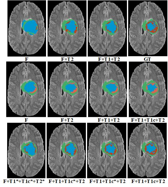
Modality Completion via GP for Multi-Modal Glioma Segmentation          3

imputation for any scenario of missing sequences as input in one model. Second,
we adapt a kernel function to capture multiple levels of correlation between sub-
modalities and subjects in our Multi-modal Gaussian Process Prior Variational
Autoencoder (MGP-VAE). Finally, we show that our model outperforms HVED
and HeMIS in terms of DSC from multi-modal brain tumor MRI scans with any
configuration of the available sub-modalities.

2   Multi-modal Gaussian Process Prior Variational
    Autoencoder
Assume we have a collection of 3D MRI scans to visualize brain and tumor tissue
in different contrasts in MR pulse sequences, each scan coupled with auxiliary
data: patient/subject IDs and sub-modality entities. Individually, we consider
BraTS’19 datasets with MRI scans of the brain with glioma tumors in four
sub-modalities per patient: F, T1, T1c, and T2, each of which contributes differ-
ing tissue contrast views and spatial resolutions. Each unique patient and sub-
modality is assigned to a feature vector, which we define as a subject feature
vector and a modality feature vector, respectively. Subject and modality fea-
tures refer to elements of kernel covariance function calculated based on present
data. Subject feature vectors might contain brain features such as brain tissue,
tumor location, or dimension, while modality feature vectors may contain con-
trast information or tumor sub-region features of each modality. Fig. 1 shows an
overview of the MGP-VAE for synthesizing missing modalities. We assume that
at least one sub-modality of each test subject is present during training.
    Formulation. Let P denote the number of subjects, M the number of
sub-modalities, N = P × M denote the number of all samples, and consider
                    N ×K
Y = {yn }N n=1 ∈ R        for all N input samples and K denotes k-dimensional
                                                      P ×Q
representation for N samples; let X = {xp }P  p=1 ∈ R       denote Q-dimensional
                                                                            M ×R
patient feature vectors for the P patients, and let W = {wm }M     m=1 ∈ R
denote R-dimensional modality feature vectors for the M sub-modalities. Four
sub-modalities provide complementary information about the brain tissue, W =
                                                                             N ×L
{w1 = F, w2 = T 1, w3 = T 1c, w4 = T2 }. Finally, let Z = {zn }N    n=1 ∈ R
denote the L-dimensional latent representations which abstract input samples
through GP, fGP . We examine the following process for the available input data:
 – The latent representation of MRI scan pn in sub-modality mn is generated
   from the subject feature vector xpn and modality feature vector wmn as:
   zn = fGP (xpn , wmn ), where fGP is a GP prior to compute sample covariances
   as a function of subject and modality feature vectors in the latent space, z.
 – Reconstructed output ŷn is created from its latent representation zn as: ŷn =
   fd (zn ), where fd is a convolutional neural network with decoder architecture
   to map latent representation, z, into the reconstruction space, ŷ.
The marginal likelihood of the MGP-VAE model is:
                                 Z
          p(Y|X, W, θd , θGP ) = p(Y|Z, θd ) p(Z|X, W, θGP ) dZ               (1)
4 M. Hamghalam et al.
Fig. 1. Overview of the proposed MGP-VAE. Each sub-modality volume is mapped to
a 1024-dimensional (L) space and decoded to the initial space. Covariances among input
volumes are formed through a GP Prior to each column of the latent representation
matrix Ze . The subject and modality correlations are modeled in the latent space due
to its compact superiority.
where θd denotes the parameters of the decoder and θGP indicates the parameters
of GP’s kernels. Eq.1 cannot be optimized straightforwardly as the integral is
not tractable. Thus we resort to variational inference, which requires introducing
an approximate posterior distribution.
2.1 Proposed Kernel Functions for MGP-VAE
The GP defines a set of random variables on z l , l−th column of Z, so any
finite number of them have a multivariate Gaussian distribution. In case z l =
fGP (X, W) is a GP, then given L observations, the joint distribution of the
random variables, z 1 = fGP (X, W), z 2 = fGP (X, W), ..., z L = fGP (X, W), is
Gaussian. For our L−dimensional latent representation, we have:
L
Y
p(Z|X, W, θGP ) = GP(z l |0, KθGP (X, W)) (2)
l=1
where KθGP (X, W) is the covariance function with the kernel parameters, θGP ,
which comprises a modality kernel and a patient kernel. The former models
covariance among sub-modalities, while the latter models covariance between
patients. KθGP (X, W) can be factorized into [3]:
KθGP (X, W) = K(xp , x0 p ) ⊗ K(wm , w0 m )
| {z } | {z } (3)
patient kernel modality kernelModality Completion via GP for Multi-Modal Glioma Segmentation 5
where xp and x0 p are feature vectors of two patients, wm and w0 m are corre-
sponding modality feature vectors. Also, ⊗ is the Kronecker product of these
two matrices to make the dimensions match between the P × P and M × M
matrix. These features are extracted from the latent space during training. We
define L = 1024 as the latent space dimension. We set a full-rank covariance
as a modality covariance (K(wm , w0 m )) for our limited sub-modalities (F, T1,
T1c, and T2) and a linear covariance (K(xp , x0 p ) = xTp .x0 p ) to measure similarity
among the subjects with Q = 64.
Loss Function and Optimization. As a standard VAE, we approximate the
latent variables by a Gaussian distribution whose mean and diagonal covariance
are defined by two functions, µe (yn ) and diag(σe2 (yn )). Thus, we have:
N
Y
N zen |µe (yn ), diag(σe2 (yn )) ,
q(Ze |Y) = (4)
n=1
which approximates the true posterior on Ze . In Eq. 4, θe denotes the weights
of the encoder in auto-encoder neural network architecture. Latent represen-
tations Ze = [ze1 , ze2 , ...., zeN ] ∈ RN ×L are also sampled employing the re-
parameterization method [12], zen = µe (yn ) + υn σe (yn ), where denotes
the element-wise product and υ is a random number drawn from a normal dis-
tribution. We compute the resulting evidence lower bound (ELBO) as:
" #
X
log p(Y|X, W, θd , θGP ) ≥ EZ∼qθe log N (yn |fd (zn )) + log p(Ze |X, W, θGP )
n
1X
+ log(σq2 (yn )l ) + const.
2
nl
(5)
To increase the ELBO as much as possible, we apply stochastic backpropa-
gation [12]. Individually, we approximate the expectation by sampling from a
reparameterized variational posterior over the latent representations, achieving
the resulting loss function:
L(θd , θe , θGP ) =
X kyn − fd (ze )k2 1X
2
n
− log p(Ze |X, W, θGP ) + log(σz2e (yn )l )
n
2σ y | {z } 2 (6)
nl
| {z } GP | {z }
L2 reconstruction loss regularization
+ NK log σy2
where we optimize regarding θd , θe , and θGP . We optimize loss function through
Adam optimizer with a learning rate of 0.001. We experimentally noted that
minimizing loss function was developed by first training the encoder and the
decoder within the VAE, next optimizing the GP weights by frozen encoder and
decoder for 100 epochs (the learning rate of 0.01), last, optimizing all parameters
jointly in our MGP-VAE model.6 M. Hamghalam et al.
2.2 Missing Modality Imputation
We derive an approximate predictive posterior for MGP-VAE that enables miss-
ing modality predictions of high-dimensional samples. Specifically, given training
samples Y , subject feature vectors X, and modality feature vectors W , the pre-
diction for the missing data yt of subject pt in modality mt is given by:
p(yt |xt , wt , Y, X, W) =
Z
p(yt |zt ) p(zt |xt , wt , Ze , X, W) q(Ze |Y) dzt dZe (7)
| {z } | {z } | {z }
decoding missing data GP prediction of zt encoding all training data
where xt and wt are feature vectors of subject pt and sub-modality mt , respec-
tively. The approximation in Eq. 7 is achieved by substituting the exact posterior
on Ze with the variational distribution q(Ze |Y) (see [4]). According to Eq. 7, the
missing sub-modality can be computed by the three steps. First, we encode all
training image data in the latent space by the encoder, Ze . Next, predict latent
representation zt of image yt through the GP model using m, X, and W. Lastly,
latent representation zt is decoded to the high-dimensional image space through
the decoder as missing modality imputation.
3D Variational Encoder-Decoder Network Architecture. The en-
coder part employs four spatial levels, where each level consists of two convolu-
tion layers with 3 × 3 × 3 kernel and ELU. The first convolution layer is without
downsampling (stride = 1), while the second one applies strode convolution for
downsizing. We follow a typical VAE approach to downsize image dimensions by
two progressively, but with fixed feature size equal to 32 except encoder endpoint
with 16 feature maps. The encoder endpoint has size 16 × 4 × 4 × 4, followed by
a fully connected layer, and is 16 times spatially smaller than the input volume
of 64 × 64 × 64. The decoder structure is similar to the encoder one, but each
level begins with volumetric upsampling using the nearest neighbor algorithm.
3 Experiments and Results
Data. We assess our method on the training set of BRATS’19 [1, 2, 14], which
includes the scans of 335 patients. Each subject is scanned with four T1, T1c,
T2, and F sequences. All scans are skull-striped and re-sampled to an isotropic
1mm resolution, and four sequences of each patient have been co-registered.
Radiologists provided the ground truth labels. The segmentation comprise the
following tumor tissue labels: 1) non-enhancing tumor, 2) edema, 3) enhancing
core. Implementation of the MGP-VAE is available1 .
3.1 Missing Modality Imputation
Fig. 2(a) illustrates a qualitative evaluation of each sub-modality reconstruction
with one missing sub-modality (second column) and two missing sub-modalities
1
https://github.com/hamghalam/MGP-VAEModality Completion via GP for Multi-Modal Glioma Segmentation 7 Fig. 2. (a) Example of modality completion (each row corresponds to a particular sub- modality) given a subset of sub-modalities as input. The 4-bit strings on top of each slice determine present and absent sub-modality with 1 and 0, respectively (bit order from left to right F, T1, T1c, and T2). (b) Covariances between sub-modalities and subjects are modeled through a GP prior model. (c) We compared our method with CVAE and VAE, the baseline of HVED and other well-known imputation methods. (third column). Our model proposes to reconstruct the brain and tumor tissue even when the tumor information is missing or not clear by coupling informa- tion from available samples. Comparing reconstructions using two sub-modalities and three sub-modalities confirms that the reconstructed volumes preserve high- frequency details. This suggests that the MGP-VAE model can effectively learn relations between available sub-modalities in different subjects (Fig. 2(b)). The PSNR values for imputation are F = 27.95, T1 = 27.80, T1c = 29.43, and T2 = 27.99 based on three available sub-modalities. Similarly, we have F = 22.36, T1 = 22.56, T1c = 24.86, and T2 = 22.66 with two available sub-modalities. Besides, Mean Squared Error (MSE) for each sub-modality is considered as an evaluation metric to compare MGP-VAE with CVAE and linear interpolation. The latter applies linear interpolation between available sub-modalities of a subject in the latent space learned through VAE to predict the missing sequence (Fig. 2(c)). The CVAE indeed improves VAE in image generation by conditioning the en- coder and decoder to the desired input. However, when we have missing input data, CVAE has a confined ability to create the latent variable for unseen data compared to VAE. This might be because CVAE is more restricted to learn par- ticular data features (latent representation) from observed input data. Therefore it has dedicated latent variables with limited features from missing data. The latent space of VAE contains more general characteristics which can be used to predict missing data. 3.2 Glioma Segmentation To assess the MGP-VAE, we examine it on the brain tumor segmentation frame- work and compare it with two state-of-the-art methods for all the possible subset
8            M. Hamghalam et al.

of sub-modalities. The first, HVED [5] is the state-of-the-art method based on
VAE for brain tumor segmentation with missing sub-modalities. The second ap-
proach, HeMIS [11], combines the available sub-modalities based on feature maps
moments. We adopt the 3D U-Net architecture [?, 15] to segment glioma where
the available sub-modalities and imputed ones are concatenated as input multi-
modal MRI scans. We use the Dice score to measure segmentation accuracy in
clinically significant glioma subregions: WT, TC, and ET in Table 1. We have
almost the same performance in Table 1 if all sub-modalities are available (with-
out imputation). Our method is designed and optimized to address problems
where either one, two, or three of four sub-modalities may be missing. When all
the sub-modalities are available, this is a different scenario [6–10, 18]. Moreover,
Fig. 3 shows comparative segmentation results of the BraTS’19 dataset with-
out (first row) and with (second row) imputation through MGP-VAE. The last
column (first row) is the ground truth.

Table 1. Comparison of MGP-VAE model with HeMIS and HVED model (Dice %)
for all subset of available sub-modalities. Sub-modalities present are denoted by 1, the
missing ones by 0. The IQR is the interquartile range (IQR) and * indicates significant
improvement by a Wilcoxon test (p < 0.05).
Modalities                WT                       TC                      ET
F T1 T1c T2 HeMIS HVED Ours HeMIS HVED Ours HeMIS HVED Ours
0   0    0     1   78.0    79.9    81.1*   49.3    52.8    56.2*   22.1    29.9     30.8
0   0    1     0   57.6    61.7    63.2*   57.7    65.8    68.1*   59.5    65.1    66.4*
0   1    0     0   53.3    51.5     53.2   37.0    36.2    39.9*   11.3    13.2     14.2
1   0    0     0   78.9    81.0    83.3*   48.8    49.9    52.7*   24.2    24.1     25.2
0   0    1     1   80.1    81.8    83.1*   68.3    73.1    75.7*   67.5    69.2    70.5*
0   1    1     0   62.6    66.0    67.6*   63.4    69.1    71.8*   64.3    66.6    68.0*
1   1    0     0   82.9    83.2    84.7*   56.0    54.4     55.3   28.2    24.1     25.1
0   1    0     1   79.8    81.1     82.0   52.5    56.6    58.4*   27.1    30.3    31.8*
1   0    0     1   84.8    86.5    87.9*   58.0    58.9     61.0   26.9    33.7    35.0*
1   0    1     0   82.2    84.8     85.8   66.7    72.6    74.6*   67.0    70.4     70.2
1   1    1     0   83.9    85.6    87.4*   69.9    73.6    75.8*   68.8    70.2    71.5*
1   1    0     1   85.9    87.1     88.3   60.1    61.0    63.0*   32.3    33.3    34.5*
1   0    1     1   85.9    87.5    89.0*   71.6    75.2    77.4*   68.9    70.3    71.4*
0   1    1     1   81.3    82.3     83.6   69.9    74.5    76.4*   68.7    70.4    71.6*
    Median         80.7    82.05   83.45   59.05   63.4    65.55    45.9   49.4     50.7
     IQR           9.98     9.40    9.80    17.0   19.23   19.75   41.58   41.78   41.33

4       Conclusion

We have introduced MGP-VAE to predict missing data of subjects in specified
MRI sub-modalities using a specialized VAE. Our model incorporates a GP prior
Modality Completion via GP for Multi-Modal Glioma Segmentation 9 Fig. 3. The first row explains the effects of different combinations of input sub- modalities overlaid on the FLAIR(F) slice without the imputation. The second row illustrates the HVED results. The last row represents the segmentation with imputa- tion based on MGP-VAE method. The segmentation colors describe edema (green), non-enhancing (blue), and enhancing (red). * indicates imputed sub-modalities. over the encoded representation of available volumes to compute correlations among available sub-modalities and subjects. We also validated the robustness of the method with all possible missing sub-modality scenarios on glioma tumors and achieved state-of-the-art segmentation results. Finally, our method offers promising insight for leveraging large but incomplete data sets through one single model. Possible future work focuses on extending the method to various imaging modalities (MRI, PET, and CT) as well as genetic data. 5 Acknowledgements This work was funded in part by National Institutes of Health R01 CA233888.
10      M. Hamghalam et al.

References
 1. Bakas, S., Akbari, H., Sotiras, A., Bilello, M., Rozycki, M., Kirby, J.S., Freymann,
    J.B., Farahani, K., Davatzikos, C.: Advancing the cancer genome atlas glioma mri
    collections with expert segmentation labels and radiomic features. Scientific data
    4(1), 1–13 (2017)
 2. Bakas, S., Reyes, M., Jakab, A., Bauer, S., Rempfler, M., Crimi, A., Shinohara,
    R.T., Berger, C., Ha, S.M., Rozycki, M., et al.: Identifying the best machine learn-
    ing algorithms for brain tumor segmentation, progression assessment, and overall
    survival prediction in the brats challenge. arXiv preprint arXiv:1811.02629 (2018)
 3. Bonilla, E.V., Agakov, F.V., Williams, C.K.: Kernel multi-task learning using task-
    specific features. In: Artificial Intelligence and Statistics. pp. 43–50 (2007)
 4. Casale, F.P., Dalca, A.V., Saglietti, L., Listgarten, J., Fusi, N.: Gaussian process
    prior variational autoencoders. In: Advances in Neural Information Processing Sys-
    tems. pp. 10369–10380 (2018)
 5. Dorent, R., Joutard, S., Modat, M., Ourselin, S., Vercauteren, T.: Hetero-modal
    variational encoder-decoder for joint modality completion and segmentation. In:
    International Conference on Medical Image Computing and Computer-Assisted
    Intervention. pp. 74–82. Springer (2019)
 6. Hamghalam, M., Lei, B., Wang, T.: Convolutional 3d to 2d patch conversion for
    pixel-wise glioma segmentation in mri scans. In: International MICCAI Brainlesion
    Workshop. pp. 3–12. Springer (2019)
 7. Hamghalam, M., Lei, B., Wang, T.: High tissue contrast mri synthesis using
    multi-stage attention-gan for segmentation. vol. 34, pp. 4067–4074 (Apr 2020).
    https://doi.org/10.1609/aaai.v34i04.5825
 8. Hamghalam, M., Wang, T., Lei, B.: High tissue contrast image synthesis via mul-
    tistage attention-gan: application to segmenting brain mr scans. Neural Networks
    132, 43–52 (2020)
 9. Hamghalam, M., Wang, T., Qin, J., Lei, B.: Transforming intensity distribution of
    brain lesions via conditional gans for segmentation. In: 2020 IEEE 17th Interna-
    tional Symposium on Biomedical Imaging (ISBI). pp. 1–4. IEEE (2020)
10. Hatami, T., Hamghalam, M., Reyhani-Galangashi, O., Mirzakuchaki, S.: A ma-
    chine learning approach to brain tumors segmentation using adaptive random forest
    algorithm. In: 2019 5th Conference on Knowledge Based Engineering and Innova-
    tion (KBEI). pp. 076–082 (2019). https://doi.org/10.1109/KBEI.2019.8735072
11. Havaei, M., Guizard, N., Chapados, N., Bengio, Y.: Hemis: Hetero-modal im-
    age segmentation. In: International Conference on Medical Image Computing and
    Computer-Assisted Intervention. pp. 469–477. Springer (2016)
12. Kingma, D.P., Welling, M.: Auto-encoding variational bayes. arXiv preprint
    arXiv:1312.6114 (2013)
13. Li, R., Zhang, W., Suk, H.I., Wang, L., Li, J., Shen, D., Ji, S.: Deep learning based
    imaging data completion for improved brain disease diagnosis. In: International
    Conference on Medical Image Computing and Computer-Assisted Intervention.
    pp. 305–312. Springer (2014)
14. Menze, B.H., et al.: The multimodal brain tumor image segmentation bench-
    mark (BraTS). IEEE Transactions on Medical Imaging 34(10), 1993–2024 (2015).
    https://doi.org/10.1109/TMI.2014.2377694
15. Ronneberger, O., Fischer, P., Brox, T.: U-net: Convolutional networks for biomedi-
    cal image segmentation. In: International Conference on Medical image computing
    and computer-assisted intervention. pp. 234–241. Springer (2015)
Modality Completion via GP for Multi-Modal Glioma Segmentation 11
16. Sharma, A., Hamarneh, G.: Missing mri pulse sequence synthesis using multi-modal
generative adversarial network. IEEE transactions on medical imaging 39(4), 1170–
1183 (2019)
17. Sohn, K., Lee, H., Yan, X.: Learning structured output representation using deep
conditional generative models. Advances in neural information processing systems
28, 3483–3491 (2015)
18. Soleymanifard, M., Hamghalam, M.: Segmentation of whole tumor using localized
active contour and trained neural network in boundaries. In: 2019 5th Conference
on Knowledge Based Engineering and Innovation (KBEI). pp. 739–744. IEEE
19. Varsavsky, T., Eaton-Rosen, Z., Sudre, C.H., Nachev, P., Cardoso, M.J.: Pimms:
permutation invariant multi-modal segmentation. In: Deep Learning in Medical
Image Analysis and Multimodal Learning for Clinical Decision Support, pp. 201–
209. Springer (2018)You can also read