A Case of Reversible Cerebral Vasoconstriction Syndrome in a Healthy Adult Male - Cureus
←
→
Page content transcription
If your browser does not render page correctly, please read the page content below
Open Access Case
                                         Report                                              DOI: 10.7759/cureus.8374

                                         A Case of Reversible Cerebral
                                         Vasoconstriction Syndrome in a Healthy
                                         Adult Male
                                         Alysha Roberts 1 , Nicholas Sowers 2

                                         1. Emergency Medicine, Dalhousie University, Halifax, CAN 2. Emergency Medicine, Queen Elizabeth II
                                         Health Science Center, Halifax, CAN

                                         Corresponding author: Alysha Roberts, aroberts@dal.ca

                                         Abstract
                                         Reversible cerebral vasoconstriction syndrome (RCVS) represents a potentially under-
                                         recognized cause of thunderclap headache in patients presenting to the ED. While a rarely
                                         made diagnosis in emergency medicine practice, RCVS may be as common as subarachnoid
                                         hemorrhage (SAH). RCVS typically presents as a sudden onset, excruciating headache that may
                                         be associated with nausea, vomiting, photophobia, or other features with overlap in the clinical
                                         presentation of both SAH and migraine headaches. As a result of historical features overlapping
                                         the presentation of SAH, particularly the rapidity of onset and peak of severity, these patients
                                         are typically investigated for SAH and when that workup ultimately is reassuring, clinicians
                                         may often misattribute RCVS symptoms as migrainous.

                                         We present a case of a 35-year-old healthy male who presented with a severe, sudden onset
                                         headache, nausea, and photophobia to the ED on four occasions within a nine-day period. He
                                         was initially investigated appropriately for SAH; receiving an unenhanced head CT and lumbar
                                         puncture, which were both unremarkable. Following this initial workup, he was assessed on
                                         several other occasions, treated symptomatically as a migraine, and discharged home. On the
                                         fourth ED visit a CT angiogram (CTA) was completed that demonstrated the characteristic
                                         “string of beads” appearance of the middle cerebral artery (MCA) diagnostic of RCVS. This case
                                         describes the key features and investigations of a patient with RCVS and highlights the
                                         importance of early and accurate diagnosis of thunder clap headache in which SAH has been
                                         excluded.

                                         Categories: Emergency Medicine, Neurology
                                         Keywords: rcvs, reversible cerebral vasoconstriction syndrome, emergency department

Received 08/20/2019                      Introduction
Review began 11/19/2019
Review ended 05/19/2020                  In the ED, patients with severe headaches are evaluated for red flags, risk factors, and any
Published 05/31/2020                     symptoms that may support the diagnosis of a serious cause of headache [i.e., subarachnoid
                                         hemorrhage (SAH), meningitis]. For patients in which SAH has been ruled out, reversible
© Copyright 2020
Roberts et al. This is an open access    cerebral vasoconstriction syndrome (RCVS) represents an underdiagnosed presentation of
article distributed under the terms of   severe, sudden onset (thunder clap) headache [1]. Though a majority of RCVS cases result in a
the Creative Commons Attribution         benign clinical course, a proportion of patients may go on to develop serious complications
License CC-BY 4.0., which permits
                                         such as intracranial hemorrhage, seizures, or cerebral infarcts secondary to RCVS [1-3].
unrestricted use, distribution, and
reproduction in any medium, provided
the original author and source are       Reversible cerebral vasoconstriction syndrome is more common in women, typically in their
credited.                                40s-50s who have a history of migraine [4-5]. In the majority of cases, there exists an

                                         How to cite this article
                                         Roberts A, Sowers N (May 31, 2020) A Case of Reversible Cerebral Vasoconstriction Syndrome in a
                                         Healthy Adult Male. Cureus 12(5): e8374. DOI 10.7759/cureus.8374
identifiable trigger such as exertion, coughing, defecation, or the use of vasoactive
substances [3-4]. Several scoring systems have been developed to help with timely diagnosis [5-
6], as patients endure an estimate of 9.3 days of symptoms and may seek care up to six times
before receiving a diagnosis [7-8]. Once a diagnosis is made, the treatment of RCVS is largely
supportive [9-10]. While the use of oral calcium channel blockers such as nimodipine and
verapamil has been studied, there remains limited evidence of their effectiveness [11-12].
In this case, we present a healthy young male who was diagnosed with RCVS in the ED after
four visits and initially receiving a diagnosis of migraine. This case illustrates the common
clinical features of RCVS, including nausea, vomiting, photophobia, and recurrent severe
headaches, as well as the pertinent investigations required to make the diagnosis in an
otherwise healthy and uncommon population.
Case Presentation
Patient information
The patient was a 35-year-old Caucasian male who presented to the ED with a chief complaint
of a sudden onset, severe headache recurring over several days and leading to significant
functional impairment. His medical history was significant for painless ocular migraines and
otherwise unremarkable. He described a paternal history of a SAH leading to a generalized
seizure several years ago. The patient smokes 8-10 cigarettes per day in addition to using
approximately 0.5 g of cannabis daily.
In the week leading up to the visit described in this case, the patient had presented to the ED
three times with a chief complaint of a severe and sudden onset headache. Over the nine-day
period of headaches, the pain became progressively worse. The headaches were associated with
neck pain, vomiting, and photophobia without other visual abnormalities. The patient denied
fever, chills, and any neurological deficits. Serial neurological examinations in the ED were
unremarkable.
Initially, the patient presented to a peripheral ED. On this occasion, he received
metoclopramide, ketorolac, and acetaminophen with codeine for pain management. An
unenhanced head CT was performed approximately 24 hours after symptom onset, which
revealed no significant findings.
On his second presentation with a recurring headache five days later, the patient received
similar treatment for pain and was discharged with a diagnosis of migraine. A lumbar puncture
performed on his third presentation the following day (seven days since symptom onset) was
unremarkable. Finally, at his fourth presentation (nine days since initial onset), his progressive
headache remained typical for a SAH and was not in keeping with a post-lumbar puncture
headache. As such, a CT angiogram (CTA) was conducted, which confirmed a diagnosis of
RCVS. See Table 1 for a detailed timeline.
2020 Roberts et al. Cureus 12(5): e8374. DOI 10.7759/cureus.8374 2 of 5Date Investigations/Treatment
07/01/2019 Unenhanced CT, pain management
12/01/2019 Pain management
13/01/2019 Lumbar puncture, pain management
15/01/2019 Second unenhanced CT, CT angiogram, pain management
TABLE 1: Timeline of presentation and investigations leading to RCVS diagnosis.
RCVS, reversible cerebral vasoconstriction syndrome
Clinical findings
On exam, the patient was in no apparent distress with stable vital signs. His Glasgow Coma
Scale (GCS) score was 15/15. His neurological exam was unremarkable, with no apparent cranial
nerve abnormalities. His pupils were equal and reactive to light. Power and sensation were
normal and equal bilaterally in the upper and lower extremities. He had no gait abnormalities
or cerebellar signs. He had no signs of meningeal irritation.
Diagnostic assessment
The patient received an unenhanced head CT on his initial presentation to rule out SAH. The
CT scan showed no intracranial hemorrhage, edema, mass, or signs of herniation. No acute
infarct was evident. A lumbar puncture was performed on the third visit to rule out meningitis.
Cerebrospinal fluid (CSF) analysis indicated normal appearing CSF with protein and glucose
levels within normal range. Table 1 illustrates the sequence of presentation and investigations
the patient received.
A second CT scan performed on his fourth visit revealed no changes from his initial
presentation. At this point, a CTA was ordered to investigate potential vascular causes of his
recurrent headaches, including arterial dissection. The results of the angiogram confirmed the
absence of dissection, stenosis, intracranial aneurysm, or occlusion. Multiple focal narrowings
were present in the posterior branches of the right middle cerebral artery territory, which were
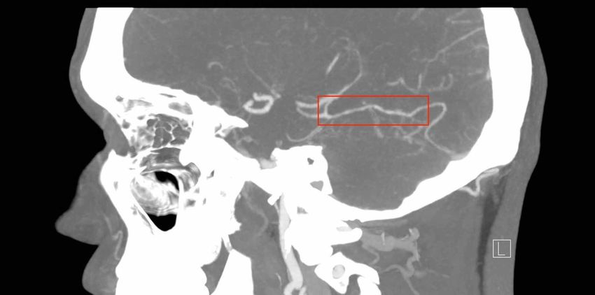
consistent with a RCVS. No other areas of focal narrowing were present. Figure 1 highlights the
area of vasoconstriction seen on CTA that was diagnostic in this case.
2020 Roberts et al. Cureus 12(5): e8374. DOI 10.7759/cureus.8374 3 of 5FIGURE 1: Sagittal view of the "string of beads" appearance
indicative of vasospasm, as seen on CTA.
CTA, CT angiogram
Neurology was consulted to assist in patient management. He was subsequently treated with
supportive management and made an uneventful recovery.
Discussion
This case illustrates the key features of RCVS in an otherwise healthy young male and the
challenges of making a timely diagnosis. In this case, the time from first symptoms to diagnosis
was consistent with previous literature suggesting an average time of nine days to receive a
diagnosis [7-8]. As RCVS presents similarly and with equal frequency to SAH [9], it is important
to consider it during the early stages of workup for SAH in the ED.
In this case, features including nausea, vomiting, and photophobia were present. Though these
are also often seen in migraines, RCVS is distinct in that the presenting headache is
excruciating and abrupt in nature [6]. Other features that should prompt the consideration of
RCVS include one or more recurrent thunderclap headaches, or a sudden increase in headache
intensity [4]. Early differentiation from migraine is particularly important. Common migraine
treatments such as triptans may worsen symptoms, and in some cases have been documented
as inducing RCVS [13].
Additionally, the patient had a history of background migraines and use of vasoactive
substances (i.e., daily cannabis use), both of which have been documented as factors associated
with RCVS [3, 6-7]. This patient did not fit with the typical demographic of RCVS, which is most
commonly reported in women in their 40s-50s [3, 5, 8]. This case demonstrates an earlier age of
presentation than is typically seen in RCVS. Further documentation of cases in younger males
is important to determine whether the features of RCVS differ by gender.
Although this patient had some historical features that are documented to be associated with
RCVS (e.g., vasoactive substance use, migraine), it is not clear whether they were temporally
related to the headache. Another important feature that has not been documented in the
literature is the patient's family history of a parental thunderclap headache and suspected SAH.
While the full details surrounding this incident could not be recounted by the patient, it is
2020 Roberts et al. Cureus 12(5): e8374. DOI 10.7759/cureus.8374 4 of 5possible that this positive family history was also contributory to our patient's presentation.
Future studies should explore whether patients with a family history of thunderclap headache
are at higher risk of RCVS.
Conclusions
Reversible cerebral vasoconstriction syndrome remains an underdiagnosed presentation of
thunderclap headache to the ED, but is an important consideration in cases where SAH has
been ruled out. The present case highlights the importance of considering RCVS in all
demographics in cases where other risk factors such as vasoactive substance use or a significant
family history have been documented.
Additional Information
Disclosures
Human subjects: Consent was obtained by all participants in this study. Conflicts of interest:
In compliance with the ICMJE uniform disclosure form, all authors declare the following:
Payment/services info: All authors have declared that no financial support was received from
any organization for the submitted work. Financial relationships: All authors have declared
that they have no financial relationships at present or within the previous three years with any
organizations that might have an interest in the submitted work. Other relationships: All
authors have declared that there are no other relationships or activities that could appear to
have influenced the submitted work.
References
1. Walling A: Headache: headache emergencies. FP Essent. 2018, 473:21-25.
2. Katz BS, Fugate JE, Ameriso SF, et al.: Clinical worsening in reversible cerebral
vasoconstriction syndrome. JAMA Neurol. 2014, 71:68. 10.1001/jamaneurol.2013.4639
3. Miller TR, Shivashankar R, Mossa-Basha M, Gandhi D: Reversible cerebral vasoconstriction
syndrome, Part 1: epidemiology, pathogenesis, and clinical course. Am J Neuroradiol. 2015,
36:1392-1399. 10.3174/ajnr.A4214
4. Sattar A, Manousakis G, Jensen MB: Systematic review of reversible cerebral vasoconstriction
syndrome. Expert Rev Cardiovasc Ther. 2010, 8:1417-1421. 10.1586/erc.10.124
5. Valença MM, Andrade-Valença LPA, Bordini CA, Speciali JG: Thunderclap headache
attributed to reversible cerebral vasoconstriction: view and review. J. Headache Pain. 2008,
9:277-288. 10.1007/s10194-008-0054-6
6. Rocha EA, Topcuoglu MA, Silva GS, Singhal AB: RCVS2 score and diagnostic approach for
reversible cerebral vasoconstriction syndrome. Neurology. 2019, 92:639-647.
10.1212/WNL.0000000000006917
7. Chen S-P, Fuh J-L, Wang S-J: Reversible cerebral vasoconstriction syndrome: an under-
recognized clinical emergency. Ther Adv Neurol Disord. 2010, 3:161-171.
8. Grooters GS, Sluzewski M, Tijssen CC: How often is thunderclap headache caused by the
reversible cerebral vasoconstriction syndrome?. Headache. 2014, 54:732-735.
9. Kim T, Ahn S, Sohn CH, Seo DW, Kim WY: Reversible cerebral vasoconstriction syndrome at
the emergency department. Clin Exp Emerg Med. 2015, 2:203-209. 10.15441/ceem.15.099
10. Santos L, Azevedo E: Reversible cerebral vasoconstriction syndrome - a narrative revision of
the literature. Porto Biomed J. 2016, 2:65-71. 10.1016/j.pbj.2016.04.002
11. Cho S, Lee M, Chung CS: Effect of nimodipine treatment on the clinical course of reversible
cerebral vasoconstriction syndrome. Front Neurol. 2019, 10:644. 10.3389/fneur.2019.00644
12. Cappelen-Smith C, Calic Z, Cordato D: Reversible cerebral vasoconstriction syndrome:
recognition and treatment. Curr Treat Options Neurol. 2017, 19: 10.1007/s11940-017-0460-7
13. Kato Y, Hayashi T, Mizuno S, et al.: Triptan-induced reversible cerebral vasoconstriction
syndrome: two case reports with a literature review. Intern Med. 2016, 23:3525-3528.
10.2169/internalmedicine.55.7185
2020 Roberts et al. Cureus 12(5): e8374. DOI 10.7759/cureus.8374 5 of 5You can also read