A Compendium of Transfusion Practice Guidelines - Red Cross Blood Donation
←
→
Page content transcription
If your browser does not render page correctly, please read the page content below

A Compendium of Transfusion Practice Guidelines
Edition 4.0 January 2021

Table of Contents
Introduction
Red Blood Cells
Platelets
Low Titer Group O Whole Blood
Plasma
Cryoprecipitated AHF
Testing Serices
Therapeutic Apheresis
Blood Component Modifications
Hospital Transfusion Committee
Patient Blood Management
Appendices

Introduction
Transfusion of blood products is one of the most common medical procedures, used for patients with a wide range of medical
conditions to improve tissue oxygenation, achieve hemostasis, and/or fight infections. Moreover, transfusion therapy enables
many complex procedures, such as organ transplantation, cardiac and other surgeries, and stem cell transplantation. Yet the
curriculum of academic medical and nursing programs provide limited exposure and training towards helping providers to
understand the attributes of the different types of blood products, appreciate the risks of transfusion, and raise awareness of
the accumulating body of evidence that is helping to refine our understanding of clinical indications for each type of transfusion.

While the approach to transfusion medicine has historically been based on personal experience, local practice, expert opinion,
and consensus conference recommendations, the availability of hemovigilance data that document the adverse effects of
transfusion, randomized controlled trials (RCTs) demonstrating both the benefits and risks of transfusion, and growing debates
regarding alternate therapies provide a good foundation to develop evidence-based resources to aid in transfusion care of
today and the future.

There is a growing belief that transfusion therapy, like many other types of drug therapies, can be tailored or personalized to
address specific patient and disease contexts. One example is in the care of the actively hemorrhaging patient, particularly in
the prehospital time frame. Though not yet supported by RCTs, there is a small but growing practice of utilizing cold-stored
low titer group O whole blood containing functional platelets in lieu of balanced component therapy. The utility of other new
products, such as cold-stored platelets, are also being actively debated in the actively bleeding population. In fact, clinical trials
will soon be initiated to assess the feasibility and efficacy of low titer O whole blood and cold stored platelets in prehospital
trauma and post complex cardiac surgery patients, respectively. In parallel, there are recent data to support reevaluating
transfusion triggers for neonatal platelet transfusions and red cell transfusion triggers in outpatient settings for patients with
chronic symptomatic anemia due to marrow failure. These examples highlight a shift in transfusion practice towards one in
which specific use case scenarios (context of active bleeding, age, and/or inpatient vs. outpatient) define transfusion triggers or
choice of blood product to ensure that transfusion therapy is optimized.

Transfusion safety continues to be the highest priority for our society and efforts to identify methods to reduce the risk of
both known and emerging pathogens in all blood products are being investigated in current clinical trials. Pathogen reduction
technology can successfully address infection risk from viral and bacterial pathogens. FDA approval of one such system, the
INTERCEPT® System, for platelets and plasma has led to both increased availability and adoption of pathogen reduced (PR)
platelets by blood centers and hospitals. Growth in the manufacturing of PR platelets has resulted in a concomitant increase in
the use of platelet additive solutions such as PAS-C.

The concepts of patient-centered blood management (PBM) continue to be a major force in the health care community,
with the focus of ensuring the right product for the right patient at the right dose and time. Optimum patient care and PBM
principles require that the medical staff agree to a set of practice guidelines for ordering and administering blood products.
Practice guidelines now can be grounded in well-designed clinical trials that clearly establish the safety, and in some cases
superiority, of restrictive red cell transfusion practices. This is supported by the results of the latest National Blood Collection
and Utilization Surveys documented a continued decline of greater than 13% in both RBC and platelet transfusions between
2013 to 2015 but remained relatively unchanged between 2015 and 2017. However, variability in patient and laboratory
parameters defining transfusion triggers between and within hospitals is still common, often reflecting hospital tradition as well
as local and community practice. Moreover, despite the continued decline in overall RBC use, the use of group O negative
RBCs continues to climb, putting pressure on a limited supply. In some hospitals, the use rate of O negative RBCs can be
as high as 17-20%, suggesting a significant opportunity to standardize transfusion practices. National and local practice
guidelines around appropriate transfusion triggers and use of universal blood products, such as group O negative RBCs, are
powerful tools to minimize this variation and optimize clinical practice.

The importance of optimum transfusion practice is now under the purview of accrediting and regulatory agencies, such as The
Joint Commission and AABB. Blood transfusion is acknowledged to be a therapy that involves risks, so that each organization’s
performance monitoring and improvement program must address the use of blood and blood components, requiring that
hospitals institute a cross-functional group of medical and support staff charged with the responsibility of oversight. Regulatory
agencies are also turning their attention to sustainability of the blood supply, specifically

This compendium is a review of the current blood usage guidelines published in English in peer-reviewed journals. Whenever
possible, RCTs are included, but where lacking, the discussion is informed by expert panels and retrospective cohort studies.
We have, when possible, avoided single institution studies and controversial retrospective studies whose analysis and
conclusions appear to be confounded, until prospective RCT data are available (for example, fresh versus old blood). This
edition also highlights and expands on the emerging new products, such as low titer whole blood. Other changes include
discussion on laboratory diagnostic testing support for transfusions and an overview of therapeutic apheresis services. The
authors, all of whom are physicians or laboratory staff for the American Red Cross, have made every attempt to fairly reproduce
the advice and lessons contained in these publications. Our hope is that this document will be a valuable resource to hospital
staff who obtain blood components, diagnostic testing and therapeutic apheresis services from the American Red Cross as
they develop and update their blood usage guidelines to help improve patient care.

Red Blood Cells
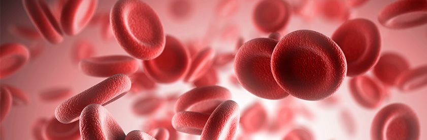
Red Blood Cells

Components
Approved name:
• Red blood cells

Commonly used names:
• Packed cells
•   Red cells
•   Packed red blood cells
•   RBCs

Description (1-3)
Red Blood Cells (RBCs) consist of erythrocytes concentrated from a whole blood donation or collected by apheresis. They
contain citrate anticoagulant and usually one of several types of preservative solutions. Depending on the preservative-
anticoagulant system, the hematocrit (Hct) of RBCs is about 55-65% for additive solutions (AS), AS-1, AS-3, AS-5, AS-7
and about 65-80% for citrate-phosphate-dextrose-adenine solutions, CPDA-1, CPD, CP2D. RBCs contain 20-100 mL of
donor plasma, but usually
populations. Anti-D is an incidental finding in a small percentage of blood donors. Studies have shown that 20-30% of
Rh-negative hospitalized patients develop and have an anti-D after transfusion with a unit of RBCs(6-8). However, the risk of
an reaction due to emergently receiving an Rh incompatible unit is less than 1%, and even then the hemolysis is usually mild
since it is extravascular(4). Rates of alloimmunization in patients with AIDS and in transplant recipients is also lower. These
immunosuppressed populations should be considered for switching to D positive blood when the need arises(4,9,10). The same
can be said of ER patients of non-child bearing potential who need urgent transfusion, as their chance of alloimmunization is as
low as 3-6%(4).

For women of childbearing potential for whom the Rh type is inconclusive or unknown, RHD genotyping is recommended to
resolve the Rh(D) type. In the majority of Caucasians with a serologic weak D phenotype, RHD genotyping will reveal that
the patient carries a weak D type 1, 2 or 3, for which the risk of alloimmunization to Rh(D) is known to be very low(11) and that
Rh(D) positive blood may be used. In pregnant patients who are found to carry one of these three RHD variant alleles, Rh
immunoprophylaxis is not necessary(12,13). In 2016, a joint statement of the AABB, American Red Cross, College of American
Pathologists , America’s Blood Centers, American College of Obstetricians and Gynecologists, and the Armed Services Blood
Program recommended RHD genotyping be used in women of childbearing potential to resolve serologic weak D phenotypes,
or discrepant D types(14). Some transfusion services have created criteria to identify patients that would benefit from RHD
genotyping(15).

Extended storage preservative-anticoagulant preparations such as AS-1 and AS-3 are appropriate for nearly all patients and
extend the shelf-life of RBCs to 42 days. Physicians concerned about preservative-anticoagulant for large volume transfusion
to neonates may request that excess supernatant in transfusion aliquots be removed prior to administration, such as by
centrifugation and volume reduction or by washing(2,16).

Age of Blood
The controversy over whether the transfusion of fresher RBCs results in fewer patient complications, compared to the use of
older blood, has gone on for some time. But the debate was reinvigorated when a single-center retrospective study garnered
intense media interest after publication by the New England Journal of Medicine in 2008(17). This study by Koch et al, involving
6002 cardiac surgery patients, showed that recipients of red cells stored for more than 14 days had an increased incidence
of adverse outcomes compared to those receiving cells stored for 14 days or less. Changes that can occur during storage of
older blood were thought to be the cause(17). However, other retrospective studies investigating the effect of red-cell storage
duration on patients undergoing cardiac surgery showed no significant differences in outcomes.

Since release of the Koch paper, there have been several prospective, randomized clinical trials (RCTs) also looking at
questions surrounding the age of stored RBCs. In developing their recent practice guidelines, AABB identified 13 RCTs
involving more than 5000 patients and none of these studies suggested the use of older blood resulted in poorer patient
outcomes(18). For example, . a study of 1098 patients undergoing cardiac surgery in more than 30 North American hospitals
showed no statistically significant differences in multiple-organ dysfunction syndrome, adverse events, or 28-day mortality in
groups receiving blood
serious hemoglobinopathies, nor anyone receiving massive or exchange transfusions or intrauterine transfusions. Hence, no
recommendations were made for any of these groups(18).

General Information
Dosing
RBCs should be transfused according to clinical need, including signs and symptoms, Hgb level, and the results of
hematologic assays. In the absence of acute hemorrhage, RBCs should be given as single units followed by appropriate
evaluation to justify additional units(27). Transfusion of an RBC unit should be completed within four hours. If more time is
required, smaller aliquots can be prepared and transfused sequentially.

Response
In a non-bleeding, non-hemolyzing adult transfused with compatible RBCs, the hemoglobin level should equilibrate within 15
minutes of transfusion. One unit should increase the Hgb in an average-size patient (70-80 kg) by approximately one g/dL and
the hematocrit by 3%(28).

In neonates, a dose of 10-15 mL/kg is generally given, and additive solution red cells with a hematocrit of approximately 60%
will increase the Hgb by about 3 g/dL.

Transfused red cells have a half-life of approximately 30 days in the absence of blood loss, hemolysis, or other processes that
might affect in vivo survival. Seriously ill adult or pediatric patients may lose significant amounts of blood from phlebotomy for
laboratory analysis(29). In addition, when active bleeding is taking place, the anticipated post-transfusion hemoglobin level may
be impacted by the dilutional effect of volume replacement with crystalloid or colloid.

Indications and Contraindications
RBCs are indicated for patients with symptomatic deficiency of oxygen-carrying capacity, or tissue hypoxia due to inadequate
circulating red cell mass. They are also indicated for exchange transfusions (e.g. in hemolytic disease of the fetus or newborn or
for acute chest syndrome in sickle cell anemia etc.). Patients must be evaluated individually to determine the proper transfusion
therapy, with care to avoid under- or over -transfusion. Transfusion decisions should be based on careful clinical assessment as
well as Hgb levels(30).

RBCs may be used for patients with acute blood loss whose symptoms or conditions may not improve with administration of
crystalloid solutions. RBCs should not be used to treat anemia that can be corrected with therapies other than transfusion,
such as pharmacologic agents. RBCs should also not be used as a means of increasing blood volume, to increase oncotic
pressure, improve wound healing, or to improve a patient’s sense of well-being.

For side effects and hazards, please see the Appendices.

Utilization Guidelines
Perioperative/Periprocedural
The function of an RBC transfusion is to augment O2 delivery to the tissues. Hemoglobin levels during bleeding are imprecise
measures of tissue oxygenation. Intravenous fluid resuscitation and the time needed for equilibration can significantly alter the
Hgb concentration(31). A number of factors besides blood Hgb level must be considered, such as pulmonary oxygenation, blood
flow, Hgb O2 affinity and tissue demands for O2(18,31,32). The Hgb level and clinical status of the patient should both be
considered in assessing the need for RBC transfusion.

The adequacy of oxygen delivery must be assessed in individual patients, particularly in patients with limited cardiac reserve
or significant atherosclerotic vascular disease. If available, mixed venous O2 levels, O2 extraction ratios, or changes in oxygen
consumption may be helpful in determining tissue oxygenation. Other factors to consider include anticipated degree and rate of
blood loss, as well as the effect of body temperature or drugs and anesthetics on oxygen consumption. The American Society
of Anesthesiologists Task Force (ASATF) recommends that RBCs should usually be administered when the Hgb concentration
is low (for example, < 6g/dL in a young healthy patient), especially when the anemia is acute. Furthermore, the ASATF
states RBCs are usually unnecessary when the Hgb concentration is >10 g/dL. These guidelines may be altered in cases of
anticipated blood loss(32).
The decision to transfuse should be based on any indication of organ ischemia, potential bleeding or the rate and magnitude
of actual ongoing bleeding, the patient’s intravascular volume status, and the risk factors for complications of inadequate
oxygenation. These risk factors include a low cardiopulmonary reserve and high oxygen consumption. The 2016 AABB
guidelines state a restrictive transfusion threshold of 8 g/dL may be warranted in patients undergoing cardiac surgery or
orthopedic surgery(18).

Preoperative assessment and efforts to reduce RBC transfusion requirements in the perioperative period include the evaluation
and treatment of anemia prior to surgery(31). A recent international consensus conference on patient blood management
recommended that screening for anemia be done well in advance of major elective surgery so that there is enough time to
potentially manage it medically, thereby possibly reducing the need for perioperative transfusions(33). The use of alternative
measures to reduce allogeneic red blood cell use should also be considered. These include acute normovolemic hemodilution,
intraoperative and postoperative autologous blood recovery, along with operative and pharmacologic measures that reduce
blood loss.

The Society of Thoracic Surgeons and the Society of Cardiovascular Anesthesiologists blood conservation clinical practice
guidelines recommend the following be performed:
• Preoperative assessment to identify patients at elevated risk from bleeding(e.g. those of advanced age, or with decreased
    preoperative RBC volume, and those undergoing emergent or complex procedures), who may subsequently require blood
    transfusions
•   Effective treatment of preoperative anemia and minimizing hemodilution during cardiopulmonary bypass (CPB) to preserve
    red blood cell volume,
•   Appropriate management of preoperative antiplatelet and anticoagulant drug therapy, and the use of anti-fibrinolytic agents
    such as epsilon-aminocaproic acid or tranexamic acid to reduce total blood loss(34).

Liberal vs. Restrictive Transfusion Thresholds
Recent RCTs have consistently demonstrated the safety of using a restrictive threshold of 7 to 8 g/dL for most patients. As
part of creating the 2016 AABB transfusion guidelines ,a review and meta-analysis of 31 RCTs, involving a total of 12,587
subjects in various clinical settings( ranging from acute coronary syndrome to hematologic malignancies to orthopedic surgery,
etc.) concluded that restrictive thresholds of 7-8 g/dL, when compared to 9-10 g/dL, were not associated with higher rates
of adverse clinical outcomes such as 30-day mortality, cardiac morbidity and infection, with the exception of two small trials in
subjects with acute myocardial infarction(35). Furthermore, the lower transfusion threshold decreased the likelihood of receiving
a PRBC exposure by 43% overall, and some studies showed survival was higher in certain patients who were transfused less:
such as those with acute upper GI bleeding(36).

However, caution is warranted before extrapolating this research on restrictive transfusion strategies to other patient groups
that have yet to be studied, or where the data are extremely limited. Additional investigation is needed for those individuals with
hematologic malignancies, or acute coronary syndromes or strokes before hemoglobin thresholds of 7 g/dl transfusions can be
more routinely used in these patients(37).

Cardiac Surgery
Transfusions in cardiac surgery have been evaluated in randomized trials and suggest a Hgb threshold of 7.5 g/dL – 8 g/
dL is acceptable in most cases(37,38). The Transfusion Indication Threshold reduction (TITRe2) trial randomized 2007 patients
undergoing coronary artery bypass graft (CABG), valve surgery, or both, to either a restrictive post-operative transfusion
threshold of
from any cause, myocardial infarction, stroke, or new-onset renal failure requiring dialysis. A follow-up to this study showed the
restrictive strategy was still noninferior up to 6 months after surgery(41).

Another study of CABG and valve surgery patients randomized 354 subjects to be transfused at a hematocrit of 28% and
to withhold PRBCs from another 363 enrollees until their hematocrit dropped to 24%. No difference in the postoperative
complications and lengths of stay was observed between the two groups, but there was greater blood product exposure to
patients in the liberal arm(42).

General Critical Care
Individualization of red cell transfusion applies to critical care patients as well as perioperative patients. To the degree possible,
the effects of anemia should be differentiated from those of hypovolemia, although both can impede tissue O2 delivery.
Blood loss greater than 30% of blood volume generally causes significant clinical symptoms and signs, but in younger
healthy patients, resuscitation with crystalloids alone may be successful with blood loss of up to 40% of the patient’s volume
(approximately 2 liters of blood loss in an average adult male). Beyond that level of acute blood loss, even with adequate
volume replacement, a normovolemic anemia will exist and the need to prevent a dilutional coagulopathy becomes even
more important.

In otherwise healthy adults, adequate O2 delivery is maintained at Hgb levels of 6-7 g/dL(32). RBC transfusion should be
strongly considered in critically ill trauma patients if, after adequate fluid replacement, the Hgb is < 7g/dL(30). Tranexamic acid,
an antifibrinolytic agent, may be helpful in trauma or surgical patients whose anemia is related to ongoing blood loss(43, 44). RBC
transfusion is indicated in patients with hemorrhagic shock and should be considered in patients with a Hgb < 7 g/dL who are
on mechanical ventilation(30).

A restrictive RBC transfusion strategy (Hgb < 7-8 g/dL) is recommended for hemodynamically stable, hospitalized, adult
patients(18). Several prospective studies demonstrated a higher mortality rate in patients receiving RBCs than in those not
receiving them(45). The TRICC (Transfusion Requirements in Critical Care) trial, published in 1999 was a multicenter RCT,
comparing a transfusion trigger of 7g/dL with one of 9 g/dL in normovolemic critically ill patients(46). Overall, 30-day mortality
was similar in the two groups and in the subsets of more seriously ill patients, but the restrictive group received significantly
fewer RBCs. For younger patients or those with a lower acuity level, the restrictive strategy resulted in lower 30-day mortality
while decreasing RBC transfusions. The Transfusion Requirements in Septic Shock (TRISS) trial demonstrated that a Hgb
threshold of 7g/dL was safe in patients with septic shock(47).

In the setting of acute upper gastrointestinal (UGI) bleeding, a prospective, randomized, controlled trial comparing a liberal
transfusion threshold (Hgb

Pediatric Critical Care
Infants may require simple or exchange transfusion for hemolytic disease of the fetus and newborn (HDFN), or for symptomatic
anemia in the first months of life. The American Academy of Pediatrics has published guidance on specific indications for
exchange transfusion in newborn infants at 35 or more weeks of gestation with hyperbilirubinemia, including that caused by
HDFN(51). Infants with jaundice caused by HDFN are at greater risk of bilirubin-related encephalopathy and are treated more
intensively than infants with physiologic jaundice at any given unconjugated bilirubin level.

Apart from HDFN, neonatal anemia occurs mainly in preterm infants because of iatrogenic blood loss for laboratory testing,
concurrent infection or illness, and inadequate hematopoiesis in the first weeks of life. Transfusion thresholds for preterm infants
and critically ill children have been widely debated for years, however recent randomized studies support the use of a restrictive
strategy(52-54). In the multicenter PINT (Premature Infants in Need of Transfusion) study, 451 very low birth-weight infants were
assigned to receive red cell transfusions using either restrictive or liberal criteria. Infants in the restrictive group had lower
mean Hgb levels than those in the liberal group, and more infants avoided transfusion completely in the restrictive group (11%)
compared to the liberal group (5%)(54). There was no difference between the two groups in composite outcomes of death, severe
retinopathy, bronchopulmonary dysplasia, and brain injury, thus supporting the use of restrictive transfusion criteria. In a smaller,
single center trial, Bell et al., randomized 100 preterm infants to either restrictive or liberal transfusion criteria and found a
reduction in the number of transfusions in the restrictive group(52). However, infants in the restrictive group were found to have
more episodes of apnea and neurologic events than infants in the liberal group. A comparison of these studies suggests that
the documented benefits of a restrictive transfusion practice are a decrease in the number of transfusions and exposure to
fewer RBC donors. It is possible that the higher Hgb values maintained in the liberal transfusion group in Bell’s study compared
to the similar group in the PINT study may have decreased the risk of apnea and brain injury(54).

A more recent meta-analysis of clinical trials comparing outcomes between restrictive vs. liberal hematocrit thresholds in
neonates suggested that transfusion thresholds could be lowered, but identified the need for additional clinical studies to clarify
the impact of this practice on long-term outcomes(55). As for all pediatric patients, transfusion must take into consideration an
infant’s cardiorespiratory status, and transfusion decisions should be individualized for each patient.

General Guidelines for Small-Volume (10-15 mL/kg) Transfusion in Infants(56)
•   Severe cardiopulmonary disease with, e.g., mechanical ventilation with FiO2 > 0.35: Hct < 40-45% (must be defined
    by institution)
•   Moderate cardiopulmonary disease, e.g., less intensive assisted ventilation, such as nasal continuous positive airway
    pressure (CPAP)or supplemental O2 therapies: Hct < 30-35%.
•   Major surgery: Hct < 30-35%.
•   Stable anemia, especially if unexplained poor growth or unexplained breathing disorder: Hct < 20-30%.

For older children, more likely to be treated in a pediatric ICU (PICU) setting, the Pediatric Critical Care Transfusion and
Anemia Expertise Initiative (TAXI) recommended transfusing when the hemoglobin was less than 5g/dl for patients not in
hemorrhagic shock; and between 5- 7 g/dl clinical judgement should guide transfusion decisions with a reasonable post-
transfusion target being a hemoglobin of 7-9.5 g/dl(57).

In hemodynamically stable children with hemoglobins exceeding 7 g/dl, transfusions were generally considered unnecessary,
but might be beneficial in the following settings:
• Acute brain injury
•   Acute respiratory distress syndrome
•   Allo- or auto-immune mediated hemolytic anemia
•   Cancer or hematopoietic stem cell transplant
•   Cardiac disease
•   Extracorporeal membrane oxygenation
•   Ventricular assist devices
Chronic Anemia
Asymptomatic Chronic Anemia
Based on the specific diagnosis, treat with pharmacologic agents, for example, vitamin B12, folic acid, erythropoietin, iron.

Symptomatic Chronic Anemia
Transfuse to minimize symptoms and risks associated with anemia. Transfusion is usually required when the Hgb is
The National Heart, Lung, and Blood Institute (NHLBI) 2014 Evidence Based Management of Sickle Cell Disease Guidelines(61)
recommend exchange transfusion for symptomatic severe acute chest syndrome (ACS) in patients with SCD. For other
conditions such as acute splenic sequestration with severe anemia, aplastic crisis, and simple anemia, “simple” transfusion is
recommended. Automated or manual RCE was also preferred over simple transfusions by the ASH panel for treating ACS but,
like the most of their recommendations, this one was also conditional, based upon their review of the evidence(62).

In preparation for surgery requiring general anesthesia, simple transfusion to increase the Hgb towards  9 g/dL and Hgb S < 30%. maintaining a Hgb
S level < 30%. The standard group received neither blood nor hydroxyurea therapy for silent infarcts. Transfused children had
significantly fewer cerebral episodes and fewer other complications such as priapism, acute chest syndrome, vaso-occlusive
pain and avascular necrosis of the hip, but no differences in cognitive ability were noted(77). As expected, there were more
transfusion reactions in the group that received blood and, longer-term, there may be higher rates of alloimmunization and
iron accumulation.

The NHLBI and ASH recommendations for transfusion in SC reflect evidence-based reconsideration of previous practices
or recommendations. (61, 62) In some clinical situations, the revised recommendations do not support automatic transfusion,
for example, in uncomplicated painful crises, priapism, asymptomatic anemia, acute kidney injury without multi-system organ
failure, and splenic sequestration. However, some patients may have better outcomes with exchange transfusion, which
reduces the circulating volume of Hgb S erythrocytes and the potential for hyperviscosity. The decision to manage SCD with
red cell exchange rather than simple transfusion should be made in consultation with an SCD clinical specialist. In contrast to
simple transfusion, exchange transfusion utilizing cytapheresis prevented tissue iron accumulation and reduced iron overload in
chronically transfused patients(78).

The use of hydroxyurea, an oral alkylating agent which has been used in adults with SCD to promote increased fetal Hgb F
levels, remains unclear in developing children. Hgb F retains higher levels of O2 in the circulation, resulting in lower sickling
rates in red cells. Additional investigation would be warranted to further define the management of SCD and how best to
handle the multiple problems associated with SCD and transfusion(79).

Evidence-based Recommendations for Transfusion in SCD(46)
•   Preoperative prophylaxis: children and adults, transfuse to 10 g/dl prior to general anesthesia:
•   In SCD patients with Hgb > 8.5 g/dl on long term hydroxyurea or facing high-risk surgery (neurosurgery, cardiac bypass,
    prolonged anesthesia, for example), consult an SCD specialist.
•   For patients not on long-term treatment with hydroxyurea and/or transfusion therapy who may have higher Hgb S and are at
    risk for hyperviscosity: avoid transfusion to hemoglobin >10g/dl.
•   Severe, symptomatic acute chest syndrome (O2 sat. < 90% despite supplemental O2 therapy)
•   Acute splenic sequestration with severe anemia
•   Children or adults with acute stroke (begin prophylactic transfusion regimen)
•   Hepatic sequestration
•   Intrahepatic cholestasis
•   Multisystem organ failure
•   Aplastic crisis
•   Symptomatic anemia
•   Child with transcranial Doppler reading > 200 cm/second
•   Adults and children with previous clinically overt stroke
The more recent ASH guidance on managing SCD patients did overlap somewhat with the NHLBI ‘s recommendations, (e.g. on the topics of preoperative transfusion, transfusion of pregnant women and iron overload screening)(61, 62). However, the 10 guidelines ASH developed, the only strong recommendation of the was on transfusing RBCs matched for the Rh (C, E or C/c, E/e) and K antigens. The other 9 recommendations were all conditional. By contrast NHLBI, graded the evidence regarding antigen matching as being of low quality, resulting in only a weak to moderate recommendation from their group. Of note, ASH also gave conditional recommendations (i.e. based on very low certainty in the evidence) to the use of isovolemic hemodilution with RCE, and to using immunosuppressive therapy for the prevention and treatment of hemolytic transfusion reactions. Thalassemias Patients with some forms of alpha and beta thalassemia require frequent or chronic transfusion. These patients have been found to be at increased risk of alloimmunization to red cell antigens (16.5%)(80). It is fairly common to obtain a red cell antigen phenotype by serology or HEA genotyping and select red cell products matched for RhC, RhE and K. Since peripheral blood DNA can be used to genotype recently transfused patients, without interference from donor cells, this is the preferred approach in a patient for which a baseline extended phenotype was not obtained prior to beginning their chronic transfusion protocol. Oxygen Therapeutics (“Artificial Blood”, Oxygen Carriers) The acute need for blood for war and other violent conflicts, difficulties in acquiring, storing and testing blood, and the continuing global threat of emerging infectious diseases have driven efforts to find “blood substitutes.” There are currently no substances that perform all the functions of blood. An ideal oxygen therapeutic: • Would circulate for a useful period of time • Could be issued without crossmatching • Could be easily stored for extended periods • Would be capable of off-loading O2 when required • Could be easily be transported Some of these criteria were met by products under development, but clinical trials identified adverse events. Several products were found to successfully circulate and deliver oxygen, but regulatory approval has not been given(81). Complications included vasoconstriction, shock, and myocardial and cerebral infarcts. Further, it has been difficult to develop acceptable protocols for testing of these agents in trauma situations. Several case reports have been published regarding the successful use of a conjugated, stabilized bovine hemoglobin solution in Jehovah’s Witnesses with life-threatening anemia(82). These infusions were approved by FDA on a case-by-case basis. A pegylated bovine Hgb solution appears to be of utility as a vasodilator, possibly helpful in various crises of SCD. Clinical trials are currently open. The effects of pegylated human tetrameric Hgb in vitro were reported in 2011 and further studies are underway(83). These products (82), may be available for enhanced access (“compassionate use”) in life-threatening situations in patients for whom blood transfusion is not a conscience-based option. Obtaining these oxygen therapeutics requires close coordination among the requesting hospital, FDA, and the manufacturer. The FDA maintains 24/7 access to the emergency IND department for assistance to assist in obtaining an enhanced access product (866-300-4374). Information on current clinical trials and access to the Help Desk can be found on line at www.clinicaltrials.gov. Warm Autoantibodies and Interfering Immunologic Substances Warm autoantibodies are antibodies that react with substances on the patient’s own red cells at body temperature. Presence of a warm autoantibody is usually associated with some kind of immunoregulatory dysfunction or infectious process and may or may not be associated with anemia or hemolysis. Red cells coated with autoantibodies presents challenges for antigen typing. Some facilities choose to match donor red cells for additional antigens. Immunotherapies such as anti-CD38 are showing significant efficacy in patients with multiple myeloma. While the monoclonal antibody binds to myeloma cells causing cell death, the antibody also binds to red cells interfering with antibody screening. Patients with warm autoantibodies as well as those receiving immunologic therapies that can act as interfering substances may benefit from utilization of HEA genotyping in predicting their red blood cell antigen phenotype.
Red Cell Treatment Techniques prior to Antigen Typing
Immunohematology reference laboratories may use techniques such as EGA-treatment to remove antibodies from the surface
of patient red cells followed by antigen typing. Such results should be interpreted with caution, since it has been shown that
when compared to the red cell phenotype obtained by HEA genotyping panel, there can be false negative findings(84).

Immunohematology reference laboratories may use hypotonic washing of a peripheral blood sample from a recently transfused
patient with sickle cell disease, since the treatment will lyse the Hgb A cells leaving behind the Hgb S cells for antigen
phenotyping. Such results should be interpreted with caution, since it has been shown that when compared to the red cell
phenotype obtained by HEA genotyping panel, there can be false negative findings(84).

Red Cell Antigen Discrepancies
In cases where a current serologic type is discordant with a historic type, or a serologic type obtained in one laboratory is
disagrees with another, or if one serologic reagent or method yields a result that is discordant with another reagent or method
(including molecular methods), an investigation should be performed to resolve the discrepancy. Molecular methods such as
genotyping using an HEA panel or higher resolution molecular methods such as Sanger sequencing can identify variant alleles
that may encode weakened or partial antigens that react differently based on the reagent or method used. These techniques
may also reveal that the patient carries a null allele that does not express the antigen. Resolution of the discrepancy may
eliminate the need for antigen negative red cell products and in some cases may uncover partial antigens that put the patient at
risk for alloimmunization.

References
1.    AABB, American Red Cross, America’s Blood Centers, Armed Services Blood Program. Circular of Information for the Use of Human Blood and Blood Components. Revised October
      2017. Available at: aabb.org
2.    Fung MK, Grossman BJ, et al., eds. Technical Manual. 18th ed. Bethesda, MD: AABB; 2014.
3.    Ooley P, Chair. Standards for Blood Banks and Transfusion Services. 30th ed. Bethesda, MD: AABB; 2015 .
4.    Murphy M, BenAvram D. AABB Association Bulletin 19-02. Recommendations on the Use of Group O Red Blood Cells. Release Date: June 26, 2019
5.    Garratty G, Glynn SA, et al. ABO and Rh(D) phenotype frequencies of different racial/ethnic groups in the United States. Transfusion 2004; 44:703-6.
6.    Frohn C, Dumbgen L, et al. Probability of anti-D development in D− patients receiving D+ red cells. Transfusion 2003;43:893-8.
7.    Gonzalez-Porras JR, Graciani IF, et al. Prospective evaluation of a transfusion policy of D+ red cells into D− patients. Transfusion 2008;48:1318-24.
8.    Yazer MH, Triulzi DJ. Detection of anti-D in D− recipients transfused with D+ red cells. Transfusion 2007;47:2197-2201.
9.    Casanueva M, Valdez MD, et al. Lack of alloimmunization to D antigen in D-negative immunosuppressed liver transplant recipients. Transfusion 1994;34:570-2.
10.   Boctor FN, Ali NM, et al. Absence of D-alloimmunization in AIDS patients receiving D-mismatched RBCs. Transfusion 2003;43:173-6
11.   Sandler SG, Flegel WA et al ; College of American Pathologists Transfusion Medicine Resource Committee Work Group. It’s time to phase in RHD genotyping for patients with a serologic
      weak D phenotype. College of Transfusion. 2015 Mar;55(3):680-9
12.   Pham BN, Roussel M, et al. Anti-D investigations in individuals expressing weak D Type 1 or weak D Type 2: allo- or autoantibodies? Transfusion. 2011 Dec;51(12):2679-85.
13.   Wagner FF, Frohmajer A, et al. Weak D alleles express distinct phenotypes. Blood. 2000 Apr 15;95(8):2699-708.
14.   AABB and the College of American Pathologists Interorganizational Work Group’s Joint Statement on Phasing-In RHD Genotyping for Pregnant Women and Other Females of
      Childbearing (2016). Available at http://www.aabb.org/advocacy/statements/Pages/statement150722.aspx?PF=1
15.   Luo X, Keller MA, et al. Strategies to identify candidates for D variant genotyping. Blood Transfus. 2018 May;16(3):293-30.
16.   New York State Council on Human Blood and Transfusion Services. Guidelines for Transfusion of Pediatric Patients. New York State Department of Health, 2016. Available at: https://
      www.wadsworth.org/sites/default/files/WebDoc/ped_tx_guidelines_2.pdf. Last accessed: Aug 20, 2019
17.   Koch CGH, Li L, et al. Duration of red-cell storage and complications after cardiac surgery. N Engl J Med 2008;358:1229-39.
18.   Carson JL, Guyatt G, et al. Clinical practice guidelines from the AABB: red blood cell transfusion thresholds and storage. JAMA 2016; 316: 2025-35.
19.   van de Watering L, Lorinser J, et al. Effects of storage time of red cell transfusions on the prognosis of coronary artery bypass graft patients. Transfusion 2006;46:1712-8.
20.   Yap CH, Lau L, et al. Age of transfused red cells and early outcomes after cardiac surgery. Ann Thorac Surg 2008;86:554-9.
21.   Fergusson DA, Hebert P. et al. Effect of fresh red blood cell transfusions on clinical outcomes in premature, very low birth weight infants: The ARIPI randomized trial. JAMA
      2012;308:1443-51.
22.   Steiner ME, Ness PM, et al. Effects of red-cell storage duration on patients undergoing cardiac surgery. N Engl J Med 2015;372:1419-29.
23.   Lacroix J, Hebert PC, et al. Age of transfused blood in critically ill adults. N Engl J Med 2015;372:1410-8.
24.   Heddle NM, Cook RJ, et al. Effect of short-term vs. long-term blood storage on mortality after transfusion. N Engl J Med 2016;375:1937-45
25.   Dhabangi A, Ainomugisha B. et al. Effect of transfusion of red blood cells with longer vs shorter storage duration on elevated blood lactate levels in children with severe anemia. The TOTAL
      randomized clinical trial. JAMA 2015;314:2514-23.
26.   Spinella C and Acker J. Storage duration and other measures of quality of red blood cells for transfusion. JAMA 2015;314:2509-10.
27.   New York State Council on Human Blood and Transfusion Services. Guidelines for transfusion options and alternatives. 1st ed. New York State Department of Health, 2010. Available at:
      wadsworth.org/labcert/blood_tissue/pdf/txoptsaltsfixed122811.pdf. Last accessed: Aug. 21, 2019.
28.   Wiesen ER, Hospenthal DR, et al. Equilibration of hemoglobin concentration in medical inpatients not actively bleeding. Ann Int Med 1994;121:278-80.
29.   Stefanini M. Iatrogenic anemia (can it be prevented?) Letter. J Thromb Haemost 2014;12:1591.
30.   Napolitano LM, Kurek S, et al. Clinical practice guideline: red blood cell transfusion in adult trauma and critical care. Crit Care Med 2009;37:3124-57.
31.   Skerrett G, Chiofol A, et al., for the New York State Council on Human Blood and Transfusion Services. Guidelines for transfusion of red blood cells – adults. 3rd ed. New York State
      Department of Health 2012. Available at: wadsworth.org/labcert/blood_tissue/rbcsadults0812final.pdf. Last accessed Aug. 18, 2019.
32.   Practice guidelines for perioperative blood management: an updated report by the American Society of Anesthesiologists Task Force on Perioperative Blood Management. Anesthesiology
      2015;122:241-75.
33.   Mueller MM, Van Remoortel H et al. Patient Blood Management: Recommendations From the 2018 Frankfurt Consensus Conference. JAMA. 2019 Mar 12;321(10):983-997
34. Society of Thoracic Surgeons Blood Conservation Guideline Task Force. 2011 update to the Society of Thoracic Surgeons and the Society of Cardiovascular Anesthesiologists blood
conservation clinical practice guidelines. Ann Thorac Surg 2011;91:944-82.
35. Carson JL, Stanworth J et al Transfusion thresholds and other strategies for guiding allogeneic red blood cell transfusion. Cochrane Database of Systematic Reviews 2016, Issue 10.
36. Villanueva C, Colomo A, et al. Transfusion strategies for acute upper gastrointestinal bleeding. N Engl J Med 2013;368:11-21
37. Carson JL, Triulzi DJ, et. al Indications for and Adverse Effects of Red-Cell Transfusion. N Engl J Med. 2017 Sep 28;377(13):1261-1272
38. Hajjar LA, Vincent SL, et al. Transfusion requirements after cardiac surgery: the TRACS randomized controlled trial. JAMA 2010;304:1559-67.
39. Murphy GJ, Pike K, et al. Liberal or restrictive transfusion after cardiac surgery. N Engl J Med 2015; 372:997-1008.
40. Mazer CD, Whitlock RP, et al. Restrictive or Liberal Red-Cell Transfusion for Cardiac Surgery. N Engl J Med. 2017 Nov 30;377(22):2133-2144.
41. Mazer CD, Whitlock RP, et al. Six-Month Outcomes after Restrictive or Liberal Transfusion for Cardiac Surgery. N Engl J Med. 2018 Sep 27;379(13):1224-1233.
42. Koch CG, Sessler DI, et al. A Randomized Clinical Trial of Red Blood Cell Transfusion Triggers in Cardiac Surgery. Ann Thorac Surg. 2017 Oct;104(4):1243-1250.
43. CRASH-2 Trial Collaborators. Effects of tranexamic acid on death, vascular occlusive events and blood transfusion in trauma patients with significant hemorrhage (CRASH-2): A
randomised placebo-controlled trial. Lancet 2010;376:23-32.
44. CRASH-2 Trial Collaborators. The importance of early treatment with tranexamic acid in bleeding trauma patients: an exploratory analysis of the CRASH-2 randomized controlled trial.
Lancet 2011; 377:1096-1101.
45. Gould S, Cimino MJ, et al. Packed red blood cell transfusion in the intensive care unit: limitations and consequences. Am J Crit Care 2007;16:39-48.
46. Hebert PC, Wells G, et al., for the Transfusion Requirements in Critical Care Investigators for the Canadian Critical Care Trials Group. A multicenter, randomized, controlled clinical trial of
transfusion requirements in critical care. N Engl J Med 1999;340:409-17.
47. Holst LB, Haase N, et al. Lower vs. higher hemoglobin threshold for transfusion in septic shock. N Engl J Med 2014;371:1381-91.
48. Carson JL, Terrin ML, et al., for the FOCUS Investigators. Liberal or restrictive transfusion in high risk patients after hip surgery. N Engl J Med 2011;365:2453-62.
49. Brunskill SJ, Millette SL, et al. Red blood cell transfusion for people undergoing hip fracture surgery. Cochrane Database Syst Rev. 2015 Apr 21;(4).
50. Kansagara D, Dyer E, et al. Treatment of anemia in patients with heart disease: a systematic review. Ann Int Med 2013;159:746-57.
51. American Academy of Pediatrics Subcommittee on Hyperbilirubinemia. Management of hyperbilirubinemia in the newborn infant 35 or more weeks of gestation. Pediatrics 2004;
114:292-316.
52. Bell EF, Strauss RG, et al. Randomized trial of liberal vs. restrictive guidelines for red blood cell transfusion in preterm infants. Pediatrics 2005;115:1685-91.
53. Bell EF. Transfusion thresholds for preterm infants: how low should we go? J Pediatrics 2006;149: 287-9.
54. Kirpalani H, Whyte RK, et al. The Premature Infants in Need of Transfusion (PINT) study: a randomized, controlled trial of a restrictive (low) vs. liberal (high) transfusion threshold for
extremely low birth-weight infants. J Pediatrics 2006;149:301-7.
55. Whyte R, Kirpalani H. Low vs. high hemoglobin concentration for blood transfusion for preventing morbidity and mortality in very low birth-weight infants. Cochrane Database Syst Rev.
2011 Nov 9;(11):
56. Strauss R. RBC transfusion and/or recombinant EPO therapy of the anaemia of prematurity. ISBT Science Series (2006).1, 11–14
57. Valentine SL, Bembea MM, et. al. Consensus Recommendations for RBC Transfusion Practice in Critically Ill Children From the Pediatric Critical Care Transfusion and Anemia Expertise
Initiative. Pediatr Crit Care Med. 2018 Sep;19(9):884-898.
58. Rodgers GM, Becker PS, et al. Cancer and chemotherapy-induced anemia. J Natl Compr Canc Netw 2012;10:628-55.
59. Estcourt LJ, Malouf R, et al. Restrictive versus liberal red blood cell transfusion strategies for people with haematological malignancies treated with intensive chemotherapy or radiotherapy,
or both, with or without haematopoietic stem cell support. Cochrane Database Syst Rev 2017; 1:Cd011305.
60. National Comprehensive Cancer Network (NCCN) clinical practice guidelines in oncology. Cancer and chemotherapy-induced anemia. nccn.org. Version 2.2019. Last accessed:
August 7, 2019
61. National Heart, Lung, and Blood Institute. NHLBI Expert Panel Report 2014: Evidence-Based Management of Sickle Cell Disease. Available at: https://www.nhlbi.nih.gov/health-topics/
evidence-based-management-sickle-cell-disease
62. Chou ST, Alsawas M, et al.; American Society of Hematology 2020 guidelines for sickle cell disease: transfusion support. Blood Adv 2020; 4 (2): 327–355.
63. Vichinsky EP, Haberkern CM, et al., for the Preoperative Transfusion in Sickle Cell Disease Study Group. A comparison of conservative and aggressive transfusion regimens in the
perioperative management of sickle cell disease. N Engl J Med 1995;333:206-13.
64. Chou ST, Jackson T, et al. High prevalence of red blood cell alloimmunization in sickle cell disease despite transfusion from Rh-matched minority donors. Blood. 2013 Aug 8; 122(6):1062-
71).
65. Chou ST, Friedman DF. Transfusion practices for patients with sickle cell disease at the Children’s Hospital of Philadelphia. Immunohematology. 2012;28(1):27-30
66. Winkler AM, Josephson CD. Transfusion practices for patients with sickle cell disease at major academic medical centers participating in the Atlanta Sickle Cell Consortium.
Immunohematology. 2012;28(1):24-6.
67. Vichinsky EP. The prevention and management of alloimmunization in sickle cell disease: the benefit of extended phenotypic matching of red blood cells. Immunohematology. 2012;28(1):20-
3.
68. Sloan SR. Transfusions for patients with sickle cell disease at Children’s Hospital Boston. Immunohematology. 2012;28(1):17-9.
69. Fasano RM, Paul W, et al.. Transfusion protocol for patients with sickle hemoglobinopathies at Children’s National Medical Center. Immunohematology. 2012;28(1):13-6.
70. Roberts DO, Covert B, et al. Directed blood donor program decreases donor exposure for children with sickle cell disease requiring chronic transfusion. Immunohematology. 2012;
28(1):7-12..
71. Karafin MS, Shirey RS, et al. Antigen-matched red blood cell transfusions for patients with sickle cell disease at The Johns Hopkins Hospital. Immunohematology. 2012;28(1):3-6.
72. Fasano RM, Monaco A, et al. RH genotyping in sickle cell disease patient contributing to hematopoietic stem cell transplantation donor selection and management. Blood. 2010 Oct
14;116(15):2836-8.
73. Nance ST, Keller MA. Comments on: molecular matching of red blood cells is superior to serological matching in sickle cell disease patients. Rev Bras Hematol Hemoter. 2013;35(1):9-11.
74. Biller E, Zhao Y, et al. Red blood cell exchange in patients with sickle cell disease-indications and management: a review and consensus report by the therapeutic apheresis subsection of
the AABB. Transfusion. 2018 Aug;58(8):1965-1972.
75. Adams RJ, Brambilla D, for the Optimizing Primary Stroke Prevention in Sickle Cell Anemia (STOP 2) Investigators. Discontinuing prophylactic transfusions used to prevent stroke in sickle
cell disease. N Engl J Med 2005;353:2769-78.
76. Lee MT, Piomelli S, et al., for the STOP Study Investigators Trial in Sickle Cell Anemia (STOP): extended follow-up and final results. Blood 2006;108:847-52.
77. DeBaun MR, Gordon M, et al. Controlled trial of transfusions for silent cerebral infarct in sickle cell anemia. N Engl J Med 2014;371:699-710.
78. Kim HC, Gugan NP, et al. Erythrocytapheresis therapy to reduce iron overload in chronically transfused patients with sickle cell disease. Blood 1994;83:1136-42.
79. Steinberg MH. More blood for sickle cell anemia? (Editorial) N Engl J Med 2014;371:775-6
80. Thompson AA, Cunningham MJ,et al.. Red cell alloimmunization in a diverse population of transfused patients with thalassaemia. Br J Haematol. 2011 Apr;153(1):121-8.
81. Chloe WH, Baek EJ. Red blood cell substitutes: from the past to the future. ISBT Science Series 2015;10:150-3.
82. Posluzny J, Napolitano LM. How do we treat life-threatening anemia in a Jehovah’s Witness patient? Transfusion 2014;54:3026-34.
83. Cole RH, Vandegriff KD. MP4, a vasodilatory PEGylated hemoglobin. Adv Exp Med Biol 2011;701: 85-90.
84. Horn T, Hamilton J, et al.. Assessment of common red blood cell pretreatments to yield an accurate serologic antigen phenotype compared with genotype-predicted phenotype.
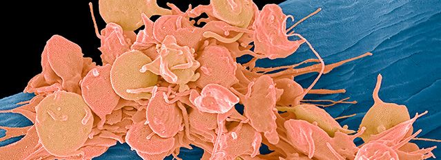
Immunohematology. 2017 Dec;33(4):147-151.Platelets
General Information
Components
Approved names:
• Platelets
• Platelets Pooled Platelets (Platelets Pooled)
• Platelets Leukocytes Reduced
• Pooled Platelets Leukocytes Reduced (Platelets Leukocytes Reduced, Pooled)
• Apheresis Platelets (Platelets Pheresis)
• Apheresis Platelets Leukocytes Reduced (Platelets Pheresis Leukocytes Reduced)
• Apheresis Platelets Platelet Additive Solution Added Leukocytes Reduced (Platelets Pheresis Platelet Additive Solution
Added Leukocytes Reduced)
Commonly used names:
• Platelets
• Single Donor Platelets (SDP)
• Platelet Additive Solution (PAS) Platelets/PAS Platelets
• Random Donor Platelets (RDP)
• Pooled Platelets
• PSP
• Pathogen-Reduced (PR) Platelets
Description of Basic Components
Apheresis platelets are collected from a single donor using automated devices known as cell separators. These products are
often called Single Donor Platelets (SDPs) and contain ≥3.0 x 10¹¹ platelets (average 3.5–4.0 x 10¹¹) per unit in approximately
100–500 mL of plasma or plasma with platelet additive solution (PAS). The anticoagulant used is acid citrate dextrose (ACD).
Approximately 93% of platelet transfusions are apheresis platelets(1).
Platelets derived from whole blood contain ≥5.5 x 10¹⁰ platelets per bag (unit) in 40–70 mL of plasma. The anticoagulant is
the same as that used for whole blood collection, usually citrate phosphate dextrose (CPD) or citrate phosphate 2 dextrose
(CP2D). They are often referred to as random donor platelets (RDPs) to distinguish them from SDPs. Four to six units are often
pooled by the blood center or hospital to make an adult dose that can be estimated by multiplying the number of RDPs in the
pool by the required minimum number of platelets, ≥5.5 x 10¹⁰, in each whole blood-derived unit. They may be used as single
units for pediatric patients.
Platelets in platelet additive solution (PAS) are Apheresis Platelets Leukocytes Reduced and are suspended in a mixture of
plasma and proprietary additive solutions. In two additive solutions that have been cleared for use in the United States, the
platelet suspension contains approximately 35% residual plasma and 65% PAS(2). Plasma proteins, including ABO isoagglutinins,
coagulation factors, and allergenic substances, are diluted in proportion to the PAS added. The shelf life of Apheresis Platelets
Leukocyte Reduced in PAS is 5 days, and they may be further processed (e.g., irradiated or aliquoted). Retrospective clinical
comparison of PAS platelets to those suspended in 100% plasma demonstrated a reduction in allergic transfusion reactions(3).
In this study, circulation of transfused platelets in PAS, as measured by the corrected count increment (CCI) immediately after
transfusion, was lower compared to those suspended in 100% plasma, but not significantly different when measured 12 to
24 hours after transfusion. In another retrospective study, patients transfused with platelets suspended in additive solution
experienced fewer febrile reactions and allergic reactions than those transfused with platelets in 100% plasma.(4)A psoralen and ultraviolet light-based pathogen reduction process forleukocyte reduced Apheresis Platelets in PAS and leukocyte reduced Apheresis Platelets in plasma have been implemented in several US blood centers. The shelf-life of pathogen reduced apheresis platelets is 5 days. Pathogen reduced apheresis platelets do not require testing for bacteria (5) , CMV (6), Babesia (7), or Zika (8), and do not require gamma irradiation as prophylaxis against transfusion-associated graft versus host disease in susceptible recipients (6). A meta-analysis of clinical trials of psoralen-based pathogen reduced platelets revealed no significant differences in mortality or bleeding of platelets receiving pathogen reduced platelets and those receiving standard platelets(9). In September 2019, the Food and Drug Administration issued Final Guidance for the industry to address bacterial contamination risk reduction(10). Several methods were provided to meet the guidance objectives including pathogen reduction, large volume—delayed sampling (LVDS) at greater than 36 or 48 hours after collection, primary culture followed by secondary culture, and point of issue testing (see FDA Final Guidance for details). The LVDS testing methodology involves holding apheresis platelets until at least 36 or 48 hours have elapsed from collection then obtaining a 16-20 mL sample from each final split product for aerobic and anaerobic culture. This longer period between collection and sampling as well as the larger aliquot drawn for testing increases the likelihood of bacterial detection(11). Leukoreduction standards are discussed in the Blood Component Modification chapter Preparation of Platelets An “apheresis platelets” product is considered to be one adult dose. To prepare an adult dose of pooled platelets, 4-6 RDPs are pooled by the blood center or hospital prior to transfusion. When prepared and pooled using an FDA-approved system, the post-collection shelf life is 5 days. ABO and Rh Compatibility Donor plasma in platelets suspended in plasma should be ABO-compatible with the recipient’s red cells. This is particularly important when transfusing infants or giving large volumes to adults, to avoid the possibility of exposure to potentially hemolyzing isoagglutinins (anti-A and/or anti-B). Rh-negative recipients should receive Rh-negative platelets when available, particularly women of childbearing age. However, apheresis platelets may contain a very small volume of RBCs (up to 0.001 mL), and it has been suggested that Rh immune prophylaxis may not be necessary when Rh positive platelets are given to Rh negative patients(5). In a study that included hematological, oncological and patients with other conditions, D alloimmunization by Rh(D) positive apheresis platelets given to Rh(D) negative recipients was reported to be approximately 1.4% after a median 77 day follow up(13). Dosing Clinical practice guidelines for prophylactic and therapeutic platelet transfusion have been recently published and are based on systematic literature review and recommendations using the Grading of Recommendations, Assessment, Development and Evaluation (GRADE) framework(14,32). To treat bleeding or prepare patients for invasive procedures, transfuse as needed to maintain hemostasis or the target platelet count, whichever is applicable. Four to ten units of RDPs, one to two units of Pooled Platelets (each containing approximately 4 to 6 whole blood platelet concentrates), or one to two SDPs are generally transfused to thrombocytopenic or thrombocytopathic adults. The Prophylactic Platelet Dose on Transfusion Outcomes (PLADO) trial concluded that prophylaxis at pre-specified triggers was accomplished with equivalent effect on hemorrhage, using three RDPs or half of an SDP in adults, provided that the minimum dose exceeded 1.1 x 10¹¹ platelets per square meter of the patient’s body surface area. This strategy, however, resulted in more frequent transfusions, usually on a daily basis(15). A higher minimum dose of 2.4 x 10¹¹ platelets per square meter for outpatients is likely to be more cost effective, minimizing the number of patient visits(16). It is recognized that in the lowest dosing arm of the PLADO study, the dose of of platelets given was
You can also read