A Modification of the Bielschowsky Silver Stain for Alzheimer Neuritic Plaques: Suppression of Artifactual Staining by Pretreatment with Oxidizing ...
←
→
Page content transcription
If your browser does not render page correctly, please read the page content below
bioRxiv preprint first posted online Mar. 6, 2019; doi: http://dx.doi.org/10.1101/570093. The copyright holder for this preprint (which
       was not peer-reviewed) is the author/funder, who has granted bioRxiv a license to display the preprint in perpetuity.
                                 It is made available under a CC-BY-NC-ND 4.0 International license.

                                                                                                                                          1

           A Modification of the Bielschowsky Silver
             Stain for Alzheimer Neuritic Plaques:
            Suppression of Artifactual Staining by
              Pretreatment with Oxidizing Agents

                                    Anthony J. Intorcia, Jessica R. Filon,
                                    Brittany Hoffman, Geidy E. Serrano,
                                    Lucia I. Sue and Thomas G. Beach

                                         Civin Laboratory for Neuropathology
                                        Banner Sun Health Research Institute
                                                  Sun City, Arizona
                                                        USA

              Corresponding author: Thomas G. Beach, MD, PhD
                                    Banner Sun Health Research Institute
                                    10515 W Santa Fe Drive
                                    Sun City, AZ 85351
                                    Ph: 623-832-5643
                                    email: Thomas. Beach@bannerhealth.com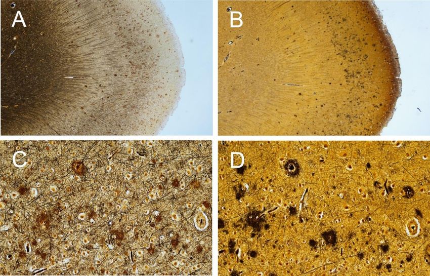
bioRxiv preprint first posted online Mar. 6, 2019; doi: http://dx.doi.org/10.1101/570093. The copyright holder for this preprint (which
       was not peer-reviewed) is the author/funder, who has granted bioRxiv a license to display the preprint in perpetuity.
                                 It is made available under a CC-BY-NC-ND 4.0 International license.

                                                                                                                                          2

      Abstract

      The Bielschowsky silver method has been the most commonly-used histological stain for

      demonstrating the neuritic plaques and neurofibrillary tangles that define Alzheimer’s

      disease (AD). The stain has been critical to providing a common measure allowing large

      clinicopathological correlation studies that have demonstrated statistically significant and

      independent contributions of both lesion types to cognitive impairment and dementia. The

      continuing relevance of neuritic plaques, and the Bielschowsky stain as the method of

      choice for their demonstration, is also indicated by its use as the US Food and Drug

      Administration (FDA) “standard of truth” for autopsy confirmation of the validity of PET

      amyloid imaging agents.                    Many modifications of the Bielschowsky stain have been

      published but it is still known to frequently generate artifactual staining that at times

      mimics the appearance of amyloid plaques. In this study we found that pretreatment with

      oxidizing agents prior to the initial incubation in silver nitrate eliminates these artifacts and

      reduces the commonly observed high background staining. This new method may be

      valuable for Alzheimer’s disease (AD) researchers and neuropathologists.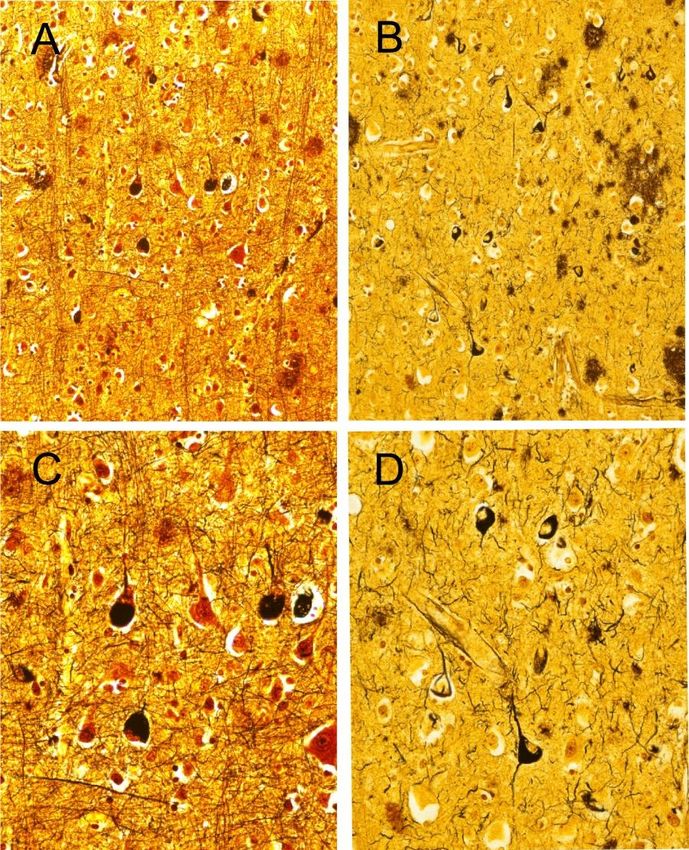
bioRxiv preprint first posted online Mar. 6, 2019; doi: http://dx.doi.org/10.1101/570093. The copyright holder for this preprint (which
       was not peer-reviewed) is the author/funder, who has granted bioRxiv a license to display the preprint in perpetuity.
                                 It is made available under a CC-BY-NC-ND 4.0 International license.

                                                                                                                                          3

      Introduction

              The Bielschowsky silver method, first published by Max Bielschowsky in 1908, has

      been the most commonly used histological stain, since the time of Alzheimer, for

      demonstrating the senile plaques and neurofibrillary tangles that define the pathology of

      the disease named for him. The evolution of subsequent modifications is described in a

      comprehensive review by Uchihara1. The method was adopted in 1991 by the Committee

      to Establish a Registry for Alzheimer’s Disease (CERAD), together with a diagrammatic

      template, to provide a standardized and reproducible density assessment for both neuritic

      plaques and neurofibrillary tangles                   2-4.   This was critical to providing a common measure

      allowing large clinicopathological correlation studies that demonstrated statistically

      significant and independent contributions of both lesion types to cognitive impairment and

      dementia 5. Most recently, an expert committee sponsored by the National Institute on

      Aging and Alzheimer’s Association (NIA-AA) have revised the histopathological

      assessment of Alzheimer’s disease (AD) by incorporating the CERAD neuritic plaque

      density assessment together with Braak neurofibrillary degeneration staging and Thal

      amyloid phases6, 7 and a subsequent study of staining methods at multiple laboratories

      has confirmed the usefulness of the Bielschowsky stain as well as its reproducibility

      between centers8. The continuing relevance of neuritic plaques, and the Bielschowsky

      stain as the method of choice for their demonstration, is also indicated by its use as the

      US Food and Drug Administration (FDA) “standard of truth” for autopsy confirmation of

      the validity of PET amyloid imaging agents                         9-11.   Many modifications of the Bielschowsky

      stain have been published but it is still known to frequently generate artifactual staining

      that at times mimics the appearance of senile plaques. This most often occurs when the

      section has few or no true plaques or neurofibrillary change. In this study we found that
bioRxiv preprint first posted online Mar. 6, 2019; doi: http://dx.doi.org/10.1101/570093. The copyright holder for this preprint (which
was not peer-reviewed) is the author/funder, who has granted bioRxiv a license to display the preprint in perpetuity.
It is made available under a CC-BY-NC-ND 4.0 International license.
4
pretreatment with oxidizing agents prior to the initial incubation in silver nitrate eliminates
these artifacts and reduces the commonly observed high background staining. This new
method may be valuable for Alzheimer’s disease (AD) researchers and
neuropathologists.
Methods
Subjects, fixation and paraffin embedding
All subjects or their legal representatives signed an Institutional Review Board-
approved informed consent. Slices of brain tissue (one cm thick) are fixed in 10% neutral-
buffered formalin for 48 hours at 4 C, dissected into standard tissue cassettes, dehydrated
in graded alcohols and xylene and then paraffin-embedded. Paraffin sections (6 m) are
deparaffinized in xylene or xylene substitute and brought through graded alcohols to
water.
A comprehensive description of the Civin Laboratory for Neuropathology, Arizona
Study of Aging and Neurodegenerative Disorders (AZSAND) and Banner Sun Health
Research Institute Brain and Body Donation Program
(www.brainandbodydonationprogram.org) is available as a free full-text document12 on
PubMedCentral.
Staining method overview
The Bielschowsky silver method was performed with “old” and “new” protocols on 6
µm sections. The old protocol was based on the most commonly-used modification,
published by Yamamoto and Hirano in 199613. Up to 24 or 48 slides (the latter when
placed in rack back-to-back) are stained in each batch, using plastic 24-slide racks andbioRxiv preprint first posted online Mar. 6, 2019; doi: http://dx.doi.org/10.1101/570093. The copyright holder for this preprint (which
was not peer-reviewed) is the author/funder, who has granted bioRxiv a license to display the preprint in perpetuity.
It is made available under a CC-BY-NC-ND 4.0 International license.
5
200 ml staining containers. All aqueous solutions are made up with reverse-osmosis-
purified water; all washes consist of reverse-osmosis purified water.
Pretreatment with oxidizing agents
The new method differs from the old protocol only in that it includes pretreatment
with 0.25% potassium permanganate (Sigma-Aldrich) and 2% oxalic acid (Sigma-
Aldrich), trialed at 2, 3 and 5 minutes for each. Slides are washed after these steps in
three successive one-minute steps with water.
Silver nitrate steps
After pretreatment and washing, slides are immersed in 150 ml of a 20% silver nitrate
(Sigma-Aldrich) solution for 15 minutes in the dark and then placed in water for five
minutes.
The same silver nitrate solution is then titrated by slowly adding 28-30% ammonium
hydroxide (Sigma-Aldrich) until the solution clears and no silver precipitate is present.
Initially 15 ml of sodium hydroxide are added, then slowly more, drop by drop, until the
precipitate that first forms disappears. Finally, 2 more drops are then added to ensure
that precipitation has completely disappeared(approximately 22 to 30 ml total). Sections
are then placed into this ammonified silver nitrate solution for 10 minutes in the dark.
Meanwhile, approximately 10 drops of ammonium hydroxide are added per 100 ml of
water to make ammoniacal distilled water. The sections are transferred into the
ammoniacal distilled water for 5 minutes prior to adding developer.bioRxiv preprint first posted online Mar. 6, 2019; doi: http://dx.doi.org/10.1101/570093. The copyright holder for this preprint (which
was not peer-reviewed) is the author/funder, who has granted bioRxiv a license to display the preprint in perpetuity.
It is made available under a CC-BY-NC-ND 4.0 International license.
6
Development and toning steps
The developing agent stock solution is composed of 0.05% formaldehyde (Fisher),
0.003% concentrated nitric acid (Sigma-Aldrich) and 14.27 M citric acid (Sigma-Aldrich).
It is made by adding 4 ml formaldehyde to 25 ml of water, then adding 10 l concentrated
nitric acid and 0.125 g citric acid, then diluting 1:10 in water. This will keep at room
temperature for up to two weeks only.
Four ml of developer stock solution are added to the ammonified silver nitrate
solution (making up 2.2% of the total volume) from the prior step and the solution quickly
stirred; the slides are then placed in this, in the dark, for 5-10 minutes until the section
turns a yellowish-tan color or until the required intensity of staining is observed ( black
fibers with a tan background). The progress may be monitored under the microscope.
Following development, the slides are washed in 3 changes of water for one minute
each, and then placed in a 5% sodium thiosulfate (Sigma-Aldrich; 7.5 g in 150 ml water)
solution for 30 seconds to remove any unreduced silver from the sections. Slides are
then given a final wash in two changes of water for 5 minutes each, dehydrated in
alcohols, cleared and coverslipped.
Comparison of old and new methods
Adjacent sections from blocks that had previously shown plaque-like artifactual
staining were stained with both old and new Bielschowsky methods and qualitatively
evaluated under the microscope. Additionally, to evaluate whether the new method
stained neuritic plaques as well as the old method, adjacent sections with abundant
plaques were compared using old and new methods.bioRxiv preprint first posted online Mar. 6, 2019; doi: http://dx.doi.org/10.1101/570093. The copyright holder for this preprint (which
was not peer-reviewed) is the author/funder, who has granted bioRxiv a license to display the preprint in perpetuity.
It is made available under a CC-BY-NC-ND 4.0 International license.
7
To quantitatively compare the ability of the new method to a different gold standard
for neuritic plaques, thioflavin S staining2, 7, adjacent or semi-adjacent sections of middle
frontal gyrus of 30 subjects with varying plaque densities from none to frequent were
assessed using the CERAD templates after staining with the new Bielschowsky method
and a standard version of the thioflavin S method 14. Semi-quantitative scores (0-3) for
both neuritic plaque and diffuse plaque density were compared using Mann-Whitney U-
tests. Spearman rank correlations were calculated, comparing each type of plaque
density estimate for both methods, i.e. the new Bielschowsky method versus the thioflavin
S method.
Results
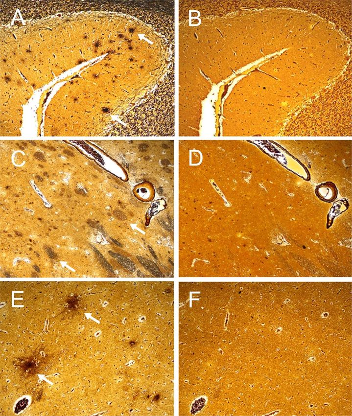
Sections of striatum and cerebellum that had previously and repetitively shown
plaque-like artifactual staining with the old method (Figure 1a, c, e) were free of artifact
when stained with the new method (Figure 1b, d, f). Pretreatment with 0.25% potassium
permanganate and 2% oxalic acid for 2 minutes each was qualitatively judged to give the
best results. Longer time trials with pretreatment solutions resulted in damage to tissue
integrity. Qualitatively, the new Bielschowsky method (Figure 2b, d) plaques were more
readily apparent than with the old method (Figure 2a, c) and background staining,
particularly that due to normal myelinated fibers, was reduced. Neurofibrillary tangles
were also more readily recognized with the new stain (Figure 3a-d), but this was not
quantitatively evaluated.
Quantitatively, plaque density estimates for either neuritic or diffuse type did not
significantly differ between the new Bielschowsky method and the thioflavin S methodbioRxiv preprint first posted online Mar. 6, 2019; doi: http://dx.doi.org/10.1101/570093. The copyright holder for this preprint (which
was not peer-reviewed) is the author/funder, who has granted bioRxiv a license to display the preprint in perpetuity.
It is made available under a CC-BY-NC-ND 4.0 International license.
8
(Table 1). Spearman correlations were greater than 0.9 and highly significant (p < 0.0001)
for both neuritic and diffuse plaques stained with the two methods.
Discussion
Pretreatment of paraffin sections with potassium permanganate and oxalic acid
eliminated troublesome artifactual staining with the Bielschowsky method. Such
artifactual staining is common in sections with little or no true AD pathology. It is possible
that the artifactual staining results from the localized presence of reducing agents in the
tissue that precipitate elemental silver. We reasoned that pretreatment with oxidizing
agents would strip electrons from these sites, preventing them from reducing silver ions
in subsequent steps. The oxidizing agents were chosen by analogy with their usage in
the Gallyas silver method1,15-17. We are not aware of any previous Bielschowsky
modifications that have employed oxidizing agent pretreatment for this purpose. Lhotka
et al (1953) experimented on the ability of five oxidizing agents to differentiate normal
from degenerating nerve fibers18. They employed varying concentrations of incubation
times with potassium permanganate, chromic acid, periodic acid, lead tetra-acetate and
sodium bismuthate, but not oxalic acid.
This modification of the Bielschowsky stain allows the confident determination of the
absence of plaques in a tissue section, by eliminating artifactual staining that mimics the
appearance of plaques. Due to reduction of the background staining from normal nerve
fibers, both plaques and tangles are more readily appreciated. As compared with the
gold standard thioflavin S method for neuritic plaques, this new Bielschowsky method
does not produce differing estimates of either neuritic or diffuse plaque densities, assuringbioRxiv preprint first posted online Mar. 6, 2019; doi: http://dx.doi.org/10.1101/570093. The copyright holder for this preprint (which
was not peer-reviewed) is the author/funder, who has granted bioRxiv a license to display the preprint in perpetuity.
It is made available under a CC-BY-NC-ND 4.0 International license.
9
that it may be substituted for the commonly-used Bielschowsky method of Yamamoto and
Hirano13.
Acknowledgements
The Civin Laboratory for Neuropathology, Arizona Study of Aging and
Neurodegenerative Disorders and Brain and Body Donation Program have been
supported by the National Institute on Aging (P30 AG19610 Arizona Alzheimer’s
Disease Core Center), the National Institute of Neurological Disorders and Stroke (U24
NS072026 National Brain and Tissue Resource for Parkinson’s Disease and Related
Disorders), the Arizona Department of Health Services (contract 211002, Arizona
Alzheimer’s Research Center), the Arizona Biomedical Research Commission
(contracts 4001, 0011, 05-901 and 1001 to the Arizona Parkinson's Disease
Consortium) and the Michael J. Fox Foundation for Parkinson’s Research.bioRxiv preprint first posted online Mar. 6, 2019; doi: http://dx.doi.org/10.1101/570093. The copyright holder for this preprint (which
was not peer-reviewed) is the author/funder, who has granted bioRxiv a license to display the preprint in perpetuity.
It is made available under a CC-BY-NC-ND 4.0 International license.
10
Table 1. Comparison of semi-quantitative estimates of neuritic and diffuse plaque
densities using the new Bielschowsky method and a standard thioflavin S method.
Means and standard deviations are given. The two methods did not differ significantly,
demonstrating that the new stain identifies both plaque types as well as a gold standard
method, the thioflavin S stain.
Neuritic Plaques Neuritic Plaques Diffuse Plaques Diffuse Plaques
New Bielschowsky Thioflavin S New Bielschowsky Thioflavin S
1.67 (1.21) 1.73 (1.26) 1.93 (1.28) 2.0 (1.36)
Mann-Whitney U-test p = 0.77 Mann-Whitney U-test p = 0.70
Spearman rho = 0.94; p < 0.0001 Spearman rho = 0.92; p < 0.0001bioRxiv preprint first posted online Mar. 6, 2019; doi: http://dx.doi.org/10.1101/570093. The copyright holder for this preprint (which
was not peer-reviewed) is the author/funder, who has granted bioRxiv a license to display the preprint in perpetuity.
It is made available under a CC-BY-NC-ND 4.0 International license.
11
Figure 1. Photomicrographs of adjacent brain sections with few or no true senile
plaques, stained with the standard Bielschowsky method of Yamamoto and Hirano 13 (A,
C, E) and the modified Bielschowsky method reported in this manuscript, using
pretreatment with potassium permanganate and oxalic acid (B, D, F). Note the
artifactual dark spots and darkly-staining normal nerve fiber bundles in the former
(arrows point to some examples) and the absence of these with the new stain.bioRxiv preprint first posted online Mar. 6, 2019; doi: http://dx.doi.org/10.1101/570093. The copyright holder for this preprint (which
was not peer-reviewed) is the author/funder, who has granted bioRxiv a license to display the preprint in perpetuity.
It is made available under a CC-BY-NC-ND 4.0 International license.
12
Figure 2. Photomicrographs of adjacent brain sections with abundant senile plaques,
stained with the standard Bielschowsky method of Yamamoto and Hirano13 (A, C) and
the modification reported in this manuscript, using pretreatment with potassium
permanganate and oxalic acid (B, D). Note that with the new method, plaques are more
readily apparent, and background staining is reduced, at both lower and higher
magnifications.bioRxiv preprint first posted online Mar. 6, 2019; doi: http://dx.doi.org/10.1101/570093. The copyright holder for this preprint (which
was not peer-reviewed) is the author/funder, who has granted bioRxiv a license to display the preprint in perpetuity.
It is made available under a CC-BY-NC-ND 4.0 International license.
13
Figure 3. Photomicrographs of semi-adjacent brain sections stained with a standard
Bielschowsky method based on that of Yamamoto and Hirano13 (A, C) and the
modification reported in this manuscript, using pretreatment with potassium
permanganate and oxalic acid (B, D). Note that with the new method, neurofibrillary
tangles and neuropil threads are more readily apparent, and background staining is
reduced, at both lower and higher magnifications.bioRxiv preprint first posted online Mar. 6, 2019; doi: http://dx.doi.org/10.1101/570093. The copyright holder for this preprint (which
was not peer-reviewed) is the author/funder, who has granted bioRxiv a license to display the preprint in perpetuity.
It is made available under a CC-BY-NC-ND 4.0 International license.
14
References
1. Uchihara T. Silver diagnosis in neuropathology: principles, practice and revised
interpretation. Acta Neuropathol 2007:113: 483-499.
2. Mirra SS, Heyman A, McKeel D et al. The Consortium to Establish a Registry for
Alzheimer's Disease (CERAD). Part II. Standardization of the neuropathologic
assessment of Alzheimer's disease. Neurology 1991:41: 479-486.
3. Hogervorst E, Barnetson L, Jobst KA, Nagy Z, Combrinck M, Smith AD.
Diagnosing dementia: interrater reliability assessment and accuracy of the
NINCDS/ADRDA criteria versus CERAD histopathological criteria for Alzheimer's
disease. Dement Geriatr Cogn Disord 2000:11: 107-113.
4. Mirra SS, Gearing M, McKeel DW, Jr. et al. Interlaboratory comparison of
neuropathology assessments in Alzheimer's disease: a study of the Consortium to
Establish a Registry for Alzheimer's Disease (CERAD). J Neuropathol Exp Neurol
1994:53: 303-315.
5. Nelson PT, Alafuzoff I, Bigio EH et al. Correlation of Alzheimer Disease
Neuropathologic changes with cognitive status: a review of the literature. J
Neuropathol Exp Neurol 2012:71: 362-381.
6. Hyman BT, Phelps CH, Beach TG et al. National Institute on Aging-Alzheimer's
Association guidelines for the neuropathologic assessment of Alzheimer's disease.
Alzheimers Dement 2012:8: 1-13.
7. Montine TJ, Phelps CH, Beach TG et al. National Institute on Aging-Alzheimer's
Association guidelines for the neuropathologic assessment of Alzheimer's disease:
a practical approach. Acta Neuropathol 2012:123: 1-11.bioRxiv preprint first posted online Mar. 6, 2019; doi: http://dx.doi.org/10.1101/570093. The copyright holder for this preprint (which
was not peer-reviewed) is the author/funder, who has granted bioRxiv a license to display the preprint in perpetuity.
It is made available under a CC-BY-NC-ND 4.0 International license.
15
8. Montine TJ, Monsell SE, Beach TG et al. Multisite assessment of NIA-AA
guidelines for the neuropathologic evaluation of Alzheimer's disease. Alzheimers
Dement 2016:12: 164-169.
9. Clark CM, Pontecorvo MJ, Beach TG et al. Cerebral PET with florbetapir compared
with neuropathology at autopsy for detection of neuritic amyloid-beta plaques: a
prospective cohort study. Lancet Neurol 2012:11: 669-678.
10. Sabri O, Sabbagh MN, Seibyl J et al. Florbetaben PET imaging to detect amyloid
beta plaques in Alzheimer's disease: phase 3 study. Alzheimers Dement 2015:11:
964-974.
11. Curtis C, Gamez JE, Singh U et al. Phase 3 trial of flutemetamol labeled with
radioactive fluorine 18 imaging and neuritic plaque density. JAMA Neurol 2015:72:
287-294.
12. Beach TG, Adler CH, Sue LI et al. Arizona Study of Aging and Neurodegenerative
Disorders and Brain and Body Donation Program. Neuropathology 2015:35: 354-
389.
13. Yamamoto T, Hirano A. A comparative study of modified Bielschowsky, Bodian
and thioflavin S stains on Alzheimer's neurofibrillary tangles. Neuropathol Appl
Neurobiol 1986:12: 3-9.
14. Vallet PG, Guntern R, Hof PR et al. A comparative study of histological and
immunohistochemical methods for neurofibrillary tangles and senile plaques in
Alzheimer's disease. Acta Neuropathol 1992:83: 170-178.
15. Braak H, Braak E. Demonstration of amyloid deposits and neurofibrillary changes
in whole brain sections. Brain Pathol 1991:1: 213-216.bioRxiv preprint first posted online Mar. 6, 2019; doi: http://dx.doi.org/10.1101/570093. The copyright holder for this preprint (which
was not peer-reviewed) is the author/funder, who has granted bioRxiv a license to display the preprint in perpetuity.
It is made available under a CC-BY-NC-ND 4.0 International license.
16
16. Gallyas F. Silver staining of Alzheimer's neurofibrillary changes by means of
physical development. Acta Morphol Acad Sci Hung 1971:19: 1-8.
17. Kuninaka N, Kawaguchi M, Ogawa M et al. Simplification of the modified Gallyas
method. Neuropathology 2015:35: 10-15.
18. Lhotka, JF, Myhre BA, Combs CM. Effects of oxidation on neurofibrillar
argyrophilia. Stain Technol 1953:28: 101-105.You can also read