A Spatiotemporal Volumetric Interpolation Network for 4D Dynamic Medical Image - Unpaywall
←
→
Page content transcription
If your browser does not render page correctly, please read the page content below
A Spatiotemporal Volumetric Interpolation Network for 4D Dynamic Medical
                                                                             Image

                                                      Yuyu Guo1 , Lei Bi2 , Euijoon Ahn2 , Dagan Feng2 , Qian Wang1* , and Jinman Kim2*
                                           1
                                               Institute for Medical Imaging Technology, School of Biomedical Engineering, Shanghai Jiao Tong
arXiv:2002.12680v2 [cs.CV] 25 Apr 2020

                                                                                      University, China
                                                                 2
                                                                   School of Computer Science, University of Sydney, Australia
                                                           {yu.guo, wang.qian}@sjtu.edu.cn, {lei.bi, euijoon.ahn, dagan.feng, jinman.kim}@sydney.edu.au

                                                                 Abstract                                      man body and are used for clinical applications, e.g., four-
                                                                                                               dimensional (4D) computed tomography (CT) for respira-
                                            Dynamic medical imaging is usually limited in appli-               tory organ motion modelling [32], 4D magnetic resonance
                                         cation due to the large radiation doses and longer image              (MR) imaging for functional heart analysis [8], and 4D ul-
                                         scanning and reconstruction times. Existing methods at-               trasound (US) for echocardiography analysis [39]. These
                                         tempt to reduce the dynamic sequence by interpolating the             4D modalities have high spatial (volumetric) and temporal
                                         volumes between the acquired image volumes. However,                  (time sequence) sampling rate to capture the periodic mo-
                                         these methods are limited to either 2D images and/or are              tion cycles of organ activities, and this information is used
                                         unable to support large variations in the motion between              for clinical decision making. However, the acquisition of
                                         the image volume sequences. In this paper, we present                 these dynamic images requires larger radiation doses which
                                         a spatiotemporal volumetric interpolation network (SVIN)              may cause harm to humans, and longer image scanning and
                                         designed for 4D dynamic medical images. SVIN introduces               reconstruction times; these factors limit the use of 4D imag-
                                         dual networks: first is the spatiotemporal motion network             ing modalities to broader clinical applications [31, 9].
                                         that leverages the 3D convolutional neural network (CNN)
                                         for unsupervised parametric volumetric registration to de-
                                         rive spatiotemporal motion field from two-image volumes;
                                         the second is the sequential volumetric interpolation net-
                                         work, which uses the derived motion field to interpolate im-
                                         age volumes, together with a new regression-based module
                                         to characterize the periodic motion cycles in functional or-
                                         gan structures. We also introduce an adaptive multi-scale
                                         architecture to capture the volumetric large anatomy mo-
                                         tions. Experimental results demonstrated that our SVIN
                                         outperformed state-of-the-art temporal medical interpola-
                                         tion methods and natural video interpolation methods that
                                         has been extended to support volumetric images. Our abla-
                                         tion study further exemplified that our motion network was
                                         able to better represent the large functional motion com-             Figure 1. The cardiac motions in two-time phases: End-Systole
                                         pared with the state-of-the-art unsupervised medical regis-           (ES) and End-Diastole (ED). The red bounding boxes highlight
                                         tration methods.                                                      the heart structure. All images showing transaxial views, cropped
                                                                                                               to enlarge the heart.

                                         1. Introduction                                                          To mitigate these factors, reducing the temporal sam-
                                                                                                               pling has been widely employed but this compromises valu-
                                            Dynamic medical imaging modalities enable the exam-                able temporal information [22, 14]. In these approaches,
                                         ination of functional and mechanical properties of the hu-            intermediary image volumes are interpolated from their ad-

                                                                                                        4321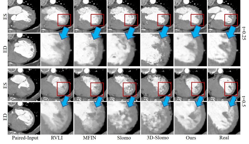
jacent volumes. Such interpolation methods are reliant on lation approaches.
either non-rigid registration [5, 27, 39] or optical flow-based
[29, 18] algorithms. Non-rigid registration approaches cal-
culate the dense image volume correspondences that oc- 2.1. Dynamic medical image interpolation
cur from one volume to another, and then uses the cal-
culated correspondences to generate the intermediary vol- Many existing medical image interpolation methods rely
umes. Such approaches, however, often generate artifacts upon optical flow-based or non-rigid registration meth-
or fuzzy boundaries and do not perform well when the vari- ods to generate a linearly interpolated image by averag-
ations in anatomy or organ activity (e.g., size and shape) ing pixel values between the adjacent image sequences
are large. An alternative approach was to use optical flow- [5, 21, 29, 27, 39, 36]. For instance, Ehrhardt et al. [13]
based methods (using deep learning) [18, 37] to estimate a presented an optical flow-based method to establish spatial
dense motion (i.e., deformation) field between image pairs. correspondence between adjacent slices for cardiac tempo-
However, these methods were limited to 2D image inter- ral image. Zhang et al. [39] used non-rigid registration-
polation and therefore did not utilize the rich spatial infor- based method to synthesize echocardiography and cardio-
mation inherent in medical image volumes. They are also vascular MR image sequences. The main advantage of these
limited when the motion between the image sequences are approaches is that they track spatiotemporal motion field,
not in linear trajectory and are not changing in a constant in a pixel-wise manner, between the neighboring images
velocity. Therefore, these approaches are not applicable to to estimate the interpolation. However, their assumption
volumetric temporal imaging modalities that exhibit large limited the spatiotemporal motion between the adjacent im-
non-linear motions in spatiotemporal space. ages to be in a linear trajectory, and thus disregarded the
In this paper we propose a spatiotemporal volumetric in- complex, non-linear motions apparent in functional organ
terpolation network (SVIN) designed for 4D dynamic med- structures. Recently, there are two CNN based methods for
ical images. To the best of our knowledge, this is the first temporal interpolation via motion field for MR images from
deep learning-based method for 4D dynamic medical im- Lin Zhang et al. [24] and Kim et al. [18]. They achieved
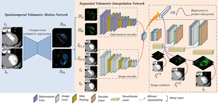
age interpolation. An overview of our model is illustrated outstanding performance compared with previous works.
in Fig. 2 which comprises of two main networks. Our first However, their method did not support full 3D volumetric
spatiotemporal motion network leverages the 3D convo- information and did not perform well when there was large
lutional neural network (CNN) for unsupervised paramet- variations in motion.
ric volumetric registration to derive spatiotemporal motion
field from two-image volumes. In the second sequential vol-
umetric interpolation network, the derived motion field is 2.2. Learning spatiotemporal motion fields from
used to interpolate the image volume, together with a new volume image sequence
regression-based module to characterize the periodic mo-
tion cycles in functional organ structures. We also propose Many studies used deformable medical image registra-
an adaptive multi-scale architecture that learns the spatial tion techniques to estimate the motion field between the in-
and appearance deformation in multiple volumes to capture put image sequences. The deformable medical image reg-
large motion characteristics. We demonstrate the applica- istration techniques can be divided into two parts: non-
tion of our method on cardiac motion interpolation, which learning based [35, 1, 19, 4, 12] and learning-based meth-
is acquired using both 4D CT and 4D MR images. These ods [20, 38, 34]. The typical non-learning based approaches
images are characterized by twisting action during contrac- are free-form deformations with B-splines [1], Demons [35]
tion to relaxation of the heart structure, and has complex and ANTs [2]. These approaches optimize displacement
changes in muscle morphology, as depicted in Fig. 1. Our vector fields by calculating the similarity of the topological
method was used to increase the temporal resolution in both structures. Deep learning-based methods, in recent years,
the CT and MR image volumes. We evaluate our method in used labelled data of spatiotemporal motion field and have
comparison to the state-of-the-art interpolation method. We shown great performances [20, 38, 34]. However, their per-
further conducted an ablation study to demonstrate the ef- formance was dependent upon the availability of large-scale
fectiveness of our motion network. labelled data. To address this, several unsupervised methods
were proposed to predict the spatiotemporal motion field
2. Related Works [11, 23, 3]. Although these methods demonstrated promis-
ing results, [11] and [23] were only useful in patch-based
We partitioned the related works into three categories volumes or in 2D slices. Jiang et al. [3] recently developed
which we deemed relevant to our research: (1) Medical dy- a CNN, VoxelMorph which used full 3D volumetric infor-
namic image interpolation; (2) spatiotemporal motion field mation. However, it was not designed for dynamic image
calculation for medical image and (3) natural video interpo- sequences where it has large variations in motion.
4322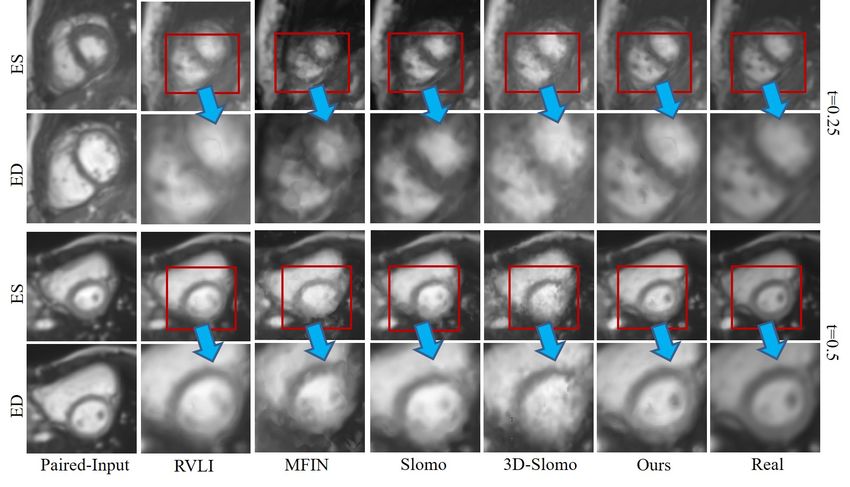
Figure 2. An overview of the proposed method which contains a motion network and an interpolation network. An adaptive multi-scale
architecure is used in both of motion and interpolation network to cover the large motion. A regression module is intergrated in our
interpolation network to constrain the intermediated motion field.

2.3. Natural video interpolation approaches                            {Ii , Ij | (i, j) ∈ T } be a pair of cardiac images indicating
                                                                       two random time points within the cardiac motion. Our aim
    Video interpolation is an active research task in natu-
                                                                       is to interpolate the intermediate image It , (t ∈ T ). For this
ral scenes, e.g., model-based tracking, patch identification,
                                                                       work, we used images at ED (denote as IED ) and ES (de-
and matching and framerate upsampling [16, 10, 26, 28].
                                                                       note as IES ) phase to interpolate the complete the cardiac
Niklaus et al. [30] developed a spatially-adaptive convo-
                                                                       motion. {φi→j , φj→i } denotes the motion field between Ii
lution kernel to estimate the motion for each pixels. Liu
                                                                       and Ij in bi-directions.
et al. [25] divided the frame interpolation into two steps,
                                                                           Fig. 2 shows the overall proposed method. Initially, spa-
optical flow estimation and image interpolation. Their net-
                                                                       tiotemporal motion network was used to learn and capture
work learnt an input pair of consecutive frames in an unsu-
                                                                       bi-directional motion fields between IED and IES in an un-
pervised manner and then refined the interpolation based
                                                                       supervised manner. Two linearly interpolated intermediate
on the outputs of the estimation. Jiang et al. [17] pre-
                                                                       images were then coarsely created using the learned spa-
sented Slomo a technique which interpolates frame motion
                                                                       tiotemporal motion fields, φED→ES and φES→ED . Using
by linearly combining bi-directional optical flows, and then
                                                                       the coarsely interpolated intermediate images and their cor-
further refining the estimated motion flow field through an
                                                                       responding deformation fields, we further refined the coarse
end-to-end CNN. Recently, Peleg et al. [33] presented a
                                                                       intermediate images by the volumetric interpolation net-
multi-scale structured architecture neural network to better
                                                                       work, where we used a regression-based module to con-
capture the local details from high resolutions frame. How-
                                                                       strain the interpolation to follow the patterns of cardiac bio-
ever, when considering the application of these methods to
                                                                       logical motion. Specifically, both of our volumetric motion
dynamic medical images interpolation, this is a challenging
                                                                       estimation and interpolation network are using an adaptive
problem as the temporal sampling in medical image volume
                                                                       multi-scale architecture which enables to capture various
sequences are much lower than that of natural scene videos.
                                                                       types motions - both small and large volume spatiotemporal
In addition, the deformation and visual differences in dy-
                                                                       deformations (see Fig. 2 and 3).
namic medical images are comparatively more complex and
non-trivial than natural scene videos.                                 3.1. Spatiotemporal volumetric motion field estima-
                                                                             tion
3. Proposed Method
                                                                           Fig. 3 presents the architecture of 3D CNN for spa-
  Let {IT , T = 1, 2..., N } be a sequence of volumetric               tiotempopral motion field estimation. We estimate a mo-
images representing the cardiac motion from end-diastole               tion field that can represent the voxel-wise motion flow of
(ED) (T = 1) to end-systole (ES) (T = N ) phase, and let               volume images at two individual time points. This can be

                                                                4323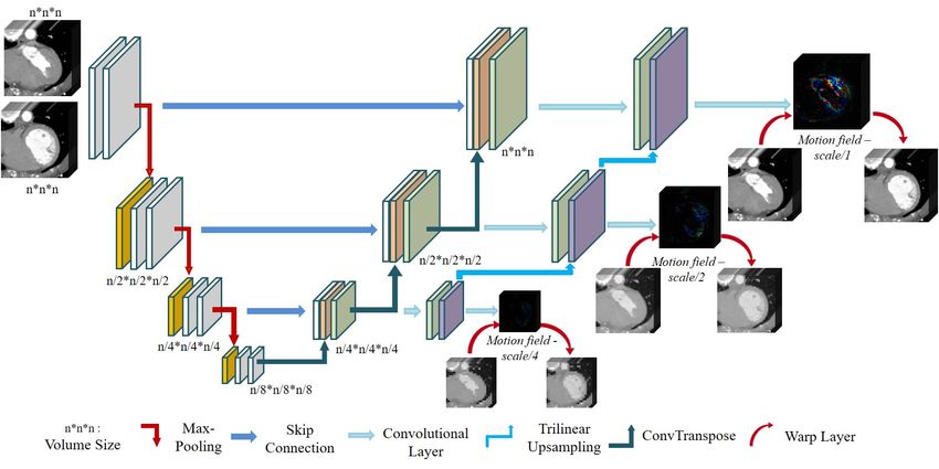
Figure 3. The architecture of our spatiotemporal volumetric motion network with an adaptive multi-scale architecture.

represented as a function Dθ (Ii , Ij ) = φi↔j (∆x, ∆y, ∆z),              in total), we define a motion field smoothness regularization
where φi↔j (∆x, ∆y, ∆z) indicates the vectors that repre-                 loss as:
sent the movement in 3D space and θ are the learnable pa-
                                                                                                               3
rameters of the network. We used an encoder-decoder archi-                                                     X
tecture with skip connections for generating φi↔j by given                            Lφ (Dθ (Ii , Ij )) =           k ∇φci→j k1           (3)
                                                                                                               c=1
Ii and Ij .
    In order to produce a volumetric motion field that can                    Where ∇ is the gradient operator. The image-wise simi-
cover various types of deformations, we propose an adap-                  larity loss was leveraged from VoxelMorph [3] and this can
tive multi-scale architecture that embeded both a global and              be defined as:
a local learning. More specifically, for global learning,
our motion field estimation network focuses on large de-                                                 3
                                                                                                         X
formation while the volumetric images in a low-scale level                    Ls (ζ(Ii |φi→j ), Ij ) =         k ζ(Iic |φci→j ) − Ijc k2   (4)
would ignore the local details while more detailed informa-                                              c=1
tion will be covered when the volumetric image in a high-
                                                                          3.2. Sequential volumetric interpolation network
scale. In addition, the global deformation from low-scale
is integrated to high-scale, which reduces the difficulty of                 Based on the derived deformation fields φED→ES and
the network for learning and constrains the network to pay                φES→ED , we used used linear interpolation approach to
more attention to detailed deformation. Our deformation                   synthesize the intermediate deformation fields, as shown in
field can be defined as:                                                  follows:

                                                                                              φED→t = tφED→ES                              (5)
      φi→j = Dθ (Ii , Ij ) or     φj→i     = Dθ (Ij , Ii )   (1)
                                                                                           φES→t = (1 − t)φES→ED                           (6)
                                                                             Based on Eqs. 3 and 4, the linear interpolation based
          Ij = ζ(Ii |φi→j ) or Ii = ζ(Ij |φj→i )             (2)          deformation field for It can be approximated as:
   where ζ(Ii )|φi→j representing the warped image by the
spatial vector field φi→j with bilinear interpolation.                        I˜t = (1 − t)ζ(IED |φED→t ) + tζ(IES |φES→t )                (7)
   For training our motion field estimation network, we
used an image-wise similarity loss and a motion field                        To improve the consistency in bi-directions, Eq. 3 and 4
smoothness regularization loss with an adaptive multi-scale               can be modified to as follows:
network architecture (as shown in Fig. 3). Given the net-
work output φni→j {n = 1, 2, 3}, where i denotes the volu-                           φED→t =t(1 − t)φED→ES
                                                                                                                                           (8)
metric images at different scales (we used 3 different scales                                   − t2 ζ(φES→ED |φES→ED )

                                                                   4324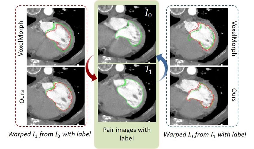
φES→t = − (1 − t)2 ζ(φED→ES |φED→ES )                               3.4. Training details for volumetric interpolation
                                                              (9)
                    + t(1 − t)φES→ED                                           For training our sequential volumetric interpolation net-
   In addition, we introduce a hyper-weight map γ to bal-                   work, our loss function is defined as a sum of an image-wise
ance the importance of using deformation from the bi-                       similarity loss Lsimilar , a regression loss Lr and a regula-
directions (forward and backward directions) and this can                   tion loss Lg :
be defined as:
                                                                                         L = λs Lsimilar + λr Lr + λg Lg            (13)
                       γES = 1 − γED                         (10)              where image-wise similarity loss Lsimilar is used to
                                                                            evaluate the similarity of the predicted synthetic interme-
   Thus, the linear image interpolation based on bi-                        diate images and the real intermediate images at multiple
directional deformation and γ can be defined as:                            image scales and is defined as:
                                                                                                      3 X
                                                                                                        N
               I˜t =(1 − t)γED ζ(IED |φED→t )                                                         X
                                                             (11)                       Lsimilar =              k I˜tck − Itck k2   (14)
                    + tγES ζ(IES |φES→t )                                                             c=1 k=1
                                                                                       P3
   As examplified in right-side of Fig. 2, we used an                          where c=1 () represents a 3-scales volumetric image
adaptive multi-scale network architecture to ensure the syn-                            N
                                                                            loss. {Itn }n=1 represents the real intermediate volumetric
thesized intermediate volumetric images will have a high                                 n oN
spatial-temporal resolution.                                                images and I˜tn        represents the predicted synthetic in-
                                                                                               n=1
                                                                            termediate volumetric images. The regression loss Lr is de-
3.3. Regression-based module for interpolation con-                         fined as the appearance difference at individual time point:
      straints
                                                                                                      N
                                                                                                      X
   Since most biological movements have a relatively fixed                                    Lr =          k t̃k − tk k1           (15)
motion pattern, especially in cardiac motion [7], we present                                          k=1
a regression-based module to model the relationship be-                       Regularization loss Lg is used to constrain the predicted
tween cardiac motion of the cardiac cycle and time phase                    motions to be consistent in bi-directions and is defined as:
(as shown in Fig. 4). Specifically, we attempted to build a
regression model representing the population-based cardiac                                    3
                                                                                              X
motion vector which indicate the shape variability at indi-                            Lg =         k ∇φcED→t + ∇φcES→t k1          (16)
vidual time point. The population-based cardiac motions at                                    c=1
individual time point was then used to constrain the appear-                  The weights λr = 1; λs = 500; λg = 50 have been set
ance of the synthetic intermediate volumetric images. Our                   empirically using a validation set.
regression estimation Rθ at time point t̃ is defined as:
                                                                            4. Experiments
            t̃ = Rθ (φED→t − tφED→ES ,
                                                             (12)           4.1. Materials and implementation details
                      φES→t − (1 − t)φES→ED )
                                                                               We demonstrate our method with two datasets: 4D Car-
                                                                            diac CT (4D-C-CT), and ACDC (4D-MR cardiac cine or
                                                                            tagged MR imaging) [6]. Fig. 5 shows a snapshot of ran-
                                                                            domly sampled cardiac sequence volume slices. The 4D-C-
                                                                            CT dataset consists of 18 patient data, each having 5 time
                                                                            points (image volumes) from ED to ES. Image volume is
                                                                            characterized by a high-resolution ranging from 0.32 to 0.45
                                                                            mm in intra-slice (x- and y-resolutions) and from 0.37mm
                                                                            to 0.82mm in inter-slice (z-resolution). The ACDC dataset
                                                                            contains 100 patients data. On average, each patient has
Figure 4. Illustration of left ventricle (LV) volume changing dur-          10.93 time points from ED to ES and it has an imaging res-
ing the cardiac contraction period. The brown curve shows the real          olution from 1.37 to 1.68 mm in x- and y-resolution and 5
motion flow of LV, and blue hidden line shows the simple linear             to 10 mm in z-resolution. All scans of 4D-C-CT were re-
assumption. The blue points and green points represent the inter-           sampled to a 128x128x96 grid and crop resulting images to
mediate time points.                                                        96x96x96. For ACDC dataset, we resampled all scans to
                                                                            160x160x10. We pad ACDC data in z-axis by 0, increasing

                                                                     4325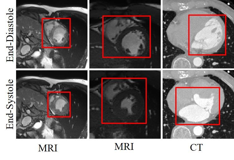
its size to 160x160x12 to reduce the border effects of 3D
convolution. We randomly selected 80 training / 20 testing
patient data and applied contrast-normalization to both of
datasets, consistent to other similar researches [15].
    We implemented all the networks using Pytorch library
and was trained on two 11GB Nvidia 1080Ti GPUs. All
models were trained with a learning rate of 0.0001. In all
our evaluations, we used 3-fold cross-validation on both the
datasets.

                                                                       Figure 6. Comparison of spatiotemporal volumetric motion esti-
                                                                       mation results. The intensity image is warped from estimated spa-
                                                                       tiotemporal motion field. The red curve represents the real seg-
                                                                       mentation results while the green color shows the warped segmen-
           Figure 5. A snapshot of our training data                   tation results (see the yellow arrows indicated). The red arrows
                                                                       indicate some organ boundaries.

4.2. Evaluation and metrics                                            Table 1. The performance of spatiotemporal motion field estima-
                                                                       tion on 4D-C-CT dataset.
    In order to evaluate the two networks in our SVIN, we
conducted an ablation study. For the unsupervised spa-                                 MSE(10−2 )    PSNR     NRMSE     SSIM      Dsc
                                                                        VoxelMorph       0.787       27.10     0.276    0.807    0.880
tiotemporal motion network, we compared it with state-                     Ours          0.197       33.17     0.138    0.918    0.944
of-the-art CNN based deformable medical image registra-
tion VoxelMorph [3]. For the interpolation network, state-
of-the-art image interpolation methods were used in the                Table 2. The performance of spatiotemporal motion field estima-
comparison including (i) RVLI [39] registration-based vol-             tion on ACDC dataset.
ume linear interpolation for medical images, (ii) MFIN [24]                            MSE(10−1 )    PSNR     NRMSE     SSIM      Dsc
CNN-based medical image interpolation (2D slice-based),                 VoxelMorph       0.194       38.06     0.132    0.912    0.920
and (iii) Slomo natural video interpolation [17] in 2D as                  Ours          0.168       38.93     0.121    0.914    0.936
well as its extension to work on medical image volumes
(3D-Slomo). For image volume interpolation, we interpo-
lated 3 intermediate volumes in between the ED-ES frames
(see Fig 5), evenly distributed across the time points.                of 33.176, NRMSE of 0.1388, SSIM of 0.9185 and MSE of
    We used the standard image interpolation evaluation                0.00197. Similarly, it also had better scores across all met-
metrics including Peak Signal-to-Noise Ratio (PSNR),                   rics on ACDC dataset. Our motion estimation architecture
Structural Similarity Index (SSIM), Mean Squared Error,                had higher improvements on 4D-C-CT dataset than that of
and Normalized Root Mean Square Error (NRMSE). We                      ACDC dataset relative to VoxelMorph. We attribute this to
used the same evaluation metrics for the spatiotemporal mo-            our robust multi-scale adaptive 3D CNN which can effec-
tion field estimation, consistent to other medical image reg-          tively learn both large and small variations in motion.
istration approaches [24]. In addition, we further used Dice               Fig. 6 shows the synthesized volumes based on the de-
Similarity Coefficient (DSC) to measure the usefulness of              rived motion field and their corresponding warped segmen-
our interpolation in medical imaging applications.                     tation results. It clearly shows that the warped segmentation
                                                                       results from the motion field learnt by our motion architec-
5. Results and Discussion                                              ture is more similar to the ground truth.

5.1. Ablation study spatiotemporal volumetric mo-                      5.2. Comparison with the state-of-the-art interpo-
      tion field estimation                                                  lation methods
    The results of motion field estimation on two datasets -              Table 3 and 4 represent the interpolation results of dif-
4D-C-CT and ACDC are shown in Table 1 and 2. Our re-                   ferent time points from ED to ES on 4D-C-CT and ACDC
sults show that motion estimation network with our adaptive            datasets, respectively. As expected, results show that the in-
architecture outperforms the recent VoxelMorph [3] across              termediate volumes that are in later time points had better
all metrics on 4D-C-CT dataset, achieving the PSNR score               performances. This is due to the fact that the earlier time

                                                                4326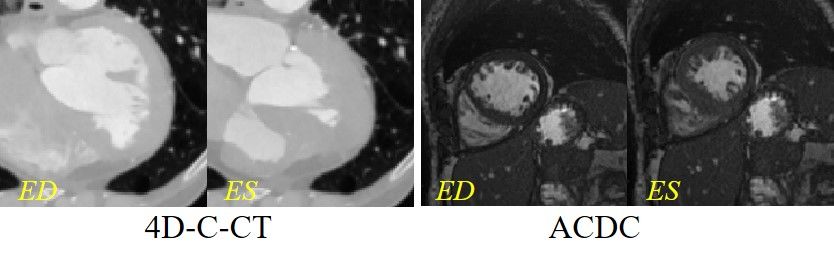
Figure 7. Visual results of two samples from 4D-C-CT. The first left column shows the paired-input volumes (ED and ES) and the last right
column shows the real intermediate volume. The rest columns show the interpolated intermediary volumes of different approaches.

points have larger motion variations, which contributed to               and learn relevant inherent functional motion patterns (see
its lower accuracy.                                                      Fig. 7 and 8). Our results show that the RVLI was the
                                                                         closest to our results. However, the RVLI was not able to
Table 3. Multi-volume cardiac sequence interpolation results on
                                                                         accurately interpolate the volumes when there were artifacts
the 4D-C-CT dataset.
                                                                         as evident in Fig. 7 and 8. MFIN and Slomo also did not
                 MSE(10−2 )        PSNR    NRMSE      SSIM               consider full 3D volumetric information, i.e., limited to 2D
    1st-point      0.45            29.45    0.211     0.830              space, which contributed to its lower scores. As expected,
    2nd-point      0.43            29.47    0.210     0.825              our implemented 3D-Slomo produced a better result rela-
    3rd-point      0.28            31.52    0.165     0.863              tive to the 2D methods. The 3D-Slomo, however, was not
                                                                         able to accurately synthesize the clear organ boundary and
                                                                         estimate the motion trajectory when there are large changes
Table 4. Multi-volume cardiac sequence interpolation results on
                                                                         of cardiac activities (see Fig. 7).
the ACDC dataset.                                                        Table 5. Performance comparisons on the 4D-C-CT dataset.
                          −2
                 MSE(10        )   PSNR    NRMSE      SSIM
    1st-point      1.22            39.34    0.109     0.934                            MSE(10−2 )    PSNR     NRMSE     SSIM      Dsc
    2nd-point      0.95            40.42    0.087     0.950                 MFIN         1.06        26.84     0.308    0.709    0.844
    3rd-point      0.28            45.86    0.052     0.977                 Slomo        1.13        26.52     0.308    0.704    0.839
                                                                           3D-Slomo      0.92        26.33     0.303    0.713    0.872
                                                                             RVLI        0.54        28.70     0.237    0.806      -
                                                                             Ours        0.39        30.15     0.196    0.840    0.917
    The comparative quantitative results for volume inter-
polation are shown in Table 5 and 6. SVIN outperformed
all other state-of-the-art interpolation method on 4D-C-CT
dataset across all measures. Similarly, it also had the best             6. Conclusion
scores across all metrics on the ACDC dataset. We attribute
                                                                         6.1. Summary
this to our adaptive multi-scale architecture capturing the
variant type of motions and regression-based module which                  We presented a novel interpolation method for 4D dy-
effectively constrains the intermediate volumetric motions               namic medical images. Our proposed two-stage network

                                                                  4327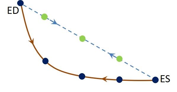
Figure 8. Visual results of two samples from ACDC. The first left column shows the paired-input volumes (ED and ES) and the last right
column shows the real intermediate volume. The rest columns show the interpolated intermediary volumes of different approaches.

    Table 6. Performance comparisons on the ACDC dataset.              plore the potential rule of functional motion. This could be
                  MSE(10−1 )    PSNR     NRMSE      SSIM               extended to the other 4D volumetric task. Also, this part
       MFIN         1.082       30.69     0.309     0.607              can be further optimized to constrain the interpolation.
       Slomo        1.001       31.08     0.296     0.630
      3D-Slomo      0.341       35.27     0.178     0.845
                                                                          Although we demonstrated our SVIN model on cardiac
        RVLI        0.331       35.66     0.173     0.860              imaging modality, there is no restriction of our method to
        Ours        0.081       41.87     0.085     0.953              be applied to other dynamic images. We suggest that our
                                                                       method is broadly applicable to other medical, and non-
                                                                       medical image volume interpolation problems where the
                                                                       motion field can be modelled.
was designed to exploit the volumetric medical images
that exhibit large variations between the motion sequences.
Experimental results demonstrated that our SVIN out-
                                                                       References
performed state-of-the-art temporal medical interpolation               [1] John Ashburner. A fast diffeomorphic image registration al-
methods and natural video interpolation methods that has                    gorithm. Neuroimage, 38(1):95–113, 2007.
been extended to support volumetric images. Our ablation                [2] Brian B Avants, Nicholas J Tustison, Gang Song, Philip A
study further exemplified that our motion network with our                  Cook, Arno Klein, and James C Gee. A reproducible eval-
adaptive multi-scale architecture was able to better repre-                 uation of ants similarity metric performance in brain image
sent the large functional motion compared with the state-                   registration. Neuroimage, 54(3):2033–2044, 2011.
of-the-art unsupervised medical registration methods.                   [3] Guha Balakrishnan, Amy Zhao, Mert R Sabuncu, John Gut-
                                                                            tag, and Adrian V Dalca. An unsupervised learning model
6.2. Extensions implementation                                              for deformable medical image registration. In Proceedings of
                                                                            the IEEE conference on computer vision and pattern recog-
   In Section 4, we discussed our general multi-scale archi-                nition, pages 9252–9260, 2018.
tecture for learning the spatial appearance volume in differ-           [4] Serdar K Balci, Polina Golland, Martha Elizabeth Shenton,
ent scales to retain the spatial information for volume syn-                and William Mercer Wells. Free-form b-spline deformation
thesis. Rather than learning a spatial transform model, in                  model for groupwise registration. Med Image Comput Com-
the future we will implement our architecture in other vol-                 put Assist Interv, 2007.
ume synthesis task.                                                     [5] Christian F Baumgartner, Christoph Kolbitsch, Jamie R Mc-
   We leverage a regression based constrain module to ex-                   Clelland, Daniel Rueckert, and Andrew P King. Group-

                                                                4328
wise simultaneous manifold alignment for high-resolution conversion using pyramid structure. IEEE Transactions on
dynamic mr imaging of respiratory motion. In International Consumer Electronics, 49(3):499–508, 2003.
Conference on Information Processing in Medical Imaging, [17] Huaizu Jiang, Deqing Sun, Varun Jampani, Ming-Hsuan
pages 232–243. Springer, 2013. Yang, Erik Learned-Miller, and Jan Kautz. Super slomo:
[6] Olivier Bernard, Alain Lalande, Clement Zotti, Freder- High quality estimation of multiple intermediate frames for
ick Cervenansky, Xin Yang, Pheng-Ann Heng, Irem Cetin, video interpolation. In Proceedings of the IEEE Conference
Karim Lekadir, Oscar Camara, Miguel Angel Gonzalez on Computer Vision and Pattern Recognition, pages 9000–
Ballester, et al. Deep learning techniques for automatic mri 9008, 2018.
cardiac multi-structures segmentation and diagnosis: Is the [18] Neerav Karani, Christine Tanner, Sebastian Kozerke, and
problem solved? IEEE transactions on medical imaging, Ender Konukoglu. Temporal interpolation of abdominal mris
37(11):2514–2525, 2018. acquired during free-breathing. In International Conference
[7] Benedetta Biffi, Jan L Bruse, Maria A Zuluaga, Hopewell N on Medical Image Computing and Computer-Assisted Inter-
Ntsinjana, Andrew M Taylor, and Silvia Schievano. in- vention, pages 359–367. Springer, 2017.
vestigating cardiac motion patterns using synthetic high- [19] Arno Klein, Jesper Andersson, Babak A Ardekani, John
resolution 3d cardiovascular magnetic resonance images and Ashburner, Brian Avants, Ming-Chang Chiang, Gary E
statistical shape analysis. Frontiers in pediatrics, 5:34, 2017. Christensen, D Louis Collins, James Gee, Pierre Hellier,
[8] Axel Bornstedt, Eike Nagel, Simon Schalla, Bernhard et al. Evaluation of 14 nonlinear deformation algorithms
Schnackenburg, Christoph Klein, and Eckart Fleck. Multi- applied to human brain mri registration. Neuroimage,
slice dynamic imaging: Complete functional cardiac mr ex- 46(3):786–802, 2009.
amination within 15 seconds. Journal of Magnetic Reso- [20] Julian Krebs, Tommaso Mansi, Hervé Delingette, Li Zhang,
nance Imaging: An Official Journal of the International So- Florin C Ghesu, Shun Miao, Andreas K Maier, Nicholas Ay-
ciety for Magnetic Resonance in Medicine, 14(3):300–305, ache, Rui Liao, and Ali Kamen. Robust non-rigid registra-
2001. tion through agent-based action learning. In International
[9] Federico Canè, Benedict Verhegghe, Matthieu De Beule, Conference on Medical Image Computing and Computer-
Philippe B Bertrand, Rob J Van der Geest, Patrick Segers, Assisted Intervention, pages 344–352. Springer, 2017.
and Gianluca De Santis. From 4d medical images (ct, [21] Gun-Ill Lee, Rae-Hong Park, Young-Seuk Song, Cheol-An
mri, and ultrasound) to 4d structured mesh models of the Kim, and Jae-Sub Hwang. Real-time 3d ultrasound fe-
left ventricular endocardium for patient-specific simulations. tal image enhancment techniques using motion-compensated
BioMed research international, 2018, 2018. frame rate up-conversion. In Medical Imaging 2003: Ultra-
[10] Byeong-Doo Choi, Jong-Woo Han, Chang-Su Kim, and sonic Imaging and Signal Processing, volume 5035, pages
Sung-Jea Ko. Motion-compensated frame interpolation us- 375–385. International Society for Optics and Photonics,
ing bilateral motion estimation and adaptive overlapped 2003.
block motion compensation. IEEE Transactions on Circuits [22] Guang Li, Deborah Citrin, Kevin Camphausen, Boris
and Systems for Video Technology, 17(4):407–416, 2007. Mueller, Chandra Burman, Borys Mychalczak, Robert W
[11] Bob D de Vos, Floris F Berendsen, Max A Viergever, Mar- Miller, and Yulin Song. Advances in 4d medical imaging
ius Staring, and Ivana Išgum. End-to-end unsupervised de- and 4d radiation therapy. Technology in cancer research &
formable image registration with a convolutional neural net- treatment, 7(1):67–81, 2008.
work. In Deep Learning in Medical Image Analysis and [23] Hongming Li and Yong Fan. Non-rigid image registra-
Multimodal Learning for Clinical Decision Support, pages tion using fully convolutional networks with deep self-
204–212. Springer, 2017. supervision. arXiv preprint arXiv:1709.00799, 2017.
[12] Ian L Dryden. Shape analysis. Wiley StatsRef: Statistics [24] Zhang Lin, Karani Neerav, Tanner Christine, and Konukoglu
Reference Online, 2014. Ender. Temporal interpolation via motion field prediction.
[13] Jan Ehrhardt, Dennis Säring, and Heinz Handels. Optical 2018.
flow based interpolation of temporal image sequences. In [25] Ziwei Liu, Raymond A Yeh, Xiaoou Tang, Yiming Liu, and
Medical Imaging 2006: Image Processing, volume 6144, Aseem Agarwala. Video frame synthesis using deep voxel
page 61442K. International Society for Optics and Photon- flow. In Proceedings of the IEEE International Conference
ics, 2006. on Computer Vision, pages 4463–4471, 2017.
[14] Kieren Grant Hollingsworth. Reducing acquisition time [26] Gucan Long, Laurent Kneip, Jose M Alvarez, Hongdong Li,
in clinical mri by data undersampling and compressed Xiaohu Zhang, and Qifeng Yu. Learning image matching by
sensing reconstruction. Physics in Medicine & Biology, simply watching video. In European Conference on Com-
60(21):R297, 2015. puter Vision, pages 434–450. Springer, 2016.
[15] Yeonggul Jang, Yoonmi Hong, Seongmin Ha, Sekeun Kim, [27] Coert T Metz, Stefan Klein, Michiel Schaap, Theo van Wal-
and Hyuk-Jae Chang. Automatic segmentation of lv and rv sum, and Wiro J Niessen. Nonrigid registration of dynamic
in cardiac mri. In International Workshop on Statistical At- medical imaging data using nd+ t b-splines and a groupwise
lases and Computational Models of the Heart, pages 161– optimization approach. Medical image analysis, 15(2):238–
169. Springer, 2017. 249, 2011.
[16] Bo-Won Jeon, Gun-Ill Lee, Sung-Hee Lee, and Rae-Hong [28] Simone Meyer, Abdelaziz Djelouah, Brian McWilliams,
Park. Coarse-to-fine frame interpolation for frame rate up- Alexander Sorkine-Hornung, Markus Gross, and Christo-
4329pher Schroers. Phasenet for video frame interpolation. In
Proceedings of the IEEE Conference on Computer Vision
and Pattern Recognition, pages 498–507, 2018.
[29] Tae-Jin Nam, Rae-Hong Park, and Jae-Ho Yun. Optical flow
based frame interpolation of ultrasound images. In Inter-
national Conference Image Analysis and Recognition, pages
792–803. Springer, 2006.
[30] Simon Niklaus, Long Mai, and Feng Liu. Video frame inter-
polation via adaptive separable convolution. In Proceedings
of the IEEE International Conference on Computer Vision,
pages 261–270, 2017.
[31] Yoshiharu Ohno, Hiroto Hatabu, Daisuke Takenaka, Shuji
Adachi, Michio Kono, and Kazuro Sugimura. Solitary pul-
monary nodules: potential role of dynamic mr imaging in
managementinitial experience. Radiology, 224(2):503–511,
2002.
[32] Tinsu Pan, Ting-Yim Lee, Eike Rietzel, and George TY
Chen. 4d-ct imaging of a volume influenced by respiratory
motion on multi-slice ct. Medical physics, 31(2):333–340,
2004.
[33] Tomer Peleg, Pablo Szekely, Doron Sabo, and Omry Sendik.
Im-net for high resolution video frame interpolation. In Pro-
ceedings of the IEEE Conference on Computer Vision and
Pattern Recognition, pages 2398–2407, 2019.
[34] Hessam Sokooti, Bob de Vos, Floris Berendsen,
Boudewijn PF Lelieveldt, Ivana Išgum, and Marius
Staring. Nonrigid image registration using multi-scale 3d
convolutional neural networks. In International Conference
on Medical Image Computing and Computer-Assisted
Intervention, pages 232–239. Springer, 2017.
[35] J-P Thirion. Image matching as a diffusion process: an
analogy with maxwell’s demons. Medical image analysis,
2(3):243–260, 1998.
[36] Erik Tryggestad, Aaron Flammang, Sarah Han-Oh, Rus-
sell Hales, Joseph Herman, Todd McNutt, Teboh Roland,
Steven M Shea, and John Wong. Respiration-based sorting
of dynamic mri to derive representative 4d-mri for radiother-
apy planning. Medical physics, 40(5):051909, 2013.
[37] Wenjun Yan, Yuanyuan Wang, Zeju Li, Rob J Van Der Geest,
and Qian Tao. Left ventricle segmentation via optical-flow-
net from short-axis cine mri: preserving the temporal coher-
ence of cardiac motion. In International Conference on Med-
ical Image Computing and Computer-Assisted Intervention,
pages 613–621. Springer, 2018.
[38] Xiao Yang, Roland Kwitt, Martin Styner, and Marc Nietham-
mer. Quicksilver: Fast predictive image registration–a deep
learning approach. NeuroImage, 158:378–396, 2017.
[39] Weiwei Zhang, J Michael Brady, Harald Becher, and J Al-
ison Noble. Spatio-temporal (2d+ t) non-rigid registration
of real-time 3d echocardiography and cardiovascular mr im-
age sequences. Physics in Medicine & Biology, 56(5):1341,
2011.
4330You can also read