In vitro multiplication, micromorphological studies and ex vitro rooting of Hybanthus enneaspermus (L.) F. Muell - a rare medicinal plant
←
→
Page content transcription
If your browser does not render page correctly, please read the page content below
Acta Bot. Croat. 77 (1), 80–87, 2018                                                                             CODEN: ABCRA 25
DOI: 10.1515/botcro-2017-0012                                                                                       ISSN 0365-0588
                                                                                                                   eISSN 1847-8476

        In vitro multiplication, micromorphological studies
        and ex vitro rooting of Hybanthus enneaspermus (L.)
        F. Muell. – a rare medicinal plant
        Mahipal S. Shekhawat1*, M. Manokari2
        1
            Biotechnology Laboratory, Department of Plant Science, M. G. G. A. C. Mahe, Puducherry, India
        2
            Department of Botany, Kanchi Mamunivar Centre for Postgraduate Studies, Puducherry, India

        Abstract – Hybanthus enneaspermus is a rare medicinal plant. We defined a protocol for micropropagation, ex
        vitro rooting of cloned shoots and their acclimatization. Surface-sterilized nodal segments were cultured on Mu-
        rashige and Skoog (MS) medium with different concentrations of 6-benzylaminopurine (BAP) and kinetin
        (Kin). Medium supplemented with 1.5 mg L–1 BAP was found optimum for shoot induction from the explants
        and 6.4±0.69 shoots were regenerated from each node with 97% response. Shoots were further proliferated max-
        imally (228±10.3 shoots per culture bottle with 7.5±0.43 cm length) on MS medium augmented with 1.0 mg L–1
        each of BAP and Kin within 4–5 weeks. The shoots were rooted in vitro on half strength MS medium containing
        2.0 mg L–1 indole-3 butyric acid (IBA). The cloned shoots were pulse-treated with 300 mg L–1 of IBA and cul-
        tured on soilrite® in a greenhouse. About 96% of the IBA-pulsed shoots rooted ex vitro in soilrite®, each shoot
        producing 12.5±0.54 roots with 5.1±0.62 cm length. The ex vitro rooted plantlets showed a better rate of survival
        (92%) in a field study than in vitro rooted plantlets (86%). A comparative foliar micromorphological study of H.
        enneaspermus was conducted to understand the micromorphological changes during plant developmental pro-
        cesses from in vitro to in vivo conditions in terms of variations in stomata, vein structures and spacing, and
        trichomes. This is the first report on ex vitro rooting in H. enneaspermus and the protocol can be exploited for
        conservation and large-scale propagation of this rare and medicinally important plant.
        Key words: conservation, ex vitro rooting, Hybanthus enneaspermus, micromorphology, micropropagation, rare
        medicinal plant

Introduction
     Hybanthus enneaspermus (L.) F. Muell. (Formerly Ionid-            burning sensations, urinary infections, leucorrhoea, dysuria
ium suffruticosum Ging.), belongs to the family Violaceae. It          and sterility (Tripathy et al. 2009). The plant is also valued
is a rare multipotent herb, endemic to the Deccan Peninsula            for its antimicrobial (Retnam and Britto 2007), antiplasmo-
in India with various invigorating properties (Prakash et al.          dial, antiarthritic (Subramoniam et al. 2013), antimalarial,
1999, Sudeesh 2012). It is a small suffrutescent perennial herb        antirheumatic, emmenagogic, sedative, antispasmodic, an-
found in India, Sri Lanka, Tropical Asia, Africa and Austra-           tiasthmatic, anti-infertility (Nathiya and Selvi 2013), anti-
lia (Anand and Gokulakrishnan 2012). The plant grows up                bacterial, anticonvulsant, antidiabetic, antifungal (Arumu-
to 15–30 cm in height with many diffused branches (Kirtikar            gam et al. 2011), anti-allergic and analgesic, antinociceptive,
and Basu 1991). H. enneaspermus is traditionally known as              antioxidant and aphrodisiac properties (Kumar et al. 2013).
Padmavati, Lakshmisheshta, Padmacharini or Purusharathna               This plant contains various important phytochemicals like
in India and considered a valuable healing herb in the Indian          dipeptide alkaloids, aurantiamide acetate, isoarborinol and
systems of medicine (Satheeshkumar 2011). It is well docu-             β- sitosterol, flavonoids, steroids, triterpenes, phenols, tan-
mented in the folklore medicine of India for its aphrodisiac           nin, glycosides etc. (Krishnamoorthy et al. 2014).
and stimulant activity (Awobajo et al. 2009). H. enneasper-                There is no commercial cultivation of H. enneaspermus
mus has therapeutic applications. Moreover, the whole plant            and the plants are collected from wild sources. It is facing
is used to treat diarrhea, painful dysentery, and strangury,           genetic threat due to sporadic distribution, poor germina-

* Corresponding author, e-mail: smahipal3@gmail.com

80                                                                                                      ACTA BOT. CROAT. 77 (1), 2018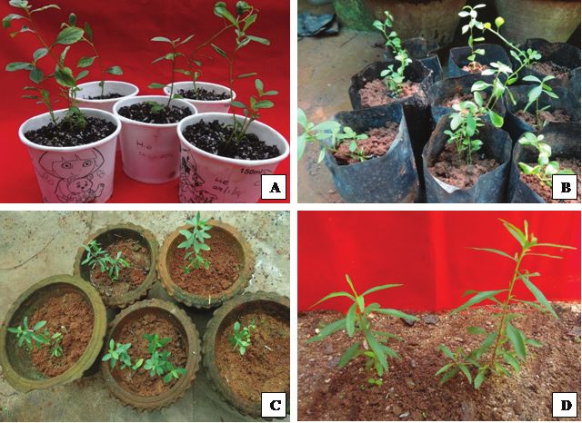
MICROPROPAGATION OF HYBANTHUS ENNEASPERMUS

tion of seeds, anthropogenic activities, overgrazing and over       as the source of explants. The nodal segments (approximate-
exploitation by herbal drug manufacturers (Arunkumar and            ly 3.0 cm in length) were harvested from two months old
Jayaraj 2011, Verma and Singh 2011). It has been disappear-         field-grown plant using sterilized surgical scissors. These ex-
ing from the large area of the Western Ghats of India due to        plants were sterilized with a systemic fungicide (0.1% Bavis-
widespread cultivation of rubber in the natural habitat of          tin; BASF India Ltd., India) and then under laminar air flow
this plant (Joseph et al. 2000). H. enneaspermus is conven-         bench with 0.1% HgCl2 (w/v) for 4–5 min. The sterilized ex-
tionally propagated through seeds. The seeds show poor vi-          plants were washed with autoclaved double distilled water
ability and germination in the wild (Arunkumar and Jayaraj          5–6 times to remove the adhered traces of HgCl2.
2011). Conventional propagation methods are unable to
meet the demand of the pharmaceutical industries and drug           Medium and culture conditions
research. Therefore, it is necessary to develop a non-conven-
                                                                        Murashige and Skoog medium (Murashige and Skoog
tional method for propagation to fulfill the demands of the
                                                                    1962) augmented with 3% sucrose as carbon source and 50
drug market (Rathore et al. 2008). In vitro propagation meth-
                                                                    mg L–1 of ascorbic acid and 25 mg L–1 each of arginine, ade-
ods offer a powerful tool for conservation of germplasm and
                                                                    nine sulphate and citric acid were incorporated in the culture
mass-multiplication of threatened plant species (Murch et al.
                                                                    medium as additives to initiate the cultures. Culture medi-
2000). They can support the in situ and ex situ conservation
                                                                    um was solidified by 0.8% Agar (Hi-Media, India) to sup-
of this rare genotype. Since natural propagation is unable to
                                                                    port the proper position of the plant material in the medium.
support the demand, in vitro methods could be viable op-
                                                                    The pH of the medium along with plant growth regulators
tions. Some in vitro work on H. enneaspermus is available in
                                                                    was adjusted to 5.8±0.02 prior to autoclaving. The cultures
the literature (Arunkumar and Jayaraj 2011, Velayutham et
                                                                    were maintained at 25±2 °C under a 12 h photoperiod light
al. 2012, Premkumar et al. 2013, Sudharson et al. 2014). The
                                                                    regime with a light intensity of 40–50 μmol m−2 s−1 photo-
present work is more effective in terms of number of mul-
                                                                    synthetic photon flux density (PPFD) implemented by cool
tiple shoots regenerated per explant.
                                                                    white fluorescent lamps (Philips, India).
     Survival of plantlets in field conditions is the major con-
straint in the micropropagation of H. enneaspermus. An ex
                                                                    Culture initiation and multiple shoot induction
vitro rooting method could help in better acclimatization
which increases the chances of field adaptation of plantlets            To establish cultures in vitro, stout, green nodal explants
in the natural environment. Improved rooting and acclima-           were inoculated on MS medium containing different con-
tization can be achieved simultaneously with ex vitro root-         centrations of cytokinins (6-benzylaminopurine, BAP; ki-
ing of in vitro propagated shoots (Baskaran and Van Staden          netin, Kin) (Hi-Media, India) ranging from 0.5–3.0 mg L–1
2013).This was found to reduce time, labor, energy involved         to induce bud break.
and the cost factor of micropropagated plantlets (Patel et al.          Cultures showing bud break were further multiplied by
2014). Therefore, the aim of the present study is to establish      subsequent transfer of in vitro regenerated axillary shoot
in vitro methodologies for mass production of this rare plant       clumps (5–7 shoots) with mother explants by subcultur-
species using ex vitro rooting and to evaluate the optimum          ing onto fresh MS medium. The medium was supplement-
conditions for in vitro development of plantlets. This is the       ed with additives, BAP and Kin (0.1 to 2.0 mg L–1) alone
first report on ex vitro rooting of in vitro regenerated shoots     or combinations of optimized concentrations. Subculturing
in H. enneaspermus.                                                 was performed at 4 weekly intervals.
     The widespread application of in vitro regeneration tech-
nologies is restricted by the difficulties during transfer of       In vitro rooting
plantlets to the field conditions (Pospíšilová et al. 1999). This
                                                                        Shoots can be harvested after 3–4 subcultures for root-
is due to the sudden change in the culture environment to
                                                                    ing experiments. For root induction under in vitro condi-
relatively harsh environments. The ultimate success of micro-
                                                                    tions, multiplied shoots longer than 4–5cm were separated
propagation depends on successful hardening and field trans-
                                                                    individually from shoot clumps and transferred to different
fer of plantlets. The plants micropropagated in a culture vessel
                                                                    strengths of MS media (full MS, ½ MS and ¼th MS) with
are partially heterotrophic; they acquire some developmen-
                                                                    various concentrations of auxins (indole-3 acetic acid, IAA;
tal changes to make them fully autotrophic after being trans-
                                                                    indole-3 butyric acid, IBA; α-naphthalene acetic acid, NAA
ferred to the field. The present study also aimed to investi-
                                                                    and naphthoxy acetic acid, NOA) (Hi-Media, India) (1.0 to
gate the foliar epidermal micromorphological changes during
                                                                    4.0 mg L–1). The cultures were initially incubated under dif-
transfer of plantlets from an in vitro to a field environment.      fused light conditions (20–25 μmol m−2 s−1) for 2–3 days for
                                                                    in vitro root induction and thereafter transferred to an in vi-
Materials and methods                                               tro culture environment, and maintained at a light intensity
Plant material and surface sterilization                            of 40–45 μmol m−2 s−1 PPFD with a 12 h photoperiod per day.

    Hybanthus enneaspermus was selected from the Coro-
                                                                    Ex vitro rooting of in vitro regenerated shoots
mandel Coast (Kanchipuram, Villupuram, Puducherry,
Cuddalore, Nagapattinam and Karaikal districts) of India for           Experiments were carried out for ex vitro root induction
the present study. Slender young emerging stems were used           from in vitro-produced shoots. Basal end (4–6 mm) of in vi-

ACTA BOT. CROAT. 77 (1), 2018                                                                                                   81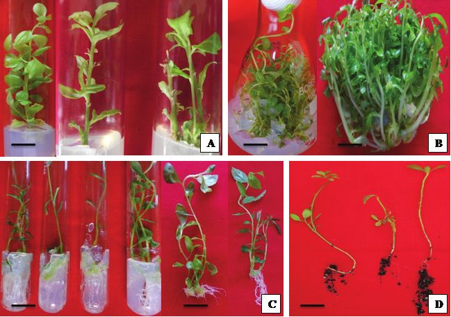
SHEKHAWAT M. S., MANOKARI M.
tro-raised shoots were treated with different concentrations ysis of variance and the significance of differences was cal-
of auxins (IAA, IBA, NAA and NOA) (50–400 mg L–1) for 5 culated by Duncan’s multiple range test using SPSS software
min and transferred to eco-friendly paper cups containing (version 16.0). Observations were noted at 4 weeks interval.
sterile soilrite® (a combination of perlite with peat moss and
exfoliated vermiculite procured from KelPerlite, Bangalore, Results
India) and moistened with one fourth strength of MS basal Establishment of cultures and multiplication of cultures
salts. The cups were kept in the greenhouse for maintenance in vitro
at 25±2 °C with 80–90% relative humidity (RH). After 4–5
weeks, the rooted plantlets were carefully taken out from The fresh and light green colored nodal segments re-
the paper cups and transplanted to the nursery polybags in sponded better than old and dark colored explants. Cultures
a greenhouse. were initially placed in diffused light (20–25 μmol m−2 s−1)
to induce bud breaking, and further transferred to a culture
Acclimatization and field transfer of regenerated room with a higher light intensity (40–50 μmol m−2 s−1) for
plantlets proper establishment of culture. The MS medium supple-
mented with 1.5 mg L–1 BAP was observed suitable for bud
In vitro rooted plantlets were taken out cautiously from breaking and 97% of the explants responded with 6.4±0.69
the culture tubes and rinsed with distilled water to remove shoots from each nodal explants (Fig. 1A, Tab. 1). Maximum
adhered nutrients and agar. They were transferred to auto-
claved soilrite® in bottles, moistened with 1/4th strength of
MS basal salts and maintained in the greenhouse. The ex vi-
tro rooted plantlets were acclimatized in paper cups which
were covered with transparent polythene cups to provide
enough space for gas exchange. The in vitro rooted plantlets
were acclimatized by gradual loosening and then complete-
ly removing the transparent cup of the bottles. These plant-
lets were subsequently transferred to nursery polybags con-
taining soilrite®, garden soil and organic manure (1:1:1) in
the greenhouse for further acclimatization process. Plantlets
were transferred to the field after 5 weeks of acclimatization.
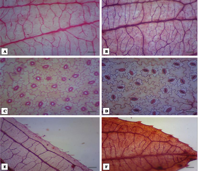
Foliar micromorphological studies of in vitro and field
transferred plantlets
Fig. 1. Micropropagation of Hybanthus enneaspermus: induction
Experiments were conducted to study foliar micromor- of shoots from the nodal segments with 6-benzylaminopurine (A);
phological developments of veins (vein density and venation multiplication of shoots on MS medium (B); in vitro rooting of the
pattern), stomata types and density, and trichomes in leaves excised shoots on MS medium with indole-3 butyric acid (C); ex
of plants grown in vitro after 4th subculture in multiplica- vitro rooted plantlets (D), scale bars = 2 cm.
tion phase and in those transferred to the field after 6th week.
Plants were randomly selected from both the environments. Tab. 1. Effect of different concentrations of cytokinins, 6-benzyl-
The entire foliar apparatus (leaves) (10 from each stage of aminopurine (BAP) and kinetin (Kin) on bud breaking from nodal
plantlets) third to seventh leaves from the base were excised explants of Hybanthus enneaspermus. Significant variation between
manually for all the experiments. To observe the changes in the concentrations was studied using Duncan’s multiple range test
structure and functioning of developing stomata, epidermal at 0.5% level, SD – standard deviation.
peels were separated manually by the traditional method (Jo- Cytokinins Response Number of shoots Shoot length (cm)
hansen 1940) from the leaves. The leaves were fixed primar- (mg L–1) (%) (mean ± SD) (mean ± SD)
ily in formalin–acetic acid–ethyl alcohol, FAA (1:1:3) and 0.00 0 0.0±0.00a 0.00±0.00a
cleared in 70% ethanol (v/v) until the chlorophyll was re- BAP
moved (12–24 h), bleached with 5% (w/v) NaOH for 24–48 0.50 32 2.3±1.06abc 3.12±0.31bcd
h, and rinsed three times in distilled water (Sass 1940). The 1.00 56 4.2±0.28bcd 3.40±0.18cd
1.50 97 6.4±0.69d 5.60±0.49e
leaves were then stained with 1% safranine (Loba chemie, In-
2.00 76 4.9±1.02cd 4.16±0.38de
dia) aqueous solution for 4–8 min and rinsed carefully in wa- 2.50 50 4.1±0.57bcd 3.28±0.33cd
ter to remove excess stain and then mounted in distilled water 3.00 37 2.3±1.09abc 2.06±0.12bc
and examined under microscope (Labomed iVu 3100, USA). Kin
0.50 26 2.1±0.54abc 1.32±0.43ab
Experimental design, data collection and statistical 1.00 41 2.9±0.76bc 2.07±0.64bc
analysis 1.50 68 3.0±0.43ab 2.78±0.21bcd
2.00 59 3.6±0.47bc 3.08±0.30bcd
The experiments were performed with 20 replicates per 2.50 50 2.0±0.53ab 3.12±0.36bcd
treatment and repeated thrice. Data were subjected to anal- 3.00 40 2.1±0.49ab 2.17±0.17bc
82 ACTA BOT. CROAT. 77 (1), 2018MICROPROPAGATION OF HYBANTHUS ENNEASPERMUS
response of the explants was observed on MS medium aug- Tab. 3. Effect of auxins indole-3 acetic acid (IAA), indole-3 butyric
mented with BAP rather than with Kin in present study. acid (IBA), α-naphthalene acid acid (NAA), and naphthoxy acetic
acid (NOA) on in vitro root induction, number and length of roots
The maximum number of shoots (228±10.3 shoots with on half strength MS medium. Significant variation between the
7.5±0.43 cm length) was regenerated on a subculture of the concentrations was studied using Duncan’s multiple range test at
in vitro regenerated shoot clumps on MS medium fortified 0.5% level, SD – standard deviation.
with 1.0 mg L–1 each of BAP and Kin within 4–5 weeks (Tab. Auxins Response Number of roots Length of root (cm)
2). The shoot number and shoot length was increased by (mg L–1) (%) (mean±SD) (mean±SD)
repetitive subculturing up to 3–4 passages onto fresh me- 0.00 0 0.00±0.00a 0.0±0.00a
dium (Fig. 1B). Six fresh shoots from a single explant trans- IAA
ferred to fresh medium yielded a maximum of 228 shoots 1.0 26 12.9±0.16d 2.98±0.83h
(228/6=38) within 4–5 weeks. The rate of shoot multiplica- 2.0 40 16.6±10.75 h
3.00±0.38h
tion increased more than 35 fold in 4–5 weeks. Cream col- 3.0 34 16.0±1.56 gh
2.35±0.33f
ored callus was observed from the basal part of the cultures 4.0 31 14.8±0.40 f
2.05±0.20de
if the medium was augmented with auxins (IAA and IBA) IBA
along with cytokinins. 1.0 63 18.5±3.43i 2.15±0.26e
2.0 98 25.7±3.90 k
6.21±0.78k
3.0 71 21.0±2.37 j
4.18±0.39j
In vitro rooting of the shoots
4.0 59 13.7±1.70 e
3.24±0.49i
Among the different strengths of MS medium and aux- NAA
ins experimented for in vitro root induction, ½ strength MS 1.0 29 10.5±0.80b 1.78±0.13c
medium supplemented with 2.0 mg L–1 IBA was observed 2.0 33 13.6±0.75 de
2.90±0.23h
best for in vitro rooting (Fig. 1C). Maximum number of roots 3.0 49 15.9±0.23 gh
1.90±0.91cd
(25.7±3.90) with highest length (6.21±0.78 cm) were record- 4.0 32 11.5±0.10 c
1.95±0.26cd
ed with this medium combination (Tab. 3). Poor response NOA
with smaller root number and root length was reported on 1.0 30 11.9±1.43c 2.71±0.83g
media fortified with IAA, NAA and NOA. 2.0 42 13.6±1.90de 3.32±0.38i
3.0 54 15.3±2.37fg 2.93±0.33h
4.0 50 10.5±1.20b 1.43±0.20b
Tab. 2. Effect of different concentrations and combinations of cy-
tokinins 6-benzylaminopurine (BAP), kinetin (Kin), and combina- Ex vitro rooting of in vitro produced shoots
tion of BAP + Kin on shoot multiplication of Hybanthus enneasper-
The basal end of in vitro-produced micro-shoots was
mus. Significant variation between the concentrations was studied
using Duncan’s multiple range test at 0.5% level, SD – standard treated (5 min) with root-inducing growth regulators and
deviation. transferred to a greenhouse environment. The lower part (4–
6 mm) of in vitro-regenerated shoots evaluated with 300 mg
Cytokinins Number of shoots Shoot length (cm)
(mg L–1) (mean±SD) (mean±SD)
L–1 IBA exhibited about 96% rooting (Tab. 4). A maximum
of 12.5±0.54 roots per shoot with 5.10±0.62 cm length was
0.00 0.0±0.00a 0.00±0.00a
observed within 4 weeks (Fig. 1D). Poorer rooting than with
BAP
IBA was recorded with all the concentrations of IAA, NAA,
0.10 14±0.10ab 4.0±0.54b and NOA in this study.
0.50 25±0.93abc 6.4±0.32hi
1.00 42±1.51d 6.6±0.23i Acclimatization and field transfer of regenerated
1.50 40±7.37d 5.3±0.17ef plantlets
2.00 34±6.24cd 6.4±0.62hi
In vitro and ex vitro rooted plants were acclimatized effi-
Kin
ciently in a greenhouse (Figs. 2A–2C). A profusely branched
0.10 19±8.91bc 5.0±0.44de
root system was observed in ex vitro rooted plantlets during
0.50 22±6.74bc 4.3±0.33bc transfer to the field. It resembled the conventional root sys-
1.00 36±5.13cd 4.6±0.63cd tem obtained under natural conditions. The hardened plant-
1.50 29±7.11abc 5.1±1.70def lets were successfully transferred to the field with 92% sur-
2.00 20±8.83bc 4.9±0.54de vival rate (Fig. 2D) but the survival rate of in vitro rooted
BAP + Kin plantlets was only 86%.
0.10 98±8.76e 5.6±1.30fg
0.50 172±9.27g 5.9±1.05gh Micromorphological studies of micropropagated
1.00 228±10.3h 7.5±0.43j plantlets
1.50 121±10.1f 6.1±0.79ghi The plants developed under in vitro conditions possessed
2.00 93±9.22e 5.1±0.43def normal leaves with hairs and denticulate margins. The mid-
ACTA BOT. CROAT. 77 (1), 2018 83SHEKHAWAT M. S., MANOKARI M.
rib was fairly prominent projecting equally on both the sides,
but bluntly conical on the adaxial side and hemispherical
on the abaxial side. Veins and vein-islets were fewer in in vi-
tro than in field transferred plantlets (Figs. 3A and 3B). The
vein density and distinct vein-islets were increased during
the hardening period, and became distinct, rhomboidal and
rectangular in shape after field transfer of the plantlets.
The stomata were more frequent in the inter-coastal ar-
eas than in the coastal areas, facing all directions with ir-
regular distribution. The stomatal frequency was greater in
the in vitro environment than in the field transferred plants
with anisocytic stomata predominating, but these were non-
functional as they were always in open condition (Figs. 3C
and 3D). Anisocytic (cruciferous), paracytic (rubiaceous),
Fig. 2. Hardening of Hybanthus enneaspermus plantlets in the diacytic (caryophyllaceous), anomocytic (rununculaceous),
greenhouse (A-C), and in vitro raised plantlets under field condi- anisotricytic, isotricytic, tetracytic, staurocytic, desmocyt-
tions (D). ic and pericytic stomata were observed in the in vitro pro-
duced leaves. Anomocytic and pericytic stomata were oc-
casionally observed in these leaves. The field-transferred
plants possessed the aforementioned stomatal types except
Tab. 4. Effect of auxins indole-3 acetic acid (IAA), indole-3 butyric for the anomocytic and pericytic. Anisocytic and paracytic
acid (IBA), α-naphthalene acid acid (NAA), and naphthoxy acetic
stomata were prominent but diacytic and desmocytic sto-
acid (NOA) on ex vitro root induction from in vitro raised shoots.
mata were rare in this plant. Trichomes were simple, unicel-
Significant variation between the concentrations was studied using
Duncan’s multiple range test at 0.5% level, SD – standard deviation. lular or uniseriate emerging from the epidermis. Under in
vitro conditions, the trichomes were unicellular, less frequent
Auxins Response Number of roots Length of root (cm) and underdeveloped but these were fully developed in field
(mg L–1) (%) (mean±SD) (mean± SD)
transferred plants after 6 weeks (Fig. 3E). The uniseriate and
0.00 2 0.43±0.13a 1.21±0.32b unicellular hairs were frequent but the bicellular and tricel-
IAA lular hairs were occasionally observed after plantlets were
50 30 0.45±0.30a 1.19±0.54b transferred to the field (Fig. 3F). Adaxial surface possessed
numerous shaggy trichomes, and the trichome density was
100 45 0.62±0.11e 2.56±0.36h
found maximum in field transferred plantlets compared to
200 51 1.93±0.15f 4.01±0.44m
300 50 3.22±0.32 f
3.92±0.19l
400 43 2.07±0.28cd 2.05±0.30g
IBA
50 35 0.96±0.21c 3.15±0.73k
100 42 2.78±0.59 h
3.84±0.61l
200 56 4.89±0.41i 4.10±0.49n
300 96 12.5±0.54l 5.10±0.62p
400 84 5.83±0.49j 4.25±0.53o
NAA
50 23 0.39±0.33a 1.03±0.69a
100 35 0.73±0.21b 1.41±0.74d
200 47 2.73±0.19h 2.05±0.21g
300 44 6.21±0.48l 3.91±0.28l
400 39 6.10±0.30 k
3.01±0.35j
NOA
50 33 0.11±0.45c 1.33±0.30c
100 47 0.29±0.29d 1.49±0.49e Fig. 3. Micromorphological studies of Hybanthus enneaspermus:
venation pattern in leaves of in vitro shoots (A); and field plant
200 56 2.39±0.16g 1.90±0.24f (B); stomatal pattern in leaves of in vitro shoots (C), and field plant
300 41 3.26±0.72h 2.94±0.92j (D); and trichomes in leaves of in vitro shoots (E), and field plant
(F). Tissues were stained with 1% safranine aqueous solution. Scale
400 38 2.30±0.64d 2.70±0.62i
bars = 100 µm.
84 ACTA BOT. CROAT. 77 (1), 2018MICROPROPAGATION OF HYBANTHUS ENNEASPERMUS
the in vitro grown leaves. Mucilaginous cells were also ob- 2015a). About 96% shoots were rooted with IBA with maxi-
served in field transferred plants but these were totally absent mum 12.5 roots per shoot in this report. IBA is more effec-
in the in vitro grown plantlets. tive than NAA and NOA in ex vitro root induction in many
plant species, and applied economically worldwide (Debergh
Discussion et al. 1992, Yan et al. 2010, Ranaweeraa et al. 2013, Shekha-
wat et al. 2015b). This is the first report on ex vitro rooting
The success of tissue culture experiments basically de- of in vitro regenerated shoots in H. enneaspermus with maxi-
pends on the selection of starting material. The mature ex- mum rate of survival under natural conditions. The rooted
plants responded later than the fresh and light green col- plantlets were hardened in greenhouse with development of
ored nodal segments under diffused light conditions in the profusely branched root system in ex vitro rooted plantlets.
present experiment. BAP induced more shoots on MS me- About 92% ex vitro rooted and 86% in vitro rooted plantlets
dium than Kin. Similar results were reported by many re- survived in the field conditions. Ex vitro rooting reduced the
searchers recently in a number of plant species (Panwar et time, energy of production of plantlets and mortality during
al. 2012, Premkumar et al. 2013, Rathore et al. 2013a, Sud- hardening and field transfer. Normal flowering and fruiting
harson et al. 2014). Shoot multiplication was achieved by re- was observed in the field transferred plantlets.
petitive transfer of mother explants with regenerated shoots
These micromorphological studies of micropropagated
onto fresh medium and by subculturing of freshly regener-
plantlets were performed to understand the developmental
ated shoots isolated from the mother explants. This approach
changes in the leaves of plantlets, when they were transferred
of shoot multiplication has been used in several plant spe-
to field conditions. The stomata were present on both surfac-
cies (Rai et al. 2010, Patel et al. 2014, Shekhawat and Mano-
es of the leaf but the frequency was less on the adaxial sur-
kari 2016). The higher rate of shoot multiplication during face (Narayanaswamy et al. 2006, Retnam and Britto 2007)
repeated transfer may be due to inhibition of apical domi- therefore, the abaxial surface was further considered for the
nance which stimulates the basal dormant meristematic cells study. The specific artificial conditions in vitro are respon-
to produce young shoots (Phulwaria et al. 2013). A maxi- sible for the structural changes occurring in micropropagat-
mum of 228 shoots were induced per culture vessel within ed plantlets. The lesser stomatal density under field condi-
4–5 weeks in this study. Premkumar et al. (2013) induced tions may help to check the rate of transpiration and prevent
the most (52.3) shoots, when the regenerated shoots were water loss (Singh et al. 2003). Transitional types of stomata
subcultured on MS medium containing IAA along with Kin between anisocytic and paracytic are also present in H. en-
and BAP. Contrary to this report, callus formation was ob- neaspermus (Inamdar 1969). Anisotricytic and isotricytic
served when the medium was supplemented with IAA and stomata could be the transitional form between anisocyt-
IBA along with BAP and Kin in the present study. Sudhar- ic and paracytic types of stomata. Unicellular, less frequent
son et al. (2014) reported maximum of 11.8 shoots, when the and underdeveloped trichomes were observed under in vitro
cultures of H. enneaspermus were inoculated on MS medium conditions but fully developed trichomes were reported in
supplemented with 2.0 mg L–1 BAP. Maximal 90 shoots were field-transferred plants. The mucilaginous cells were not ob-
reported in this plant by Velayutham et al. (2012) from the served with the in vitro leaves but found in field-transferred
callus cultures on MS medium augmented with BAP and plants. Our findings are supported by the results of various
Kin. This supports our findings where the most shoots were researchers (Chandra et al. 2010, Rathore et al. 2013b, Lodha
regenerated on BAP and Kin, but our results were far better et al. 2015). Understanding the changes in foliar micromor-
than the earlier reports in multiple shoot formation. phology of in vitro grown and hardened plantlets could be
The shoots were rooted maximally on half strength MS useful in improvement of in vitro clonal propagation proto-
medium augmented with IBA. The half strength MS salts cols and for large scale production of plants.
and sucrose in medium was appropriate for in vitro root-
ing and supports many authors’ findings in different plant
Conclusion
species (Rai et al. 2010, Premkumar et al. 2013, Patel et al.
2014). We report more roots (25.7±3.90) per shoot in this An efficient in vitro propagation protocol has been de-
study than were found in earlier works on H. enneaspermus. veloped using various plant growth regulators for successful
Maximum 2.8 roots per shoot was reported by Prakash et conservation of this rare plant species. Excellent rate of shoot
al. (1999), 5–8 roots by Velayutham et al. (2012) and 21.3 multiplication was achieved in vitro. Ex vitro rooting has
roots by Premkumar et al. (2013) in this plant species. The been successfully demonstrated in H. enneaspermus, which
superiority of IBA over other auxins for root induction has could save time, labor and energy in production of plantlets.
been recognized by several researchers in a number of plants The hardened plantlets were successfully transferred to the
(Barreto and Nookaraju 2007, Rai et al. 2010, Rathore et al. field with a 92% survival rate. The results of foliar micromor-
2013b). Plants rooted under ex vitro environment were bet- phological studies could help in understanding the response
ter suited to natural conditions and reported easy to hard- of plantlets under field conditions. The data could contribute
en (Yan et al. 2010). It has been reported that ex vitro root- significantly to meeting the market demand for this multipo-
ed plants are better suited to tolerate environmental stresses tent healing herb and conservation of this valuable genotype
(Pospíšilová et al. 1999, Tiwari et al. 2002, Shekhawat et al. through biotechnological interventions.
ACTA BOT. CROAT. 77 (1), 2018 85SHEKHAWAT M. S., MANOKARI M.
Acknowledgements ment of Science, Technology and Environment, Government
of Puducherry for providing financial support to their labo-
The authors are grateful to the University Grants Com- ratory as Major Research Project and Grant–In-Aid Scheme
mission, New Delhi, Government of India and the Depart- respectively.
References
Anand, T., Gokulakrishnan, K., 2012: Phytochemical analysis of Narayanaswamy, V. B., Kumar, D. C., Setty, M. M., Shirwaikar,
Hybanthus enneaspermus using UV, FTIR and GC-MS. IOSR A., 2006: Histological and physico-chemical evaluation of Hy-
Journal of Pharmacy 2, 520–524. banthus enneaspermus (L.) F. Muell. Natural Products Science
Arumugam, N., Sasikumar, K., Malipeddi, H., Sekar, M., 2011: 12, 104–108.
Antifungal activity of Hybanthus enneaspermus on wet Nathiya, S., Selvi, S. R., 2013: Anti-infertility effect of Hybanthus
clothes. International Journal of Research in Ayurveda and enneaspermus on endosulfan induced toxicity in male rats.
Pharmacy 2, 1184–1185. International Journal of Medicine and Biosciences 2, 28–32.
Arunkumar, B. S., Jayaraj, M., 2011: Rapid In vitro callogenesis Panwar, D., Ram, K., Harish, Shekhawat, N. S., 2012: In vitro prop-
and phytochemical screening of leaf and leaf callus of Ionidi- agation of Eulophia nuda Lindl. – an endangered orchid. Sci-
um suffruticosum, Ging. – A seasonal multipotent medicinal entia Horticulturae 139, 46–52.
herb. World Journal of Agricultural Sciences 7, 55–61. Patel, A. K., Phulwaria, M., Rai, M. K., Gupta, A. K., Shekhawat,
Awobajo, F. O., Olatunji-Bello, I. I., Adegoke, O. A., Odugbemi, T. S., Shekhawat, N. S., 2014: In vitro propagation and ex vitro
O., 2009: Phytochemical and antimicrobial screening of Hy- rooting of Caralluma edulis (Edgew.) Benth. & Hook. f.: an en-
banthus enneaspermus and Paquentina nigricense. Recent Re- demic and endangered edible plant species of the Thar Desert.
search in Science and Technology 1, 159–160. Scientia Horticulturae 165, 175–180.
Barreto, M. S., Nookaraju, A., 2007: Effect of auxin types on in Phulwaria, M., Rai, M. K., Patel, A. K., Kataria, V., Shekhawat, N.
vitro and ex vitro rooting and acclimatization of grapevine S., 2013: A genetically stable rooting protocol for propagat-
as influenced by substrates. Indian Journal of Horticulture ing a threatened medicinal plant Celastrus paniculatus. AoB
64, 5–11. Plants 5, pls054.
Baskaran, P., Van Staden, J., 2013: Rapid in vitro micropropaga- Pospíšilová, J., Ticha, I., Kadleck, R., Haisel, D., Plzakova, S., 1999:
tion of Agapanthus praecox. South African Journal of Botany Acclimatization of micropropagated plants to ex vitro condi-
86, 46–50. tions. Biologia Plantarum 42, 481–497.
Chandra, S., Bandopadhyay, R., Kumar, V., Chandra, R., 2010: Ac- Prakash, E., Sha Valli, K. P. S., Sairam, R. P., Rao, K. R., 1999: Re-
climatization of tissue cultured plantlets: from laboratory to generation of plants from seed-derived callus of Hybanthus
land. Biotechnology Letters 32, 1199–1205. enneaspermus L. Muell., a rare ethnobotanical herb. Plant Cell
Debergh, P., Aitken-Christie, J., Cohen, D., Grout, B., Arnold von, Reports 18, 873–878.
S., Zimmerman, R., Ziv, M., 1992: Reconsideration of the term Premkumar, G., Arumugam N., Muthuramkumar, S., Varathara-
vitrification as used in micropropagation. Plant Cell Tissue ju, G., Rajarathinam, K., 2013: Improved micropropagation in
and Organ Culture 30, 135–140. Hybanthus enneaspermus L. Muell. American Journal of Plant
Inamdar, J. A., 1969: Epidermal structure and development of sto- Sciences 4, 1169–1172.
mata in vegetative and floral organs of Hybanthus enneasper- Rai, M. K., Asthana, P., Jaiswal, V. S., Jaiswal, U., 2010: Biotechno-
mus (Linn.) F. Muell. Biologia Plantarum 11, 248–255. logical advances in guava (Psidium guajava L.). Recent devel-
Johansen, D. A., 1940: Plant microtechnique. McGraw Hill Co., opments and prospects for further research. Trees Structure
New York. and Function 24, 1–12.
Joseph, T. S.,. Skaria, B. P., Sajithakumari, 2000: Disappearing me- Ranaweeraa, K.K., Gunasekarab, M. T. K., Eeswara, J. P. 2013: Ex
dicinal plant resources of Kottayam district of Kerala State. In- vitro rooting: a low cost micropropagation technique for tea
dian Journal of Areca Nut, Spices and Medicinal Plants 2, 79–81. (Camellia sinensis (L.) O. Kuntz) hybrids. Scientia Horticul-
Kirtikar, K. R., Basu, B. D., 1991: Indian medicinal plants. Vol I. turae 155, 8–14.
Periodical Experts Book Agency, Delhi, India. Rathore, M. S., Dagla, H. R., Singh, M., Shekhawat, N. S., 2008:
Krishnamoorthy, B. S., Nattuthurai, Logeshwari, R., Dhaslima, N., Rational development of in vitro methods for conservation,
Syedali, H., Fathima, I., 2014: Phytochemical study of Hyban- propagation and characterization of Caralluma edulis. World
thus enneaspermus (Linn.) F. Muell. Journal of Pharmacogno- Journal of Agricultural Science 4, 121–124.
sy and Phytochemistry 3, 6–7. Rathore, M. S., Rathore, M. S., Shekhawat, N. S., 2013a: Ex vivo im-
Kumar, S. B., Vijaya Kumar, J., Selvaraj, R., 2013: Aphrodisiac ac- plications of phyto-hormones on various in vitro responses in
tivity of Cycas circinalis and Ionidium suffruticosum Ging. on Leptadenia reticulata (Retz.) Wight. and Arn. – an endangered
male wister albino rat. Asian Journal of Pharmaceutical and plant. Environmental and Experimental Botany 86, 86–93.
Clinical Research 6, 215–217. Rathore, N. S., Rathore, N., Shekhawat, N. S., 2013b: In vitro prop-
Lodha, D., Pate, A., Shekhawat, N. S., 2015: A high-frequency in agation and micromorphological studies of Cleome gynandra:
vitro multiplication, micromorphological studies and ex vitro a C4 model plant closely related to Arabidopsis thaliana. Acta
rooting of Cadaba fruticosa (L.) Druce (Bahuguni): a multi- Physiologiae Plantarum 9, 2691–2698.
purpose endangered medicinal shrub. Physiology and Mo- Retnam, R. K., Britto, A. J. de, 2007: Antimicrobial activity of a
lecular Biology of Plants 21, 407. medicinal plant Hybanthus enneaspermus (L.) F. Muell. Natu-
Murashige, T., Skoog, F., 1962: A revised medium for rapid growth ral Product Radiance 6, 366–368.
and bioassays with tobacco cultures. Physiologiae Plantarum Sass, J. E., 1940: Elements of botanical microtechnique. McFraw-
15, 473–497. Hill Book Co. New York and London.
Murch, S. J., Choffe, K. L., Victor, J. M. R., Slimmon, T. Y., Raj, Satheeshkumar, D., Kottai, M. A., Manavalan, R., 2011: Antioxi-
K., Saxena, P. K., 2000: Thiazuron-induced plant regeneration dant potential of various extracts from whole plant of Ionidi-
from hypocotyls cultures of St. John’s wort (Hypericum perfo- um suffruticosum (Ging). Research Journal of Pharmaceutical,
ratum L. cv. Anthos). Plant Cell Reports 19, 576–581. Biological and Chemical Sciences 2, 286–293.
86 ACTA BOT. CROAT. 77 (1), 2018MICROPROPAGATION OF HYBANTHUS ENNEASPERMUS
Shekhawat, M. S., Kannan, N., Manokari, M., 2015a: In vitro prop- Sudharson, S., Anbazhagan, M., Balachandran, B., Arumugam,
agation of traditional medicinal and dye yielding plant Morin- K., 2014: Effect of BAP on in vitro propagation of Hybanthus
da coreia Buch. -Ham. South African Journal of Botany 100, enneaspermus (L.) F. Muell., an important medicinal plant.
43–50. International Journal of Current Microbiology and Applied
Shekhawat, M. S., Kannan, N., Manokari, M., Ravindran, C. P., Sciences 3, 397–402.
2015b: Enhanced micropropagation protocol of Morinda Tiwari, S. K., Tiwari, K. P., Siril, E. A., 2002: An improved micro-
citrifolia L. through nodal explants. Journal of Applied Re- propagation protocol for teak. Plant Cell Tissue and Organ
search on Medicinal and Aromatic Plants 2, 174–181. Culture 71, 1–6.
Shekhawat, M. S., Manokari, M., 2016: In vitro propagation, mi- Tripathy, S., Sahoo, S. P., Pradhan, D., Sahoo, S., Satapathy, D. K.,
cromorphological studies and ex vitro rooting of Alternan- 2009: Evaluation of anti-arthritic potential of Hybanthus en-
thera philoxeroides (Mart.) Griseb.: an important aquatic neaspermus. African Journal of Pharmacy and Pharmacol-
plant. Aquaculture International 1, 423–435. ogy 3, 611–614.
Singh, I. P., Parthasarathy, V. A., Handiqu, P. J., 2003: Comparative Velayutham, P., Karthi, C., Nalini, P., Jahirhussain, G., 2012: In vi-
growth of micropropagated plantlets and seedlings of citrus tro regeneration and mass propagation of Hybanthus ennea-
varieties. Agrotropica 15, 9–16. spermus (L.) F. Muell. from the stem explants through cal-
Subramoniam, A., Madhavachandran, V., Gangaprasad, V., 2013: lus culture. Journal of Agricultural Technology 8, 1119–1128.
Medicinal plants in the treatment of arthritis. Annals of Verma, S., Singh. S. P., 2011: Current and future status of herbal
Phytomedicine 2, 3–36. medicines. Vet World 1, 347–350.
Sudeesh, S., 2012: Ethnomedicinal plants used by Malyaraya tribes Yan, H., Liang, C., Yang, L., Li. Y., 2010: In vitro and ex vitro root-
on Vannapuram village in Idukki, Kerala, India. Indian Jour- ing of Sratia grosvenorii – a traditional medicinal plant. Acta
nal of Science and Technology 1, 7–11. Physiologiae Plantarum 32, 115–120.
ACTA BOT. CROAT. 77 (1), 2018 87You can also read


