Abnormal Primary Tissue Collagen Composition in the Skin of Recurrent Incisional Hernia Patients
←
→
Page content transcription
If your browser does not render page correctly, please read the page content below
Abnormal Primary Tissue Collagen Composition
in the Skin of Recurrent Incisional
Hernia Patients
BRENT WHITE, M.D., CHARLES OSIER, B.S., NANA GLETSU, PH.D., LOUIS JEANSONNE, M.D.,
MERCEDEH BAGHAI, M.D., MELANIE SHERMAN, PH.D., C DANIEL SMITH, M.D., BRUCE RAMSHAW, M.D.,
EDWARD LIN, D.O.

From the Hernia Institute, Emory Endosurgery Unit, Department of Surgery, Emory University
School of Medicine, Atlanta, Georgia

      Recurrence of incisiotial hernia may be as high as 50 per cent. Abnormal collagen I/III ratios have
      been observed within scar tissue of patients with recurrent incisional hernias. We sought to
      determine whether collagen composition in primary, nonscarred tissue was similarly affected in
      these patients. In this prospective, case-control study, nonscarred, primary abdominal wall skin
      and fascia biopsies were obtained in 12 patients with a history of recurrent incisional hernias and
      11 control subjects without any history of hernia while undergoing abdominal laparoscopic
      surgery. Tissue protein expression of collagen I and III was assessed by immunohistochemistry
      followed by densitometry analysis. The collagen I/III ratio in skin biopsies from the recurrent
      hernia group was significantly less compared with control subjects (0.88 ± 0.01 versus 0.98 ± 0.04,
      respectively, P < 0.05). Fascia biopsies from patients with recurrent hernias was not significantly
      decreased in collagen I/III ratio compared with control subjects (0.90 ± 0.04 versus 0.94 ± 0.03,
      respectively, P = 0.17). Decreased collagen I/III ratios within the skin of patients with recurrent
      hernias not involved with scar or healing tissue suggest an underlying collagen composition
      defect. Such a primary collagen defect, in addition to abnormal scar formation, likely plays a
      significant role in the pathogenesis of recurrent incisional hernias.

I   NCISIONAL HERNIAS CAN BE A major Complication of
   the 9.8 million abdominal operations that are per-
formed in the United States each year.' Estimates of
                                                                 composed of three polypeptide chains that intertwine
                                                                 to form a triple helix structure The variation in chain
                                                                 combinations account for different collagen molecules
failed primary fascial closure after laparotomy have             (collagen types I, TI. III. and so on, reviewed in Van-
been reported to be as high as 50 per cent.- Abdominal           der-Rest et al.'^). The collagen I/ill ratio of any given
incisional hernias can subsequently lead to chronic              tissue determines its tensile strength and mechanical
pain, bowel obstruction, or incarceration. Based on              stability with an increase in collagen type HI leading to
research by Luijendink et a!., up to 58 per cent of              abnormal crosslinking of fibrils.'' '^^ This abnomial
nonmesh incisional hernia repairs will recur.^ Such a            crosslinking ultimately reduces the tensile strength of
high recurrence rate has not only emphasized the im-             the abdominal wall, making it more susceptible to her-
portance of tnesh repairs and continued reseaich in              nia formation under increased stress.
development of better mesh material, but also has                   After injury, balanced collagen maturation and deg-
prompted further research on primary tissue healing              radation is a requirement for normal scar formation.
mechanisms and physiology of scar formation.                     Prior research has focused on the pathogenesis of in-
   Collagen is the principal component of the extracel-          cisional hernia as a disorder of collagen regeneration
lular matrix of both ptimary and scar tissue."* It is            during wound healing. Specifically, decreases in the
                                                                 collagen 1/IIi ratio in scar tissue from both skin and
                                                                 fascial sites have been demonstrated in several studies
                                                                 using both protein and niRNA assays when comparing
  Address correspondence and reprint requests to Edward Lin,     patients with hernias with nomial subjects.-- '^- "• '^ To
D,0., Assistant Professor of Surgery. Departmenl of Surgery,     date, comparative analysis of the collagen content of
Emory University School of Medicine, 1364 Clifton Road. H124,
Atlanta, GA 30322, E-mail: elin2@eniory.edu.                     primary, nonscarred connective tissue taken from pa-
  This research was supported in part by a research grant from   tients with recurrent incisional hernias and healthy
W,L. Gore & Associates.                                          control subjects has yet to be performed.

                                                             1254
No. 12         ABNORMAL PRIMARY TISSUE COLLAGEN OOMPOSITION                                White et at.          1255

    In this prospective, case-control study, we assessed      were performed at room temperature and sections
collagLMi biochemistry in abdominal wall fascia and           were washed with Tris-bulfered saline buffer between
skin biopsies obtained from patients with recurreni           incubations. Coverslipping was performed using the
incisional hernias and from a control group (no history       Tissue-Tek SCA (Sakura Finetek USA. Inc.. Torrance,
of incisional hernias). Taking full advantage of lapa-        CA) automatic coverslipper. After staining, regions of
roscopic surgical techniques, we focused on non-              skin (subepidermal) and fascia were imaged and cap-
scarred tissue located away from prior incisional sites       tured in a high-power field (400x) by a digital camera
to determine if there were primary defects in skin and        (Nikon Coolpix 4500. Tokyo. Japan) using Slidebook
fascial collagen that were not confounded by previous         software (Intelligent Imaging innovations. Denver.
healing. Our hypothesis was that patients with recur-         CO) based on a Zeiss Axiovert 200M inverted micro-
rent incisional hernias had inherent alterations in col-      scope (Carl Zeiss, Thornwood, NY).
lagen I and III deposition in skin and fascia compared
with a group of patients with normal wound healing.           Densitometry
                                                                 Labeled collagen types I and III were quantified
                 Materials and Methods                        through densitometry measurements using ImageJ
Patients and Specimens                                        software (National Institutes of Health. Bethesda,
                                                              MD).'** The entire image was selected for each tissue
   A total ot" 32 patients consented to participate in this   and converted to a 32-bit gray scale image (gray =
study. Informed consent was obtained as approved by           0,299 red -f- 0.587 green + 0).'^ The image was then
the institution's Institutional Review Board. Five pa-        inverted to display labeled tissue as bright (Fig. I).
tients were excluded for known connective tissue dis-         The mean gray value (the sum of the gray values of all
order, if they were undergoing initial (nonrecurrent)         the pixels in the selection divided by the number of
hernia repair, or if they were undergoing ongoing ste-        pixels) was reported in calibrated units of opticai den-
roid therapy. Four patients cancelled their surgical          sity.
procedure. All patient medical records were reviewed
for demographic data collection and to obtain relevant
surgical and medical history.                                 Statistical Analysis
   During each patient's operation, a small piece of             Data was analyzed using R statistical software (R
elliptical nonscarred skin was sharply excised at the         Foundation, Vienna. Austria). The ratio of collagen I
site of a lateral trocar incision located well away from      to collagen III density was computed for each skin and
the site of previous surgery and scar tissue. Subse-          fascia specimen. Mann-Whitney tests were used to
quently, a small piece of transversalis fascia was            compare the collagen 1/collagen III ratios between pa-
sharply excised under direct laparoscopic visualization       tient groups for both skin and fascia. Data is expressed
well away from any scar, hernia defects, or other pa-         as mean ± standard etror of mean. The significance
thology. Fascia and skin specimens were then dis-             level was set at P < 0.05.
sected free from surrounding adipose tissue and im-              Subanalysis was then performed comparing the ex-
mediately fixed in 10 per cent formaldehyde.                  perimental group witb the control patients who had
                                                              undergone prior abdominal surgery without hernia for-
hnnmnohistocliemistry and Microscopy                          mation. Mann-Whitney tests were again used to com-
                                                              pare the mean collagen I/III ratios between patient
   Specimens were then embedded in paraffin, cut to           groups for both skin and fascia. Data is expressed as
5-(jLm-thick sections, and adhered to a slide. The slides     mean ± standard error of mean. The significance level
were then deparaffinized and rehydrated. Antigen re-          was set at P < 0.05.
trieval was in a citrate buffer (pH 6) using an electric
pressure cooker for 5 minutes at 120°C with cooling
for 10 minutes before immunostaining. All tissues                                     Results
were then exposed to 3 per cent hydrogen peroxide for            Twenty-three patients were included in the final
5 minutes, collagen antibodies for 30 minutes, labeled        study analysis and divided into two categories. The
polymer, horseradish peroxidase for 30 minutes, di-           study group consisted of patients with a history of
aminobenzidine as chromogen for 5 minutes, and au-            recurrent incisional hernia undergoing surgery (n =
tomation hematoxylin (Dako, Carpinteria. CA) as               12). The control group consisted of patients who had
counterstain for \5 minutes. The primary antibody             no clinically evident or historical incisional hernia
used was a commercial peroxidase conjugated, goat             (n ^ II), Demographic data, including age, gender,
anti-human IgG for collagen types I and III (Santa            body mass index, and relevant surgical and medical
Cruz Biotechnology. Santa Cruz, CA). Incubations              history, is noted in Table 1. There were no statistically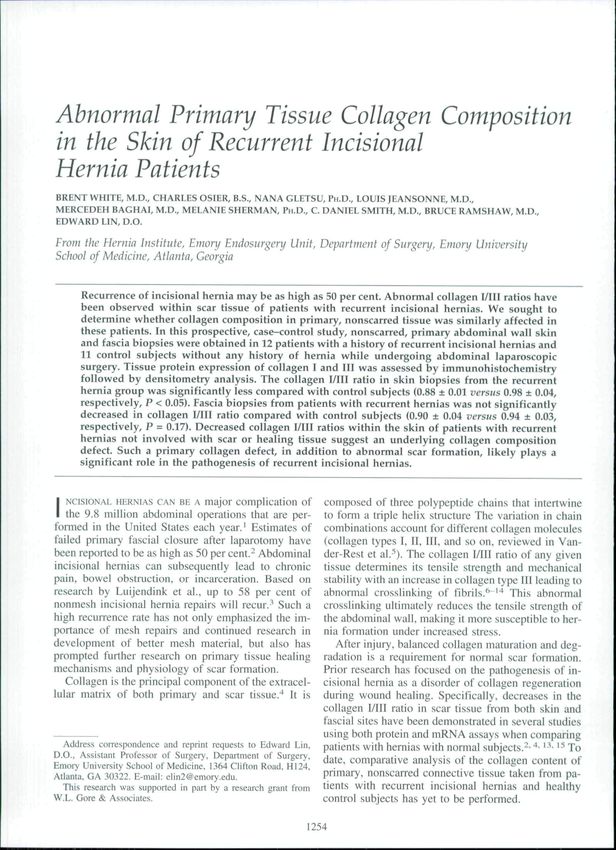
1256                                    THE AMERICAN SURGEON                        December 2007                                  Vol. 73

                                                                         evident on a microscopic level (Fig. I). Analysis of
                                                                         optical density collagen I/III ratios in skin and fascia
                                                                         samples found that skin biopsies obtained from pa-
                                                                         tients with hernias had significantly lower collagen
                                                                         I/III ratios compared with skin biopsies obtained from
                                                                         control subjects (0.88 ± 0.01 versus 0.98 ± 0.04, P <
                                                                         0.05, Fig. 2). When comparing collagen I/TTl ratios iti
                                                                         fascial samples biopsied from patients with hernias
                                                                         versus control subjects, lower ratios were found in
                                                                         samples obtained from patients with hernias, although
                                                                         this difference was not statistically significant (0.90 ±
                                                                         0.04 versus 0.94 ± 0.03, P = 0.17, Fig. 2).
                                                                            Subanalysis of the experimental group (n — 12)
                                                                         compared with the control subjects who had under-
                                                                         gone prior abdominal surgery without hernia forma-
                                                                         tion (n — 6) did not alter the findings noted in our
                                                                         main analysis or their significance for skin (0.88 ±
                                                                         0.01 versus 1.00 ± 0.05, P = 0.02) or fascia (0.90 ±
                                                                         0.04 versus 0.94 ± 0.03, P = 0.25).

    FIG. 1, IgG labeling of collagen types I and III in skin (400x.                                 Discussion
white is collagen). Note the decrease in collagen type I (A) and
 increase In collagen type III (B) in the tissue from a patient with a      This study shows a decreased collagen I/III ratio in
history of recurrenl incisional hernia compared wilh control sub-        primary skin and fascia biopsies of patients with re-
jects (C, collagen type 1. D. collagen type III).                        current incisional hernias when compared with their
                                                                         normal counterparts. Skin samples obtained from pa-
TABLE 1. Patient Demographics                                            tients with hernias had significantly lower collagen
        Variable              Control                Hernia              I/III ratios when compared with control subjects. Al-
                                                                         though not significant, fascia biopsies obtained from
   Number                        11                   12
   Men
                                                                         patients with hernias versus control subjects also had a
                                  4                    2
   WotnetT                        7                   10                 lower collagen I/III ratio.
   Age (years)                                                              In normal tissue, collagen I and collagen II maintain
      Mean                      51.4                  50                 a relatively constant ratio. Previous studies have dem-
      Range                    30-74                 37-62
   Body        mass                                                      onstrated abnormalities in the ratio of these collagen
   index (kg/m^)                                                         molecules in patients with a number of genetic condi-
      Mean                      35.4                  34.5               tions known to predispose to hernia formation such as
      Range                    22-53                 23^7
   Prior abdominal
   surgery                                                                   1.06
      Mean                       1.3                    2.6
     Range                      (M                     1-8
   Prior      hernia
   repair
      Mean                       0                     4.7
      Range                      —                    2-23
   Diabetes
   mellitus                       2                    2
   Smoke tobacco                  1                    3

significant differences in age, body mass index, gen-
der, diabetes, or tobacco use in control subjects versus
patients with hernias. As expected, patients in the ex-                                  Control   Hernia           Control   Hernia
perimental group with recurrent incisional hernias had                                    Skin Tissue              Fascial Tissue
experienced more abdominal surgery compared with                            FIG, 2. Subepidermal (skin) and transversalis fascia eollagcn
control subjects {P < 0.05).                                             i/lll ratio (ratio of optical density of eoltagen lype 1 to optical
                                                                         density of collagen type III in a high-power field). The data arc
   Differences in collagen type staining between con-                    presented as mean values wilh standard error, {*P < 0.05 versus
trol subjects and patients with hernias were visually                    control).
No. 12         ABNORMAL PRIMARY TISSUE COLLAGEN COMPOSITION                                       White et al.              1257

osteogenesis impeifecta or Ehlers-Danlos.''^ '"^ Other        strated they were not prone to hernia formation them-
prevtou.s work has also demonstrated reduced collageti        selves, because 36 per cent had no abdominal surgical
I/Ill ratios in the .skin and fa.scia taken from the scar     history before their enrollment in this study. However,
tissue of patients with incisional hernias and patients       on subanalysis of only those control subjects with
with recurrent incisional hernias compared with that of       prior abdominal surgical history and no hernia, our
heallhy tissue or scar from patients without her-             results were not significantly altered.
nias.-- '^ Therefore, the understanding of the patho-            Our findings suggest a possible pre-existing colla-
genesis of incisional and recurrent incisiona! hernia         gen defect in the primary tissues of the abdominal wall
formation is based on the concept of abnormal colla-          in patients with recurrent incisional hernias. We be-
gen metabolism either through impaired wound heal-            lieve that further research using microarray technology
ing or impaired constitutive collagen expression and          may elucidate a specific threshold or "protein signa-
formation associated with genetic disorders.                  ture" that could allow clinical screening for hernia
   To our knowledge, this study is the first to examine       formation risk. Such screening, if developed, could
nonscarred skin and fascial tissues of healthy control        allow tailoring of future patients" surgical treatment so
subjects and compare them with nonscarred skin and            as to minimize or avoid the risk of incisional hernia.
fascial tissues of patients with recurreni incisional her-
nias. This proved feasible by examining a patient                                    Acknowledgments
population undergoing laparoscopic surgery, in which
                                                                 We thank Kent van Sickle, M.D., for assistance in tissue
trocar incisions are typically made well away from the
                                                              collection and Dianne Lawson. Emory University Hospital
site of prior surgery or known pathology. This experi-        Department of Pathology, for mounting and staining our
mental design allowed us to compare primary, non-             biopsy specimens.
scarred tissue in both the experimental and control
groups. The significant differences found in skin col-                                  REFERENCES
lagen I/III ratios between the two groups strongly sug-
                                                                 1. DeFrances C, Hail M. 2005 Nationai iiospitai discharge sur-
ge.st that the abnormal collagen metabolism of patients
                                                              vey. Advance Data From Vitai and Heaith Statistics 2OO7;385:16.
with recurrent incisional hernias is not only the result
                                                                 2. Kiinge U. Si Z, Ziieng H. el al. Collagen I/III and matrix
of abnormal wound healing as suggested in prior stud-         metalioproteinases (MMP) I and i3 in liie fascia of pulieiils with
ies, but is also the result of an impaired constitutive       incisionai lieniias. J Invest Surg 20()i;13:47-54.
ct)llagen expression and formation.                              3. Luijendijk R, Hop W, van-den-Tol M. et al. A comparison of
    In the present study, significant differences were        suttire with mesh repair for incisional hernia. N Engi J Med 2(HK);
found in the collagen I/Ill ratios between our experi-        343:392-8.
mental and control groups tor skin, but not for fascial          4. Zheng H. Si Z, Kasperk R. et al. Recurrent inguinal hertiia:
tissues. Because our power calculations were based on         disease of the collagen matrix? World J Surg 2002:26:401-8.
previous work, which compared nonscarred and                     5. Van-der-Rest M. Garrone R. Collagen famiiy of proteins.
scarred tissues and found larger ratio differences.-^ it is   FASP.B J l991;5:2814-23.
possible the lack of a significant difference found in           6. Fieischmajer R, Periish JS. Burgcsoii RE, el al. Type I and
                                                              type III interactions during fihriliogenesis. Ann N Y Acad Sci
fascial tissues between groups may simply be the re-          1990:580:161-75.
sult of a type II error. However, nearly all previous            7. Friedman DQ. Boyd C. Mackenzie JW. el ai. Regulalion of
work demonstrating alteration in collagen I/IU ratios         collagen gene expression in keloids and hypcrlrophicd scars. J
in primary tissues (e.g., osteogenesis irnperfeeta) have      Surg Res i993:55:214-22.
done so using skin (issues or fibroblasts cultured from          8. Henkel W. Glanville RW, Covalent crosslinking between
dermal tissues.'^''^ Therefore, the findings in this          molecules of type I and III coliagen. Eur J BiiH:heiTi 1982:122;
study may indicate that no significant difference exists      205-13.
in the primary fascial collagen ratio between groups.            9. Uitto J, Perejda AJ, Patrick-Abergel R, et al. Altered steady-
   Possible weaknesses in our study design may have           state ratio of type I/III procollagen mRNAs correlates with selec-
potentially affected or confounded our results. Our           tively increased type I procollagen biosynthesis in cultured keloid
study population was not stratified by hernia size, lo-       llbroblasts. Proc Nail Acad Sci USA 1985:82:5935-9.
cation or number of recurrences. Also, although the              10. Romanic AM. Adachi E. Kadler KE, Hojima Y, Prockop
                                                              DJ. Copolymerization of pNcollagen III and coliagen I. pNcolla-
frequency of obesity in each group was the same, over
                                                              gen HI decreases the rale of incorporation of coiiagen I into llbrils,
60 per cent of all patients were obese (mean body mass        the amount of coiiagen I incorporated, and the diameter of the
index = 34.5 kg/m-^). Obesity is thought to be a risk         tlbrils formed. J Biol Chem i991:266:12703-9.
factor for hernia recurrence as a result of poorer               11. Biric DE, Mayne R. Legalization of coiiagen type I. Ill, and
wound healing, increased intra-abdominal pressure,            V during tendon development. Changes in collagen types 1 and MI
and a technically more difficult surgical repair.^"           are correlated with changes in fibril diameter. Eur J Ceil Biol
Moreover, not all control subjects had clearly demon-          1997:72:352-61.
1258 THE AMERICAN SURGEON December 2007 Vol, 73
12. Dale PD. Sherratt JA. Maini PK, A tnathetnatical tnodel for 17, Deak SB. Ricotta JJ. Mariani TJ. et al. Abnorrnaiities in the
collagen fibre formation during foetai and adult dermal wound biosynthiesis of type III procoilagen in cultured skin fibroblasts
healing. Proc R Soc Lond B Biol Sci i996;263:653-60. from two patients with muitipie aneurysm. Matrix i992;12:
13. Klinge U. Si Z, Zheng H. et ai. Abnormal collagen I to [II 92-100.
distribution in the skin of patients with incisional hernia, Eur Surg 18. Rowe DW. Shapiro JR. Piorier M. Schlesinger S. Dimin-
Res 20{K);32:43-8. isiied type I collagen synthesis and reduceil iiipiia 1(1) coiiagen
14. Klinge U, Ziieiig H, Si Z, et ai. Expression of the extracel- messenger RNA in euitured fibroblasts from patients with domi-
luiar matrix proteins collagen I. coilagen III and fibronectin and nantly inherited (type 1) osteogenesis imperfect. J Ciin Invest
matrix metalloproteinase-1 and -13 in tiic skin of patients with i985:76:604-Il,
inguinal hernia. Eur Surg Res iy99;31:43-8, 19, Leitn MSL. Van der Graff Y, BeemerFA. Van Vroonhoven
15. Rosch R. Junge K. Knops M. et al. Analysis of coilagen- TJMV. Increased risk for inguinal hernia in patients with Ehlers-
interacting proteins in patients witii incisional hernias. Langen- Danlos syndrome. Surgery i997;i22:il4-5,
hccivs Arch Surg 2OO3;387:427-32. 20. Sauerland S, Korenkov M, Kieinen T. et ai. Obesity is a risk
16. Rasband WS. image J. 20(H. Avaiiable at: httpiWsb.info factor for recurrence after incisionai hernia repair. Hernia 2OO4;8:
,nih,gov/ij. Accessed Aprii 1, 2007. 42-6,You can also read