Anticariogenic Effect of Selenium Nanoparticles Synthesized Using Brassica oleracea
←
→
Page content transcription
If your browser does not render page correctly, please read the page content below
Hindawi
Journal of Nanomaterials
Volume 2021, Article ID 8115585, 9 pages
https://doi.org/10.1155/2021/8115585

Research Article
Anticariogenic Effect of Selenium Nanoparticles Synthesized
Using Brassica oleracea

 1 2
 Ganapathy Dhanraj and Shanmugam Rajeshkumar
 1
 Department of Prosthodontics, Saveetha Dental College, Saveetha Institute of Medical and Technical Sciences,
 Chennai 600 077, India
 2
 Department of Pharmacology, Saveetha Dental College, Saveetha Institute of Medical and Technical Sciences,
 Chennai 600 077, India

 Correspondence should be addressed to Shanmugam Rajeshkumar; ssrajeshkumar@hotmail.com

 Received 20 April 2021; Revised 16 June 2021; Accepted 24 June 2021; Published 10 July 2021

 Academic Editor: Abdelwahab Omri

 Copyright © 2021 Dhanraj Ganapathy and Rajeshkumar Shanmugam. This is an open access article distributed under the Creative
 Commons Attribution License, which permits unrestricted use, distribution, and reproduction in any medium, provided the
 original work is properly cited.

 Selenium is a trace element in the human body present in various enzymes with antioxidant activities and several functional
 proteins. This study is aimed at synthesizing selenium nanoparticles using Brassica oleracea (broccoli) and characterizing and
 assessing the antioxidant and antimicrobial effectiveness against cariogenic microorganisms. UV-visible spectrum displayed a
 peak at 370 nm which confirms the formation of SeNPs. TEM images of synthesized selenium nanoparticles showed
 polydisperse nanoparticles, spherical. The size of the particles ranged from 10 to 25 nm, and the average particle size obtained
 was 15:2 ± 1:9 nm. SEM images of nanoparticles were spherical and ranged in size from 10 to 28 nm. The SeNPs showed
 effective antimicrobial activity against cariogenic pathogens. The SeNPs synthesized with Brassica oleracea extract can be
 incorporated in toothpaste, gums, and mouthwashes that are cost-effective and biocompatible and used for the prevention of
 dental caries.

1. Introduction Selenium is found in nature as ionic forms of selenite
 (Na2SeO3), selenate (Na2SeO4), and selenium oxide (SeO)
Owing to their characteristic attributes, nanoparticles find among the other forms. Elemental selenium nanoparticles
extensive implementation in several scientific, medicinal, manifest lesser cytotoxicity compared with selenite (SeO3)
and industrial domains [1, 2]. Among the various kinds of and selenate compounds (SeO4) and show anticancer and
nanoparticles, selenium has attracted appreciable interest several therapeutic properties that enable them to be utilized
due to its greater bioactivity, protein interactions, strong in versatile medicinal applications [7, 8]. Researchers have
absorption capability, and reduced toxicity, together with reported on the antioxidant function of SeNPs. SeNPs can
interdisciplinary applications in medicine, therapeutic sci- unswervingly scavenge the free radicals in a size-dependent
ences, nanobioinformatics, and nanobiotechnology [3]. manner within the size ranging from 5 to 200 nm [9].
 Selenium corresponds to Group VI in the periodic table Traditional physiochemical methods of SeNP synthesiz-
as an integral trace element. Numerous antioxidant enzymes ing are cumbersome, nonecological, and require prerequi-
and functional protein molecules contain selenium and sites such as high temperatures, toxic substances, reagents,
maneuver a crucial role in mitigating oxidative stress, thus precursors, and complicated procedures. Besides, in the con-
minimizing the damage in cardiovascular diseases, cancer, ventional process, the form, size, and instability of the syn-
diabetes, and hypercholesterolemia. On the other hand, the thesized particle are the main limitations [10]. This creates
intake of selenium is necessary for different metabolic pro- a great desire to synthesize nanoparticles with harnessed size
cesses and indiscriminate intake beyond acceptable concen- and nanometer morphology in an eco-friendly mode [11].
tration leads to selenium toxicity [4–6]. The size and the shape of nanoparticles are permanently
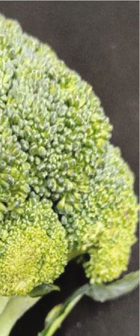
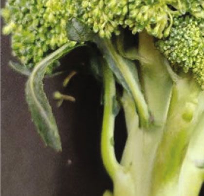
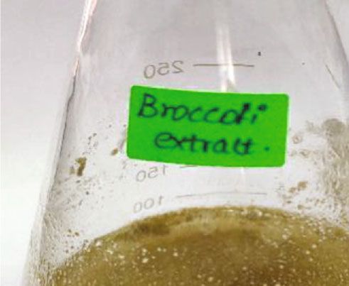
2 Journal of Nanomaterials

related to the unique characteristics, work, and application of phosphate, hydroxyapatite, and fluorohydroxyapatite are
nanomaterials [12, 13]. Because of its eco-friendliness, low used in a process called remineralization. The second method
toxicity along with increased stability, small size, and shape is to imbibe antimicrobial nanomaterials such as selenium,
distribution, emerging advances in nanoparticle generation platinum, quaternary ammonium compounds, and zinc
using green syntheses are attracting much attention today oxide nanoparticles [25, 26]. To present better results, a
[14, 15]. blend of these two methods can also be used. The anticario-
 Brassica oleracea (broccoli) has all the nutrients that genic effects of selenium nanoparticles through inhibition
prove its exceptional health benefits, including vitamins, of cariogenic microbes were not studied extensively and thus
minerals, secondary metabolites, and fiber. The active princi- need to be explored further. Hence, this study was formu-
ples for demonstrating medicinal properties are the break- lated with the aim to green synthesize selenium nanoparticles
down products of sulfur-containing glucosinolates, phenolic using Brassica oleracea and study its characterization and
isothiocyanates, antioxidants, vitamins, and dietary minerals. antioxidant and antimicrobial effectiveness against cario-
These bioactive compounds most likely mediate the medici- genic microorganisms.
nal properties of broccoli consumption by the inducement
of a range of functions such as antioxidant behavior, enzyme 2. Material and Methods
regulation, and the control of apoptosis [16].
 Organic chemicals such as S-methyl cysteine sulfoxide 2.1. Preparation of Broccoli Extract. Brassica oleracea was
and glucosinolates present in Brassica oleracea in conjunc- procured from organic farms in Poonamallee. Broccoli was
tion with other ingredients inclusive of vitamins C, K, and washed thoroughly under tap water and made into small
E and minerals like selenium, iron, and zinc and polyphenols pieces to shade dry it for 3-4 days. After that, the shade-
such as kaempferol, glucosides of quercetin, and isorhamne- dried broccoli pieces were ground into a fine powder. From
tin are likely to be accountable for the various health benefits that, finely grounded broccoli powder 0.7 g was taken and
of broccoli [17, 18]. added to 70 mL distilled water. The mixture was kept in a
 Concerning dental problems, caries is one of the most heating mantle at 60-80°C for 10 minutes. Then, the boiled
common human diseases characterized by susceptibility to mixture was filtered using Whatman No. 1 filter paper. The
enamel and dentin tissue damage due to acidic by- filtered broccoli extract was stored in the refrigerator for sele-
products generated from dietary carbohydrates owing to nium nanoparticle synthesis.
bacterial fermentation [19]. This problem is initiated by
 2.2. Preparation of Selenium Nanoparticle. 30 mM of sodium
the acid production from bacteria that ferment carbohy-
 selenite was prepared in 70 mL distilled water. To that 30 mL
drates resulting in demineralization of dental enamel and
 filtered broccoli, the extract was added and kept in a mag-
the development of caries. Supplementation of calcium
 netic stirrer for 2-3 days at 650-800 rpm to obtain uniform
and phosphate can aid in the rarefy demineralization of
 dispersion, a mandatory condition in nanoparticle synthesis.
teeth to avoid dental caries. Such ions are also present in
 The color changes in the reaction mixture were noted contin-
the saliva, and their concentration determines reminerali-
 uously using a double-beam UV-visible spectrophotometer
zation and demineralization [20, 21].
 at the different wavelength range from 250 to 650 nm. The
 Nanocarriers are presently being reasoned for their appli-
 synthesized broccoli extract-mediated selenium nanoparti-
cation in dentistry, and their particular characteristics iden-
 cles were centrifuged at 8000 rpm for 10 minutes. The
tify possible use in the delivery of antimicrobial agents in
 obtained selenium nanoparticle pellet was powdered using
the prevention and management of oral diseases caused by
 a hot air oven at 70°C for 2 hours and preserved in airtight
microbial invasion. Implementation of nanomaterials in den-
 vials for further use.
tistry may be divided into two major categories: dental pre-
ventive and restorative care [22]. According to caries 2.3. Characterization of Selenium Nanoparticles. The maxi-
research, novel approaches to prevent and manage dental mum absorbance of broccoli-mediated selenium nanoparti-
caries have been implemented through nanotechnology, in cles was measured by using a double-beam UV-vis
particular through plaque-related biofilm regulation and spectrophotometer (UV-2450, Shimadzu) in the wavelength
remineralization of carious lesions [23]. range of 250-650 nm. The morphological features such as size
 In this area, nanotechnological advances have also pro- and shape were analyzed by TEM. The functional groups
vided advantages as novel drivers of innovation. It has been present in the synthesized selenium nanoparticles were iden-
shown that natural biomineralization, which is an integral tified by using FT-IR analysis and also subjected to test the
repair mechanism, is induced by the use of nanotechnology. elemental analysis using an energy-dispersive X-ray detector
For enhancing oral health through the prevention of dental (EDX) attached to the SEM machine.
caries, the use of nanomaterials in toothpaste and other
mouth rinsing solutions can be recommended. Caries is often 2.3.1. Antioxidant Activity. The DPPH assay was used to test
arrested by nanomaterials used in polishing agents and den- the antioxidant activity of biogenic synthesized selenium
tal restorative materials. Per se, antimicrobial nanoparticles nanoparticles. Diverse concentrations (2-10 μg/mL) of broc-
can inhibit the growth of bacteria inducing dental caries [24]. coli extract-interceded selenium nanoparticle were mixed
 Dental caries can be addressed by nanotechnology in two with 1 mL of 0.1 mM DPPH in methanol and 450 μL of
primary methods. In the first method, fluoride- and calcium- 50 mM Tris HCl buffer (pH 7.4) and incubated for 30
releasing nanomaterials such as calcium fluoride, calcium minutes. Later, the reduction in the quantity of DPPH free
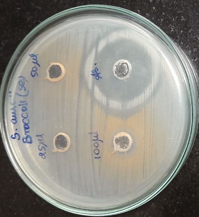
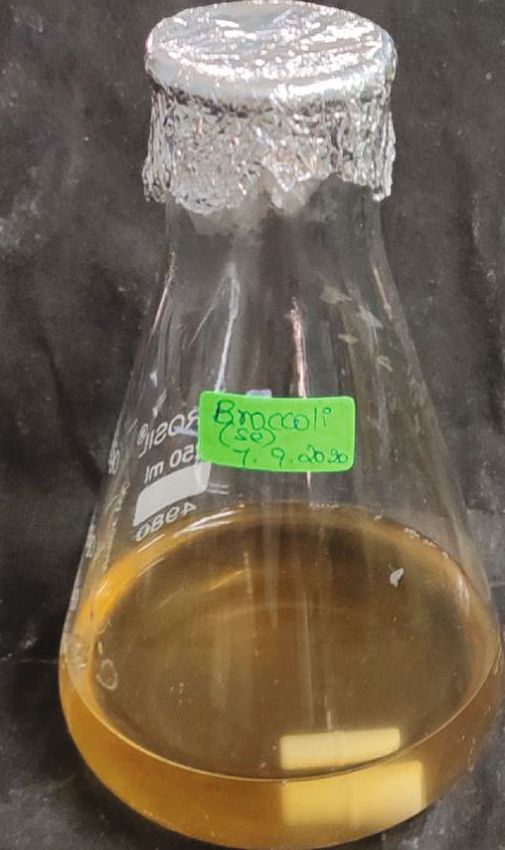
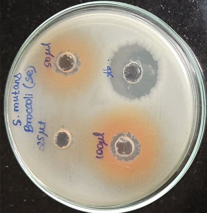
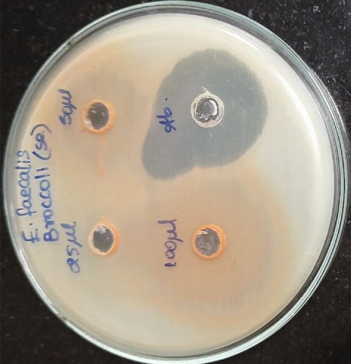
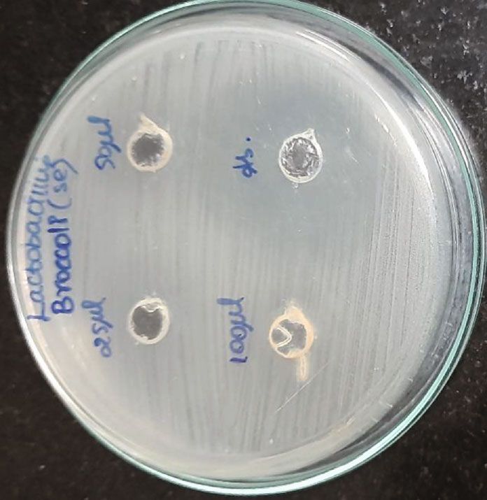
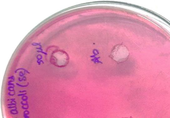
Journal of Nanomaterials 3

 (a) (b) (c)

 Figure 1: Color changes on selenium nanoparticles synthesis: (a) broccoli; (b) plant extract; (c) selenium nanoparticles.

 3.500

 3.000

 2.500
 Absorbance

 2.000

 1.500

 1.000

 0.500

 0.000
 250.0 350.0 450.0 550.0 650.0
 Wavelength (nm)

 1h 48 h
 12 h 72 h
 24 h

Figure 2: UV-vis spectroscopic analysis of broccoli-mediated
selenium nanoparticles at different time intervals and wavelength
is from 250 nm to 650 nm. Figure 3: Transmission electron microscopic image of selenium
 nanoparticles synthesized using broccoli extract and scale
radicals was assessed depending on the absorbance at measures at 100 nm.
517 nm. BHT was employed as control. The percentage of
inhibition was determined from the following equation:
 oral pathogens such as S. mutans, S. aureus, E. faecalis, Lacto-
 Absorbance of control − Absorbance of test sample bacillus, and C. albicans. Mueller Hinton agar was utilized for
%inhibition = × 100:
 Absorbance of control this activity to determine the zone of inhibition. Mueller Hin-
 ð1Þ ton agar was prepared and sterilized for 45 minutes at 120 lbs.
 Media were poured into the sterilized plates and let it stabi-
2.3.2. Anticariogenic Activity. The anticariogenic activity of lize for solidification. The wells were cut using the good cut-
broccoli-mediated selenium nanoparticles was tested against ter, and the test organisms were swabbed. The selenium
4 Journal of Nanomaterials

 Element Weight% Atomic%
 CK 24.22 39.99
 OK 31.49 39.04
 Na K 16.09 13.88
 Se L 28.21 7.09

 Na Totals 100.00

 O Se

 C

 Se

 0 0.5 1 1.5 2
 Full scale 314 cts cursor: 2.424 (6 cts) keV
 (a) (b)

 Figure 4: Selenium nanoparticles: (a) scanning electron microscopy image; (b) elemental X-ray diffraction spectrum.

nanoparticles with different concentrations were loaded, and peak amplitude has increased over time because of the reduc-
the plates were incubated at 37°C for 24 hours. After the incu- tion of SeO3 2- to Se0. No further peak increase was observed
bation time, the zone of inhibition was measured. rafter 48 h, suggesting a maximum conversion of SeO3 2- to
 Se0 (Figure 2). Transmission electron microscopy (TEM)
3. Results and Discussion images of selenium nanoparticles synthesized in nutrient
 broth supplementation with 1.0 mM selenite at 72 hours of
Compared to physical or chemical approaches of nanoparti- incubation are shown in Figure 3. The generated nanoparti-
cle synthesis, one of the effective methods is the generation of cles were polydisperse, shaped spherically; the particle size
nanoparticles utilizing green sources such as plant phyto- ranged from 10 to 25 nm, and the mean particle size that pre-
compounds, proteins, and enzymes as reducing agents in vailed from the distribution of the corresponding diameter
the case of microbes. The benefits are that they are nontoxic was approximately 15:2 ± 1:9 nm (Figure 3).
materials, requiring relatively less complicated and affordable Synthesized plant extracts, such as hydrogen bond and
equipment, biodegradable material processing, improved the electrostatic interaction, were present in the SEM image
selectivity, and high yields. of SeNPs and were mounted on the surface because of the
 Visual coloring is the first stage in the moulding of interaction with the bioorganic capping molecules attached
nanoparticles. It was observed following the incubation to the SeNPs. Synthesized SeNPs with sizes ranging from
period that the color changed to brown. The reaction mix- 10.32 nm to 25.88 nm were observed (Figure 4(a)). SEM
ture with Brassica oleracea and sodium selenite during study of the synthesized SeNPs was easily distinguishable
incubation showed a time-dependent color shift of 48 h due to their size variation. The SEM picture showed the
at 30°C, as shown in Figure 1. At the primary reaction majority of nanoparticles are spherical in size, ranging from
stage, the color of the concoction was yellow, which pro- 10 to 25 nm, and uniformly distributed. SEM analysis of
gressively changed to brown over time. After 48 hours of SeNPs was readily distinguishable due to the size difference
incubation, no further alteration in color was observed. of synthesized SeNPs. Elemental X-ray diffraction assay was
This brown color may be attributed to the arousal of the used to assess the elemental percentage and presence; sele-
surface plasmon vibrations by the selenium nanoparticles nium nanoparticles synthesized with broccoli extract had
and thus render beneficial spectroscopic evidence of their an elemental presence of selenium with a 28.02 percent
formation. weight percentage and a 7.09 percent atomic percentage
 UV-visible spectra have shown a large peak at 370 nm, (Figure 4(b)).
supporting the formation of SeNPs [27]. The peak intensity FT-IR estimation was performed to determine the possi-
increased over time. No further significant elevation in peak ble bands present in the biomolecules accountable for near
intensity was observed after 48 hours of the reaction. The capping peaks and effective stabilization of the metal NPs
Journal of Nanomaterials 5

 120

 100

 % of transmittance
 80

 60

 40

 20

 0
 600 1100 1600 2100 2600 3100 3600 4100
 Wavenumber (cm–1)

 Figure 5: Fourier-transform infrared spectroscopy of selenium nanoparticles synthesized using broccoli aqueous extract.

 Antibacterial activity
 35

 30
 Zone of inhibition (mm)

 25

 20

 15

 10

 5

 0
 25 L 50 L 100 L Antibiotic
 Concentration

 E. faecalis S. mutans
 S. aureus C. albicans
 Lactobacillus sp

Figure 6: Anticariogenic activity against caries-causing microbes (S. mutans, Lactobacillus sp., E. faecalis, S. aureus, and C. albicans) at
different concentrations and zone of inhibition was measured in millimeter.

synthesized by broccoli extract. FT-IR estimation revealed a the composition of glutathione peroxidases, thioredoxin
weak broad peak at 3235.32 cm-1 indicating the presence of reductases, and other selenoenzymes [4]. Glutathione perox-
alcohol functional group with O-H stretching, strong to idase is an enzyme that prevents tissue oxidative stress and
medium peaks at 1595.30 cm-1 indicating amine group with can thus inhibit the activity of harmful free radicals.
N-H bending, 1407.07 cm-1 indicative of fluoro compounds UV-vis spectroscopy is one of the most commonly used
with C-F stretching, and 1099.56 cm-1 indicating aliphatic methods for the structural characterization of SeNPs. The size
ether with C-O stretching (Figure 5). and shape of the tracked aqueous suspension NPs can usually
 Selenium nanoparticles displayed potent antimicrobial be analyzed by UV-vis spectroscopy. Our outcome indicates
activity against caries-causing microorganisms at all concen- that C leaf extract synthesizes SeNPs. Measurements based
trations closely comparable to the antibiotic controls. The on data and spectrophotometers range from 200 to 700 nm
maximum antimicrobial activity against S. mutans was with a 420 nm peak indicating SeNP performance.
observed (Figures 6 and 7). Figure 8 shows the antioxidant Energy-dispersive microanalysis spectroscopy was carried
activity by DPPH was also important and displayed more out using EDX techniques to gain further perception of the
efficacy than the controls at concentrations of 10 μL and SeNPs’ functionality. Our results suggest that selenium bind-
20 μL selenium nanoparticles. Selenium has a major biologi- ing energies have EDaX peaks of approximately 72.64. The
cal role in species health and is an important component of outcome indicates the presence in the pure form of SeNPs of
6 Journal of Nanomaterials

 (a) (b)

 (c) (d)

 (e)

Figure 7: Anticariogenic activity of selenium nanoparticles: (a) S. mutans; (b) Lactobacillus sp.; (c) E. faecalis; (d) S. aureus; (e) C. albicans.

the reaction product. For synthesized extracts of SeNPs, the ria perish because of the stress caused by the existence of toxic
recorded EDaX showed a strong signal of 3 keV selenium. inorganic compounds in the atmosphere, thereby reducing the
 In our study, the bacterial growth was affected by the addi- production of selenium nanoparticles [8].
tion of various sodium selenite concentrations under aerobic It has also been shown that selenium dioxide (SeO2) as
conditions to the growth medium. Elevated concentrations Na2SeO3 causes noxious effects on microbial plasmid DNA
of sodium selenite induce a lot of impairment to the genetic under stress conditions of H2O2, but with greater intensity,
configuration in microbes. Thus, significant numbers of bacte- whereas other selenium-containing compounds such as
Journal of Nanomaterials 7

 (a)
 Antioxidant activity
 120

 100

 80
 % of inhibition

 60

 40

 20

 0
 10 L 20 L 30 L 40 L 50 L
 Concentration

 Standard
 SeNPs
 (b)

Figure 8: Antioxidant activity of selenium nanoparticles: (a) color changes; (b) graph representing the antioxidant activity at percentage of
inhibition.
8 Journal of Nanomaterials

Na2SeO4 and Na2Se have some inhibitory effects on Escheri- Data Availability
chia coli plasmid DNA. Previous studies support our study of
the antibacterial activity of selenium on Staphylococcus and The data used to support the findings of this study are
suggest that selenium is capable of preventing the develop- included within the article.
ment of biofilms by bacteria and cytotoxic effect has been
demonstrated by SeNPs [28]. In live aureus assays, it was Conflicts of Interest
demonstrated that SeNPs inhibited pneumonia in cell lines
[29]. In this analysis, the antimicrobial impact of SeNPs on The authors declare that there is no conflict of interest.
microbes was observed.
 The mechanism attributed may be owed to the interrup- Authors’ Contributions
tion of the cell wall, indicating that the selenium nanoparti- DG and SR designed, carried out research, and wrote the
cles were able to penetrate the cell wall. The antibacterial manuscript.
role of these selenium nanoparticles can be distinguished by
morphological alterations in bacterial strains both intracellu-
lar and extracellular. The antimicrobial behavior of selenium
 References
nanoparticles is mainly due to the formation of reactive oxy- [1] S. al-Musawi, S. Albukhaty, H. al-Karagoly et al., “Antibacte-
gen species, which contributes to the disruption of bilayer rial activity of honey/chitosan nanofibers loaded with capsai-
phospholipids where intracellular proteins are associated cin and gold nanoparticles for wound dressing,” Molecules,
with and inactivated by SeNPs or where the sulfhydryl and vol. 25, no. 20, article 4770, 2020.
the thiol groups existing in membrane proteins can react [2] M. Jabir, U. I. Sahib, Z. Taqi et al., “Linalool-loaded
with and eventually denature them. By altering the cycle of glutathione-modified gold nanoparticles conjugated with
protein synthesis, interfering with the mechanism of respira- CALNN peptide as apoptosis inducer and NF-κB translocation
tory or food metabolism, or impairing the replication of inhibitor in SKOV-3 cell line,” International Journal of Nano-
DNA, oxidative stress caused by reactive oxygen induces cell medicine, vol. Volume 15, pp. 9025–9047, 2020.
death [30]. [3] Z. Qiao, Y. Xie, Y. Qian, and Y. Zhu, “γ-Irradiation prepara-
 Toxicological examinations have demonstrated that tion and characterization of nanocrystalline ZnS,” Materials
small selenium nanoparticles possess a higher surface area Chemistry and Physics, vol. 62, no. 1, pp. 88–90, 2000.
and those particle numbers per unit mass can induce harmful [4] S. Menon, H. Agarwal, S. Rajeshkumar, P. Jacquline Rosy, and
respiratory damage and inflammation. The larger particles V. K. Shanmugam, “Investigating the antimicrobial activities
 of the biosynthesized selenium nanoparticles and its statistical
about 100 nm are more easily engulfed by macrophages.
 analysis,” Bionanoscience, vol. 10, no. 1, pp. 122–135, 2020.
Their aggregation can, thus, decrease the toxicity of the nano-
 [5] P. Narasingarao and M. M. Häggblom, “Identification of
particles [31, 32]. anaerobic selenate-respiring bacteria from aquatic sediments,”
 Selenium nanoparticles can be used in various applica- Applied and Environmental Microbiology, vol. 73, no. 11,
tions in the prevention of caries in dental tissues. Incorporat- pp. 3519–3527, 2007.
ing selenium nanoparticles in toothpaste, chewing gums, and [6] J. Zhang, S.-Y. Zhang, J.-J. Xu, and H.-Y. Chen, “A new
mouthwashes can potentially help control the growth of car- method for the synthesis of selenium nanoparticles and the
ies causing microbes. Similarly, selenium nanoparticles can application to construction of H2O2 biosensor,” Chinese
be incorporated in luting cement, endodontic sealers, pit Chemical Letters, vol. 15, no. 11, pp. 1345–1348, 2004.
and fissure sealants, and restorative cement to control sec- [7] P. A. Tran and T. J. Webster, “Antimicrobial selenium nano-
ondary dental caries. Further extensive research in these particle coatings on polymeric medical devices,” Nanotechnol-
domains will enhance our understanding of the therapeutic ogy, vol. 24, no. 15, article 155101, 2013.
effectiveness of selenium nanoparticles in the prevention of [8] N. Srivastava and M. Mukhopadhyay, “Biosynthesis and struc-
dental caries. tural characterization of selenium nanoparticles mediated by
 Zooglea ramigera,” Powder Technology, vol. 244, pp. 26–29,
 2013.
4. Conclusion
 [9] H. Wang, J. Zhang, and H. Yu, “Elemental selenium at nano
Nanomaterials have emerged as promising therapeutic size possesses lower toxicity without compromising the funda-
agents in the prevention and treatment of dental caries. In mental effect on selenoenzymes: comparison with seleno-
 methionine in mice,” Free Radical Biology & Medicine,
this study, SeNPs were synthesized using Brassica oleracea
 vol. 42, no. 10, pp. 1524–1533, 2007.
extract, characterized using UV-vis spectroscopy which con-
 [10] A. G. al-Dulimi, A. Z. al-Saffar, G. M. Sulaiman et al., “Immo-
firmed the SeNP formation and electron microscopic images
 bilization of l-asparaginase on gold nanoparticles for novel
from TEM and SEM which showed nanoparticles less than drug delivery approach as anti-cancer agent against human
50 nm, and evaluated for the antibacterial activity against breast carcinoma cells,” Journal of Materials Research and
dental caries-causing pathogens, and it was found that SeNPs Technology, vol. 9, no. 6, pp. 15394–15411, 2020.
showed effective antimicrobial activity against cariogenic [11] M. S. Jabir, Y. M. Saleh, G. M. Sulaiman et al., “Green synthesis
pathogens. The SeNPs synthesized with Brassica oleracea of silver nanoparticles using Annona muricata extract as an
extract can be incorporated in toothpastes, gums, and inducer of apoptosis in cancer cells and inhibitor for NLRP3
mouthwashes that are cost-effective and also biocompatible inflammasome via enhanced autophagy,” Nanomaterials,
and effective for the prevention of dental caries. vol. 11, no. 2, p. 384, 2021.
Journal of Nanomaterials 9

[12] O. al Rugaie, M. Jabir, R. Kadhim et al., “Gold nanoparticles bacterial activity,” Materials Research Express, vol. 6, no. 8,
 and graphene oxide flakes synergistic partaking in cytosolic article 0850d8, 2019.
 bactericidal augmentation: role of ROS and NOX2 activity,” [29] S. Shoeibi and M. Mashreghi, “Biosynthesis of selenium nano-
 Microorganisms, vol. 9, no. 1, p. 101, 2021. particles using Enterococcus faecalis and evaluation of their
[13] G. M. Sulaiman, E. H. Ali, I. I. Jabbar, and A. H. Saleem, “Syn- antibacterial activities,” Journal of Trace Elements in Medicine
 thesis, characterization, antibacterial and cytotoxic effects of and Biology, vol. 39, pp. 135–139, 2017.
 silver nanoparticles,” Digest Journal of Nanomaterials and [30] X. Chen, K. Cai, J. Fang et al., “Fabrication of selenium-
 Biostructures, vol. 9, no. 2, pp. 787–796, 2014. deposited and chitosan-coated titania nanotubes with antican-
[14] W. J. al-Kaabi, S. Albukhaty, A. J. M. al-Fartosy et al., “Devel- cer and antibacterial properties,” Colloids and Surfaces B,
 opment of Inula graveolens (L.) plant extract electrospun/poly- Biointerfaces, vol. 103, pp. 149–157, 2013.
 caprolactone nanofibers: a novel material for biomedical [31] S. Chaudhary, P. Chauhan, R. Kumar, and K. K. Bhasin, “Tox-
 application,” Applied Sciences, vol. 11, no. 2, p. 828, 2021. icological responses of surfactant functionalized selenium
[15] W. Zhang, Z. Chen, H. Liu, L. Zhang, P. Gao, and D. Li, “Bio- nanoparticles: a quantitative multi-assay approach,” Science
 synthesis and structural characteristics of selenium nanoparti- of The Total Environment, vol. 643, pp. 1265–1277, 2018.
 cles by Pseudomonas alcaliphila,” Colloids and Surfaces. B, [32] R. Tarrahi, A. Khataee, A. Movafeghi, F. Rezanejad, and
 Biointerfaces, vol. 88, no. 1, pp. 196–201, 2011. G. Gohari, “Toxicological implications of selenium nanoparti-
[16] C. I. R. Gill, “The effect of cruciferous and leguminous sprouts cles with different coatings along with Se4+ on Lemna minor,”
 on genotoxicity, in vitro and in vivo,” Cancer Epidemiology, Chemosphere, vol. 181, pp. 655–665, 2017.
 Biomarkers & Prevention, vol. 13, no. 7, pp. 1199–1205, 2004.
[17] G. Brandi, G. F. Schiavano, N. Zaffaroni et al., “Mechanisms of
 action and antiproliferative properties of Brassica oleracea
 juice in human breast cancer cell lines,” The Journal of Nutri-
 tion, vol. 135, no. 6, pp. 1503–1509, 2005.
[18] E. H. Jeffery, A. F. Brown, A. C. Kurilich et al., “Variation in
 content of bioactive components in broccoli,” Journal of Food
 Composition and Analysis, vol. 16, no. 3, pp. 323–330, 2003.
[19] R. A. Bagramian, F. Garcia-Godoy, and A. R. Volpe, “The
 global increase in dental caries. A pending public health crisis,”
 American Journal of Dentistry, vol. 22, no. 1, pp. 3–8, 2009.
[20] R. H. Selwitz, A. I. Ismail, and N. B. Pitts, “Dental caries,” The
 Lancet, vol. 369, no. 9555, pp. 51–59, 2007.
[21] T. Walsh, H. V. Worthington, A. M. Glenny et al., “Fluoride
 toothpastes of different concentrations for preventing dental
 caries in children and adolescents,” Cochrane Database of Sys-
 tematic Reviews, vol. 1, 2010.
[22] K. J. Toumba and M. E. J. Curzon, “A clinical trial of a slow-
 releasing fluoride device in children,” Caries Research,
 vol. 39, no. 3, pp. 195–200, 2005.
[23] S. Maleki Dizaj, F. Lotfipour, M. Barzegar-Jalali, M.-
 H. Zarrintan, and K. Adibkia, “Application of Box–Behnken
 design to prepare gentamicin-loaded calcium carbonate nano-
 particles,” Artificial Cells, Nanomedicine, and Biotechnology,
 vol. 44, no. 6, pp. 1475–1481, 2016.
[24] M. Hannig and C. Hannig, “Nanotechnology and its role in
 caries therapy,” Advances in Dental Research, vol. 24, no. 2,
 pp. 53–57, 2012.
[25] L. Cheng, K. Zhang, M. D. Weir, M. A. S. Melo, X. Zhou, and
 H. H. K. Xu, “Nanotechnology strategies for antibacterial and
 remineralizing composites and adhesives to tackle dental car-
 ies,” Nanomedicine, vol. 10, no. 4, pp. 627–641, 2015.
[26] M. A. S. Melo, S. F. F. Guedes, H. H. K. Xu, and L. K. A. Rodri-
 gues, “Nanotechnology-based restorative materials for dental
 caries management,” Trends in Biotechnology, vol. 31, no. 8,
 pp. 459–467, 2013.
[27] C. M. Debieux, E. J. Dridge, C. M. Mueller et al., “A bacterial
 process for selenium nanosphere assembly,” Proceedings of
 the National Academy of Sciences of the United States of Amer-
 ica, vol. 108, no. 33, pp. 13480–13485, 2011.
[28] S. Boroumand, M. Safari, E. Shaabani, M. Shirzad, and
 R. Faridi-Majidi, “Selenium nanoparticles: synthesis, charac-
 terization and study of their cytotoxicity, antioxidant and anti-
You can also read