Approach to Fever and Drooling in Infants and Toddlers
←
→
Page content transcription
If your browser does not render page correctly, please read the page content below

CME: This peer-reviewed article is offered for AMA PRA
    Clinical                                                                         Category 1 Credit.™ See CME Quiz Questions on page 7.

Approach to Fever and Drooling
in Infants and Toddlers
Urgent message: Typical drooling is no cause for alarm in infants and toddlers. However,
excessive drooling accompanied by fever or other various red flags could be signs of
more serious concerns such as retropharyngeal abscess, Ludwig angina, or upper airway
obstruction. Recognition should trigger a thorough evaluation by the urgent care provider.

KATHERINE P. DUREAU, MD

Case
A 24-month-old previously full-term and vaccinated male
presents to an urgent care center with 24 hours of fever with
a maximum temperature of 102.5°F. The father also reports
drooling, decreased oral intake, fussiness, and no urine output
for 12 hours. There is associated diaper rash, congestion, and
rhinorrhea but no neck stiffness, neck swelling, or difficulty
breathing. The child goes to daycare and has two older
school-aged siblings.
   On examination, the patient is fussy but nontoxic appear-
ing and in no respiratory distress. He is febrile to 102.2°F
with a heart rate of 140 beats/minute and a respiratory rate
of 32 breaths/minute. He is irritable but consoles well to the
father. He has normal range of motion of his neck without
a palpable mass. He has copious clear drooling from the
mouth.

Introduction
  t is normal for infants and toddlers to drool as part of

I
                                                                                                                                                                         ©fotolia.com

  their oral-motor development, as well as with the erup-
  tion of new teeth. However, excessive drooling coupled
with fever or an ill appearance should prompt the
urgent care clinician to perform a thorough examina-                            History And Exam Pearls
tion of the head and neck, placing special emphasis on                          The majority of the diagnoses that include fever and
the mouth and oral cavity. Visualization of the oral                            drooling can be made clinically, often without the need
mucosa can help distinguish benign and self-limiting                            for additional laboratory testing or imaging.
illnesses from acute and life-threatening emergencies.                            Inquire about prodromal symptoms including nasal

Katherine P. Dureau, MD is a board-certified general pediatrician and Fellow, PGY4, in Pediatric Urgent Care, Department of Pediatrics, Division of Emer-
gency Medicine, at Emory University. The author indicated no financial relationship with a commercial interest relevant to this activity. Article contains
discussion of unlabeled/investigational uses of a commercial product.

w w w. j u c m . c o m                                            JUCM T h e J o u r n a l o f U r g e n t C a r e M e d i c i n e | J u l y -A u g u s t 2 0 1 8   11
A P P R O A C H TO F E V E R A N D D R O O L I N G I N I N FA N T S A N D TO D D L E R S

 Differential Diagnosis                                                                         within the mouth being the clue to distinguishing
                                                                                                one from the other.
 What’s Common                              • Hand-foot-and-mouth
                                              disease                                           – The lesions of HFMD and herpangina are typi-
                                            • Herpangina                                          cally located in the posterior mouth, including
                                            • Herpes gingivostomatitis                            the soft palate, tonsils, and uvula.
                                            • Thrush                                            – Herpes gingivostomatitis commonly involves
                                            • Streptococcal/viral                                 the anterior oral cavity, lips, and skin around the
                                             pharyngitis & tonsillitis                            mouth. The affected mucosa appears friable, ery-
 What Not to Miss                           • Retropharyngeal abscess                             thematous, and edematous.
                                            • Ludwig angina                                     – Herpes stomatitis has a more insidious onset and
                                                                                                  longer duration. HFMD and herpangina mostly
 What to Think About                        • Epiglottitis                                        occur in the summer and early fall, whereas her-
                                                                                                  pes stomatitis occurs year-round.
congestion, chest congestion, coryza, cough, emesis,                                         A gentle approach should be taken when examining
and diarrhea which may suggest a viral etiology. Other                                     the mouth, as the aforementioned lesions can be quite
factors to ascertain include change in voice, refusal to                                   painful to touch.
eat, dysphagia, sore throat, trismus, neck pain, neck stiff-                                 In addition to the oral exam, perform a skin assess-
ness, and difficulty breathing.                                                            ment, paying particular attention the palms, soles, and
   A child who is up to date on their immunizations has                                    diaper area as HFMD and other enterovirus variants may
protection against vaccine-preventable illnesses such as                                   be characterized by a vesiculopapular rash in these areas.
epiglottitis and diphtheria. A series of DTaP and Hib vac-                                 Involvement of the buttocks and genital area occur in
cines is recommended starting at 2 months old to protect                                   30% of cases.1
against the aforementioned diseases commonly caused
by H influenzae type B and C diphtheriae, respectively.                                    Diagnosis and Management
   The examination should begin by noting the general                                      What’s Common
appearance of the child. An ill-appearing child is more                                    ! The diagnosis of HFMD and herpangina is made with
suggestive of a bacterial etiology, including retropharyn-                                   the identification of ulcers on the posterior orophar-
geal abscess, Ludwig angina, and epiglottitis. Stridor                                       ynx. These illnesses are commonly caused by the Cox-
and/or neck extension are suggestive of upper airway                                         sackie virus (an enterovirus). When ulcers are isolated
obstruction. Children with lesions causing glottic nar-                                      to the mouth, it is called herpangina. When coupled
rowing such as epiglottitis or deep neck infection clas-                                     with lesions on the palms or soles, it is referred to as
sically prefer to sit up in the “tripod” or “sniffing                                        hand-foot-and-mouth. Some variants (eg, Coxsackie
position” to maximize airway patency.                                                        A6) are characterized by more diffuse rash, particularly
   Though it may be difficult in a fussy child, valuable                                     around the mouth and on the buttocks. The illness
information can be discovered by visualization of the                                        begins with the sudden onset of high fever along with
tongue, buccal mucosa, soft and hard palate, gingival                                        the eruption of painful oral lesions.2-4 Management
ridge, uvula, posterior pharynx, and tonsils in addition                                     includes supportive care with antipyretics and pain
to the neck and cervical area.                                                               relievers, as the illness is usually benign and self-lim-
   Specific findings may include:                                                            ited. Reinforce adequate hydration; a mouthwash con-
   ! The presence of neck swelling and stiffness in con-                                     taining equal parts Maalox and Benadryl may help
      junction with drooling and fever which may be                                          soothe oral discomfort, but data proving its efficacy
      suggestive of a deep neck infection.                                                   are lacking.5
   ! Tenderness, erythema, and fluctuance of the sub-                                      ! Herpes gingivostomatitis is the most common manifes-
      mandibular area are suggestive of Ludwig angina.                                       tation of a primary herpes simplex virus (HSV) infection
   ! White plaques on the buccal mucosa, palate,                                             of childhood. It is characterized by the onset of pro-
      tongue, or the oropharynx are characteristic of oral                                   dromal symptoms including fever, irritability, and
      candidiasis (thrush).                                                                  malaise followed by the eruption of painful mucocu-
   ! Oral ulcers are the hallmark of hand-foot-and-                                          taneous vesicular lesions. Relative to the Coxsackie
      mouth disease (HFMD), herpangina, and herpes                                           viruses, HSV-1 more typically causes ulcers in the ante-
      gingivostomatitis, with the location of these lesions                                  rior oral area (eg, gingiva, tongue, and lips). Classic

12   JUCM T h e J o u r n a l o f U r g e n t C a r e M e d i c i n e | J u l y -A u g u s t 2 0 1 8                                  w w w. j u c m . c o m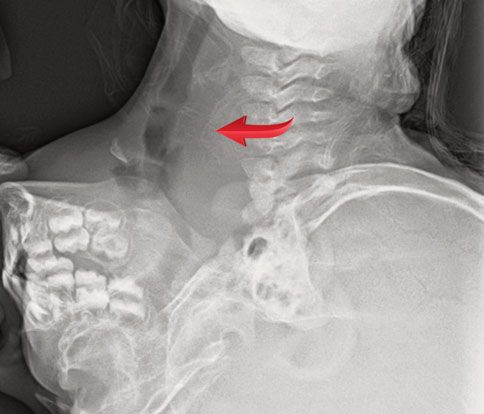
A P P R O A C H TO F E V E R A N D D R O O L I N G I N I N FA N T S A N D TO D D L E R S

  skin lesions around the lips are         Figure 1. Suspected herpangina
  vesicles that often cluster together
  and coalesce. The gingiva appear
  inflamed and bleed easily.6,7 The
  diagnosis can be made clinically
  without the use of additional lab-
  oratory techniques to confirm the
  diagnosis; however, sending a
  viral PCR test may be necessary
  in children who are immunocom-
  promised as the risk of complica-
  tions may be higher. Oral
  acyclovir may help shorten the
  duration of symptoms if initiated
  within 72-96 hours of disease
  onset.5 Keep in mind that
  neonates concerning for cuta-
  neous HSV infection require spe-
  cial consideration for escalation
  of care due to the high risk of
  morbidity and mortality associ-
  ated with HSV encephalitis and
  disseminated infections in this
  particular population.
! Thrush is an oropharyngeal Can-
  dida infection that is common in
  healthy infants. It is manifested
  as white plaques on the intraoral
  mucosa. Milk curd can be difficult
  to distinguish from thrush; a trick      Credit: Katherine P. Dureau, MD
  is to run a tongue depressor over
  the plaques, as thrush is difficult to remove. Treatment               ! As opposed to common viral illnesses that present
  is with topical nystatin suspension.8 Thrush does not                    with fever and drooling, children with deep neck
  typically cause a fever, although some infants could                     infections usually have a more dramatic presentation
  have a coexisting viral infection that causes elevation                  including ill appearance, refusal to move the neck,
  in body temperature.                                                     and, rarely, stridor.
! Acute bacterial pharyngitis is most commonly due to
  a group A streptococcus (GAS) infection. It affects chil-              What Not to Miss
  dren ages 5-15 years old. Clinical manifestations                      ! A retropharyngeal abscess (RPA) is a deep neck space
  include fever and sore throat and, when severe, can                      infection that presents in children
A P P R O A C H TO F E V E R A N D D R O O L I N G I N I N FA N T S A N D TO D D L E R S

Figure 2. Child with prevertebral space widening                                           Figure 3. Note arrow indicating prevertebral space
                                                                                           widening

 Credit: Neil Vachhani, MD, Children’s Hospital of the King’s Daughters, Norfolk VA          Credit: Neil Vachhani, MD, Children’s Hospital of the King’s Daughters, Norfolk VA

                                                                                            agement. Contrast-enhanced neck CT is the imaging
Figure 4. Child with normal prevertebral space
                                                                                            modality of choice to identify, localize, and differentiate
                                                                                            abscess from phlegmon/cellulitis. Signs of an RPA
                                                                                            should prompt referral to a specialty center for further
                                                                                            evaluation by a pediatric otolaryngologist, as man-
                                                                                            agement includes intravenous antibiotic therapy and
                                                                                            possible surgical intervention.10,11
                                                                                          ! Ludwig angina (submandibular space infection) is a
                                                                                            bacterial cellulitis of the floor of the mouth in the sub-
                                                                                            lingual and submaxillary space. In the most severe
                                                                                            cases, oropharyngeal intubation is difficult because
                                                                                            of the inability to lift the tongue, making this infec-
                                                                                            tion a potential airway emergency. Ludwig angina is
                                                                                            typically a mixed anaerobic bacterial infection that is
                                                                                            often due to spreading of a mandibular molar abscess.
                                                                                            Patients present with fever, mouth pain, stiff neck,
                                                                                            drooling, and dysphagia. They have tenderness in the
                                                                                            submandibular area and the mouth is held open by
                                                                                            lingual swelling. The diagnosis is made based on the
 Credit: Neil Vachhani, MD, Children’s Hospital of the King’s Daughters, Norfolk VA         suggestive exam findings. CT imaging may be helpful
                                                                                            to evaluate the depth and size of the infected area.
  or there is a high clinical suspicion. If the clinician                                   The treatment is empiric broad-spectrum antibiotics,
  does opt for lateral neck imaging on site, the film                                       but surgery is usually not necessary. Immediate trans-
  should be taken during inspiration and false thickening                                   port to a specialty center is indicated given the risk of
  can be caused by crying, especially in infants. Prever-                                   airway compromise from glottic swelling.12,13
  tebral space thickening on a good quality film has fair
  specificity for an RPA, but a negative film does not                                    What to Think About:
  exclude the possibility of a deep neck infection. In                                    ! Epiglottitis is an inflammation of the epiglottis that can
  addition, this is not a definitive study to guide man-                                    lead to a rapidly progressive upper airway obstruction.

14   JUCM T h e J o u r n a l o f U r g e n t C a r e M e d i c i n e | J u l y -A u g u s t 2 0 1 8                                                      w w w. j u c m . c o m
A P P R O A C H TO F E V E R A N D D R O O L I N G I N I N FA N T S A N D TO D D L E R S
“Classic epiglottitis Pearls—Fever and Drooling
• The diagnostic evaluation is mostly clinical; the use of
should be suspected laboratory and radiographic tests is rarely necessary.
• The most common causes are self-limited viral illnesses
usually requiring only supportive therapy.
in an unimmunized toddler • Red flags include: toxic appearance, respiratory distress,
stridor, neck stiffness, tripoding, decreased neck range of
with an acute onset of fever, motion, and trismus.
• Neck radiography is a reasonable screening tool to evaluate
for neck infections (eg, retropharyngeal abscess or
dysphagia, drooling, epiglottis); however, it is not sufficiently sensitive if clinical
suspicion is high.
and respiratory distress.” • Reasons for referral include dehydration, radiographic
evidence of prevertebral space widening, clinical suspicion
of deep neck infection, stridor and respiratory distress in the
absence of cough, and signs of potential airway compromise
(eg, epiglottitis, Ludwig angina).
He is given a dose of ibuprofen and a popsicle. Reexami-
With the introduction of the Haemophilus influenza nation 30 minutes later shows a playful child without drool-
type b vaccine, the incidence has dropped dramati- ing. He appears comfortable and well hydrated. No diagnostic
cally; however, the epidemiology has changed with an testing is performed. He is discharged home with education
increasing incidence secondary to Streptococcus infec- and supportive care including acetaminophen or ibuprofen
tion in older vaccinated children. Classic epiglottitis as needed for fever reduction and pain control. Return pre-
should be suspected in an unimmunized toddler with cautions were discussed, including dehydration or inability
an acute onset of fever, dysphagia, drooling, and res- to manage pain at home. !
piratory distress. The child may appear toxic and prefer
References
to sit in the “sniffing” or “tripod” position to maxi- 1. Hardy E. Hand-foot-mouth disease. Ferri’s Clinical Advisor 2018. 1st ed. Philadelphia, PA:
mize airway patency. Stridor may also be present, but Elsevier; 2018:540.
2. Centers for Disease Control and Prevention (CDC). Severe hand, foot, and mouth disease
cough is distinctly uncommon, differentiating epiglot- associated with Coxsackievirus A6–Alabama, Connecticut, California, and Nevada,
titis from tracheal diseases such as croup and bacterial November 2011-February 2012. MMWR Morb Mortal Wkly Rep. 2012;61:213-214.
3. Abzug MJ. Hand foot and mouth disease and herpangina. Nelson Textbook of Pediatrics.
tracheitis. A lateral neck radiograph may show the clas- 20th ed. Philadelphia, PA: Elsevier; 2016:1561-568.
sic “thumb sign” demonstrating a swelling of the 4. Michaels MG, Williams JV. Coxsackievirus and other enteroviruses. Zitelli and Davis’
Atlas of Pediatric Physical Diagnosis. 7th ed. Philadelphia, PA: Elsevier; 2018:455-509.
epiglottis. If the diagnosis of epiglottitis is suspected, 5. Faden H. Management of primary herpetic gingivostomatitis in young children. Pediatr
immediate transport to a specialty center is indicated Emerg Care. 2006;22:268-269.
6. Cohen BA. Herpes simplex virus. Pediatric Dermatology. 4th ed. Philadelphia, PA: Else-
and additional stresses should be avoided to prevent vier; 2013:104-125.
the risk of sudden airway obstruction.14,15 7. Schiffer JT, Corey L. Therapy for HSV Infections. Mandell, Douglas, and Bennett’s Principles
and Practice of Infectious Diseases. 8th ed. Philadelphia, PA: Saunders; 2015:1713-1730.
8. Marcdante KJ, Kliegman RM. Oral Cavity. Nelson Essentials of Pediatrics. 7th ed. Philadel-
Case Conclusion phia, PA: Saunders; 2015:429–430.
9. Yellon RF, Chi DH. Otolaryngology. Zitelli and Davis’ Atlas of Pediatric Physical Diagnosis.
Upon further examination, the patient’s oral exam shows 7th ed. Philadelphia, PA: Elsevier; 2018:868–915.
multiple erythematous oral ulcers on his soft palate. He has 10. Pappas DE, Hendley JO. Retropharyngeal abscess, lateral pharyngeal (parapharyngeal)
abscess, and peritonsillar cellulitis/abscess. Nelson Textbook of Pediatrics. 20th ed. Philadel-
scattered erythematous macules on his palms and soles. phia, PA: Elsevier; 2016:2021-2023.
Removal of his diaper reveals erythematous papules and 11. Rose E. Pediatric respiratory emergencies: upper airway obstruction and infections.
Rosen’s Emergency Medicine: Concepts and Clinical Practice. 9th ed. Philadelphia, PA: Elsevier;
vesicular lesions. The patient is diagnosed with hand-foot- 2018:2069-2080.
and-mouth disease based on the findings of oral ulcers cou- 12. Marcdante KH, Kliegman RM. Pharyngitis. Nelson Essentials of Pediatrics. 7th ed.
Philadelphia, PA: Saunders; 2015:347–349.
pled with skin lesions found on the palms, soles, and buttock. 13. Shaw J. Infections of the oral cavity. Principles and Practice of Pediatric Infectious Diseases.
His history and physical examination are absent for red flags 5th ed. Philadelphia, PA: Elsevier; 2018:193-199.
14. Marcdante K, Kliegman RM. Croup (Laryngotracheobronchitis). Nelson Essentials of
to suggest a deep neck space infection or a bacterial infection Pediatrics. 7th ed. Philadelphia, PA: Saunders; 2015:354–356.
of the floor of the mouth or epiglottis. 15. Nayak JL, Weinberg GA. Epiglottitis. Mandell, Douglas, and Bennett’s Principles and Practice
of Infectious Diseases, Updated Edition. 8th ed. Philadelphia, PA: Saunders; 2015: 785-788.
w w w. j u c m . c o m JUCM T h e J o u r n a l o f U r g e n t C a r e M e d i c i n e | J u l y -A u g u s t 2 0 1 8 15You can also read