Assessment of Chest Pains in the ER - Assessment of Chest Pains in the Emergency Room
←
→
Page content transcription
If your browser does not render page correctly, please read the page content below
3/15/2018
Assessment of Chest Pains in the
Emergency Room
Assessment of Chest Pains in the ER
Goals:
Levin’s sign
Identification or
exclusion of acute
coronary disease
Accurate risk
stratification
1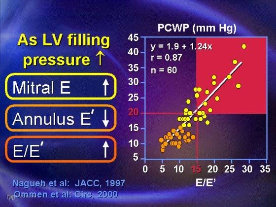
3/15/2018
Assessment of Chest Pains in the ER
ESC 2016
Emergency Echocardiography
Advantages of echocardiography as a diagnostic tool in
the emergency room:
Safe and tolerable
Easily reproducible
Readily available (portability)
Relatively low-cost
No radiation
2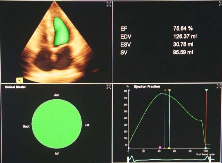
3/15/2018
Emergency Echocardiography
Philippine Heart Center Echocardiography Census (2011)
Total In-patient Echo 4,407
Bedside Echo (ER and ICU) 1,110 (25%)
Intra-operative TEE (IOTEE) 221 ( 5%)
Top 3 indications for emergency echocardiography:
Chest pains
Dyspnea
Hypotension
Emergency Echocardiography:
Acute Coronary Syndrome (ACS)
Wall thickening abnormalities
Evidence of ruptured intraventricular septum, LV
free wall or papillary muscle (significant MR)
LV systolic and diastolic functions
Cardiac filling pressures
Pulmonary arterial pressure
3
3/15/2018
Echo: Acute Myocardial Infarction
Case
Systolic Function Assessment
4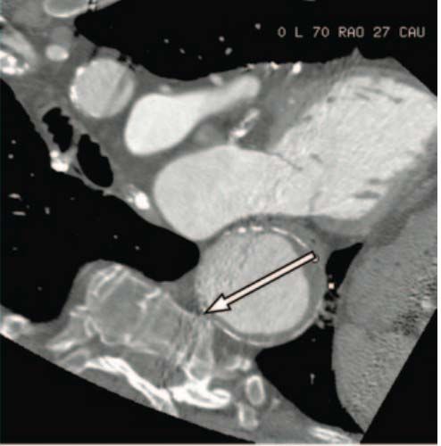
3/15/2018
3D echo for LV analysis
Full-volume reconstruction of the LV with EF computation
Doppler Tissue Imaging (DTI)
E/Ea ratio – correlates well
with LV filling pressure
E/Ea ratio > 15
Highly specific for elevated
LA pressure
E/Ea < 8
Sensitive for normal LA
pressure
Khouri et al, JASE March 2004
E/Ea ratio < 10
- PCWP < 15 mmHg
E/Ea ratio > 15
- PCWP > 20 mmHg
Nagueh et al. Circulation. 2000
5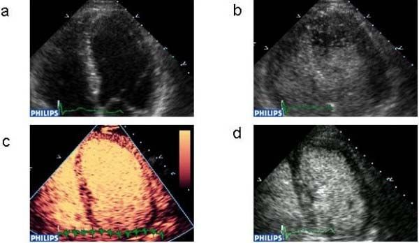
3/15/2018
Myocardial Contrast
Echocardiography in the ER setting
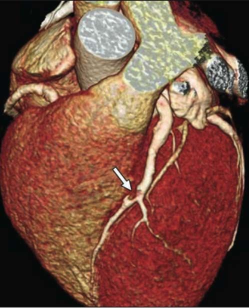
Cardiac Computed Tomography Angiography
(CTA)
Bastarrika, et al. AJR, 2009
6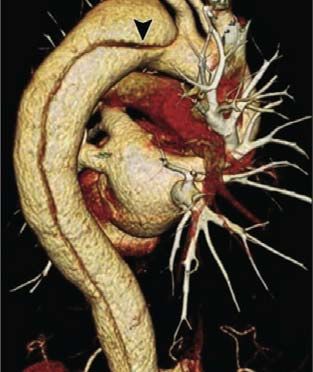
3/15/2018
Coronary CTA
Double discharge rates from ER
Shorten length of stay at the ER
Reduced the likelihood of negative coronary catheterization
Sensitivity 93.3% Specificity 89.9%
Takakuwa KM,et al. Acad Radiol, 2011 Litt HL, et
al. N Eng J Med 2012
Hoffmann U, et al. N Eng J Med 2012
Coronary CTA
is an appropriate option for the low to intermediate risk
with chest pains at ER
Negative CTA is associated with a very low event rate,
and discharge of patients from ER is safe
ACC/ACR/AHA Concensus 2010
7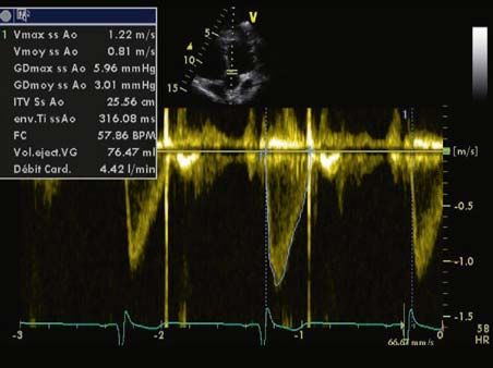
3/15/2018

Indications for CTA in ED

                     Hollander et al. Circulation 2010

                               Circulation 2010

                                                                8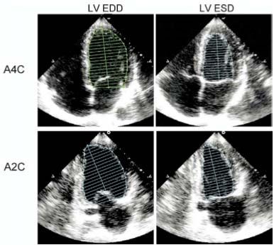
3/15/2018
Cardiovascular Magnetic Resonance Imaging
(CMR)
• CMR is highly accurate for the detection of ACS in the
ER setting, and provides incremental value to initial
risk assessment and traditional risk factors.
Cury, et al. Circulation, 2010
Chest Pains in the Emergency Room
“Triple rule-out concept”:
Acute coronary syndrome (myocardial ischemia or
infarction)
Aortic aortic syndrome (aortic dissection, intramural
hematoma and rupturing aortic aneurysm)
Pulmonary embolism
9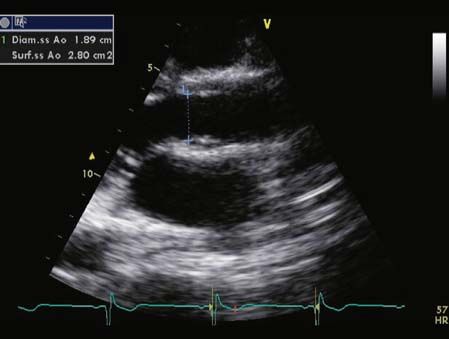
3/15/2018
Triple Rule-out by CT
Bastarrika, et al. AJR, 2009
Triple rule-out CTA
Requires higher scanning time and longer radiation
exposure
Volume of contrast used is high
Coronary CT dedicated protocol provides excellent
visualization of coronaries and proximal ascending
aorta, but not well enough the pulmonary arteries
103/15/2018
Echo: Aortic Dissection
Case
Emergency Echocardiography:
Aortic Dissection
TTE findings associated with aortic dissection
Aortic insufficiency
Enlarged aortic root (>3.5 cm at annulus or sino-tubular
junction)
Presence of pericardial effusion
Infero-posterior wall motion abnormality (RCA territory)
113/15/2018
Emergency Echocardiography:
Aortic Dissection
May require TEE to visualize distal ascending aorta,
transverse and descending aorta
Intimal flap seen on TTE would clinch a diagnosis of an
aortic dissection
Emergency Echocardiography:
Aortic Dissection
Lack of definite signs on TTE/TEE does not exclude an
aortic dissection …
CLINICAL DATA still important !!!
123/15/2018
Cardiac CT-MRI: Aortic Dissection
- Complimentary tools to emergency echocardiography in
the assessment of aortic dissection
- Cardiovascular CT-MRI “triple rule-out” capability is an
advantage over other modalities
- Disadvantages: not readily available, non-portable,
expensive, and requires highly skilled technician and staff
CT-MRI aortography
Bastarikka et al. AJR, 2009
133/15/2018
Echo: Pulmonary Embolism
Case
Pulmonary Embolism:
RV Systolic Overload
Elevated right ventricular afterload leading to pulmonary
hypertension and right heart failure
Echocardiographic features:
1. Dilated pulmonary artery, right ventricle and right atrium
2. Hypokinetic right ventricle
3. Moderate to severe TR with elevated pulmonary systolic
arterial pressure
4. Systolic septal flattening
5. No significant left heart abnormality
143/15/2018
RVEF by 3D Echocardiography
Full-volume reconstruction of the right ventricle with
EF computation
CMR: Pulmonary Embolism
Computed Tomography CMR
153/15/2018
Summary
Echocardiography is still the most commonly used and
readily available diagnostic tool for patients having
chest pains at the ER.
CTA and CMR have important roles in the “triple rule-
out” approach for patients presenting with chest pains
at the emergency room.
Use of CMR to detect ACS in patients presenting with
chest pains in ER setting may not be practical.
Thank You
16You can also read