AUTOMATIC PALATE DELINEATION IN ULTRASOUND VIDEOS - ICPHS 2019
←
→
Page content transcription
If your browser does not render page correctly, please read the page content below
AUTOMATIC PALATE DELINEATION IN ULTRASOUND VIDEOS
             Guillaume Faucher1 , Elham Karimi1 , Lucie Ménard2 and Catherine Laporte1

                  1 École   de technologie supérieure; 2 University of Québec in Montréal
   guillaume.faucher.1@etsmtl.net, elham.karimi.1@etsmtl.net, menard.lucie@uqam.ca, catherine.laporte@etsmtl.ca

                    ABSTRACT                                fers the palate surface as the upper boundary of the
                                                            space reached by the (automatically tracked) tongue
Measurements of palate location can assist ultra-           as speech is continuously elicited from a speaker.
sound (US)-based analysis of articulatory tongue            This method is appealing because it can be used in
motion by providing complementary information               real-time. However, it requires high speed imaging
about oral cavity constriction. They also provide a         to capture the instants where the tongue touches the
rigid reference frame relative to which tongue mea-         palate. It also requires eliciting such contacts over
surements from different time points can be regis-          the entire oral cavity, which can be challenging in
tered. Locating the palate in US images is challeng-        populations with speech impairments.
ing because it is generally invisible except during            The traditional approach is to directly delineate
swallowing, and even then, it is often not readily rec-     the palate in recordings where the speaker is swal-
ognizable in any single frame. This paper introduces        lowing or holding liquid in his/her mouth [13, 3, 9].
a new automated method to extract a palate con-             Then, US can reach the palate, causing a visible echo
tour from an US video acquired during swallowing.           in the US images. Edgetrak [8] offers a rudimentary
The method is based on a cumulative echo skeleton           semi-automatic tool to fit a snake to manually an-
image, which highlights structures that are consis-         notated palate points on a single image. However,
tently located over time. In experiments with 22 US         in many cases, the palate is only partially visible
videos, most of the automatically extracted palate          in any given image. Thus, Epstein and Stone [3]
traces were within 3 mm of a manual palate trace in         recommend using a short video clip acquired dur-
terms of mean sum of distances error, demonstrating         ing swallowing and manually accumulating partial
the potential of the proposed approach.                     palate traces over a series of frames as a bolus of wa-
                                                            ter or saliva travels through the mouth and different
Keywords: Ultrasound, palate, image processing
                                                            parts of the palate become visible. This is challeng-
               1. INTRODUCTION                              ing for the operator, who must simultaneously see
                                                            through time and space to delineate the palate in a
Ultrasound (US) imaging is ideally suited and               piecewise fashion from one frame to the next.
widely used to study tongue shape and motion dur-              This paper proposes a new automatic method
ing speech. However, tongue location and shape              to extract the mid-sagittal palate contour from US
alone fail to fully capture phonetically relevant vari-     videos of a swallow that overcomes the aforemen-
ables pertaining to constrictions of the oral cavity.       tioned difficulty. The method, described in Sec-
To remediate this, it is useful to measure the config-      tion 2, is based on a cumulative echo skeleton im-
uration of the tongue relative to the palate, which         age, which (1) highlights features of the image that
in turn requires locating the palate in the same ref-       are consistently located over time (and might corre-
erence frame as the tongue. Palate measurements             spond to the palate) and (2) connects the parts of the
also provide a rigid reference frame for registration       palate that are visible in different frames as a sin-
of US tongue data over time [9], and for studying the       gle structure. Section 3 describes experiments on
influence of the palate shape on articulation [2].          22 swallow videos, wherein automatically extracted
   While dental casts provide detailed palate mea-          palatal traces were compared against a manual trace,
surements (e.g., in electropalatography), measuring         with promising results. A discussion, including di-
the palate from the US images themselves is quick           rections for future work, is presented in Section 4.
and inexpensive in comparison [12]. Unfortunately,
the palate is usually invisible in US images be-                               2. METHOD
cause of the air separating it from the tongue. Re-
cently, Wrench [14] demonstrated an indirect, “vo-          The proposed palate extraction method comprises
cal tract carving” approach that automatically in-          processing at the level of (1) the individual frames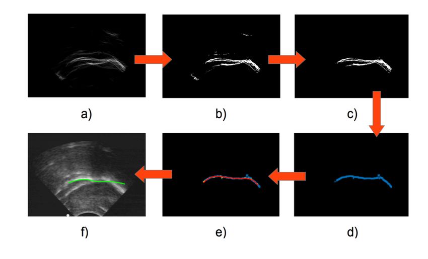
composing the swallow video sequence and (2) the         shown in Figs. 1 (right) and 2.
sequence itself. The individual image processing
step, described in Section 2.1, extracts a line draw-    2.2. Sequence level processing
ing, called a skeleton, that characterizes the shape
of the brightest ridge-like structures (echoes) in the   The echo skeletons computed from individual US
image (Fig. 1, right). At the sequence level, the        images typically contain information about parts of
skeletons from the individual images are summed          the tongue and/or palate and/or some imaging arte-
over time, leading to a cumulative echo skeleton im-     facts. One skeleton is generally insufficient to in-
age (Fig. 2), which emphasizes the structures that       fer the palate surface in a robust manner. For this,
are most persistent and consistently located over the    one must exploit the temporal information contained
duration of the video sequence. Since the palate         in the sequence of images. Thus, the skeletons ex-
is mostly rigid and immobile (unlike the tongue or       tracted from the different images in the sequence are
imaging artefacts), the cumulative echo skeleton im-     summed to form a cumulative echo skeleton image,
age carries meaningful information about its shape       as shown in Fig. 2. In this sum, each white pixel
and location. This information is extracted using        from an individual skeleton image slightly increases
thresholding, clustering and robust curve fitting op-    the intensity of the corresponding pixel in the cu-
erations prior to shape refinement using a snake fit-    mulative echo skeleton image. Immobile and per-
ted to one or more of the original US images, as         sistent structures like the palate contribute to similar
shown in Fig. 3 and detailed in Section 2.2.             locations in the cumulative echo skeleton image over
                                                         time, leading to high signal levels, whereas moving
2.1. Frame-level processing                              or non-persistent structures like the tongue or imag-
                                                         ing artefacts, though often brighter than the palate in
                                                         single images, contribute to more diverse locations
Fig. 1 shows the processing steps applied to each
                                                         and lead to weaker signals.
image in the swallow sequence. The bright ridge-
like echoes in the US image, typically the palate and        Fig. 3 shows how the cumulative echo skeleton
tongue, along with some imaging artefacts, are en-       image is processed to extract palate contours. Otsu
hanced within a phase symmetry map [7, 6]. This          thresholding [10] is applied to its non-zero pixel in-
map is obtained by first filtering the image using       tensities to remove noise arising from non-persistent
odd and even log-Gabor filters at different scales and   structures in the US images. The locations of the
orientations (5 scales and 14 orientations were used     non-zero valued pixels in the thresholded image
here). Phase symmetry is the average amplitude dif-      are clustered using DBSCAN [4], an algorithm that
ference in the responses from the even and odd filters   forms arbitrary numbers of clusters from spatial data
or, more intuitively, the degree of even symmetry or     based on thresholds ε, the maximum distance be-
“ridgeness” of the structures in the image. Fig. 1       tween points within a cluster, and MinPts, the mini-
(middle) shows a typical result.                         mum acceptable number of points within each clus-
                                                         ter. Here, ε = 20 pixels and MinPts = 10. The
   Figure 1: Processing of individual images. From       widest cluster is selected as potentially containing
   left to right: original image, phase symmetry map,    the palate. Within this cluster, the point of maximal
   skeleton of thresholded phase symmetry image.         height is retained for each position along the hor-
                                                         izontal axis. This favours points arising from the
                                                         reflection of US off the palate rather than off the
                                                         tongue. A second order polynomial is then fitted
                                                         to the resulting points using RANSAC [5], a robust
                                                         fitting algorithm that finds the least-squares fitting
                                                         curve accounting for the largest number of inliers,
                                                         while rejecting outliers. Outliers are defined by a
   The phase symmetry map is thresholded to pre-         maximum allowable distance to the curve (10 pixels
serve only the brightest and largest echoes from the     in this work). A cubic spline is then fit to the inliers,
original images. A skeleton of these structures then     which is then used to initialize a snake fit [8] to the
extracted by finding their medial axis. The medial       palate for refinement.
axis is the locus of points within a shape that are
equidistant from two or more of the shape bound-                  3. EXPERIMENTAL RESULTS
ary points [1]. Many methods exist to compute me-
dial axes; here, Rezanejad et al’s robust average out-   Automatic palate extraction was tested on US data
ward flux method [11] is used. Typical skeletons are     from 6 healthy subjects, 3 adults (A1-A3) and 3 chil-
Figure 2: Creation of the cumulative echo skeleton image. The 6 leftmost panels show sample US images from a
swallowing video sequence (top) and the skeletons extracted from each one (bottom). The rightmost panel shows
the cumulative echo skeleton image computed from the sum of the skeletons over time.
(respectively v), i ∈ {1, . . . , m} and j ∈ {1, . . . , n}.
Figure 3: Palate contour extraction from the cu-
mulative echo skeleton image. The cumulative Fig. 4 compares the automatically extracted palate
echo sekeleton image (a) is thresholded (b) to en- traces with the reference palate traces and reports
hance temporally persistent structures. The widest the MSD between them. Generally, the automati-
cluster of white pixels is extracted from the thresh- cally detected palate traces overlap fairly well with
olded image (c). Data from the earliest US echoes the manual ones. However, the automatically de-
are removed (d), and a cubic spline is robustly fit- tected traces tend to be shorter, particularly towards
ted to the remaining points (e). A snake is fitted to the back of the mouth. In several clips from child
US images to obtain palate contours (f).
subjects, the automatic palate trace matches quite
well to the location of the hard palate, which is rigid
and generally more visible in the images, whereas
the manual trace often also comprises the velum.
This suggests that the proposed method is most suc-
cessful at locating the rigid part of the palate. Ar-
guably, this is desirable for applications requiring
measurements of the relative configuration of the
tongue with respect to a rigid palate reference frame.
The method failed in a few cases. For subject
A2, it only detected a small segment of the palate
due to the relatively poor quality of the images in
this subject’s recordings, where even the tongue was
dren (C1-C3), acquired during speech. Twenty-two less visible than in other recordings. In subject
clips with swallowing, ranging in length from 48 to A3, the method detected the tongue instead of the
154 frames, were manually extracted from the full palate in two of three swallowing clips. Upon in-
recordings. A reference palate contour was manu- spection, both clips were found to depict a resting,
ally traced on one reference image in each clip. The fairly immobile tongue, for many frames before and
mean sum of distances (MSD) between the reference after the swallowing motion. Thus, the cumulative
palate trace u and the automatic palate trace v was echo skeleton image contained stronger contribu-
computed as tions from the resting tongue than from the palate.
This points to the importance of feeding quality in-
n put to the method. Ideally, this would include little
1
(1) MSD(u, v) = ( ∑ min ||vvi − u j || + but the actual swallowing motion.
m + n i=1 j
m
||uu j − v i ||), 4. CONCLUSIONS
∑ min
i
j=1
This paper presented a new method to automati-
where u i (respectively v j ) is the vector of x and y cally extract the mid-sagittal palate contour from
coordinates of the ith (respectively jth) vertex of u US video sequences of swallowing by exploiting theFigure 4: Automatic palate extraction results (solid red) and manual palate trace (dashed green) overlayed on the
reference image from each swallowing sequence. The MSD between the two traces is reported below each image.
A1, Swallow 1 A1, Swallow 2 A1, Swallow 3 A2, Swallow 1 A2, Swallow 1 A2, Swallow 3
MSD = 2.36 mm MSD = 4.24 mm MSD = 2.68 mm MSD = 4.00 mm MSD = 2.63 mm MSD = 5.49 mm
A3, Swallow 1 A3, Swallow 2 A3, Swallow 3 C1, Swallow 1 C1, Swallow 2 C1, Swallow 3
MSD = 8.35 mm MSD = 3.34 mm MSD = 2.96 mm MSD = 1.65 mm MSD = 2.84 mm MSD = 3.69 mm
C2, Swallow 1 C2, Swallow 2 C2, Swallow 3 C2, Swallow 4 C2, Swallow 5 C2, Swallow 6
MSD = 1.73 mm MSD = 2.18 mm MSD = 2.23 mm MSD = 2.79 mm MSD = 1.63 mm MSD = 1.99 mm
C3, Swallow 1 C3, Swallow 2 C3, Swallow 3 C3, Swallow 4
MSD = 1.63 mm MSD = 1.46 mm MSD = 1.81 mm MSD = 2.38 mm
persistence of the echoes generated by the palate tion (e.g. front to back) in the analysis of US record-
over time. The method was tested on 22 video se- ings in the field, where sophisticated head motion
quences with promising results in terms of accuracy. measurement devices may not be practical or avail-
In future work, ideal or near ideal swallowing se- able.
quences could probably be extracted automatically
from larger speech video recordings by searching for ACKNOWLEDGMENTS
segments with large amounts of motion and weak
acoustic signal. Automated palate extraction using This work was supported by the Natural Sciences
spontaneous swallowing in US video sequences is and Engineering Research Council of Canada, the
an important step towards facilitating more mean- Social Science and Humanities Research Council of
ingful articulatory measurements. It may also pro- Canada and the Fonds de Recherche Québécois -
vide a useful rigid reference frame which can help Nature et Technologie.
evaluate and compensate for some types of head mo-5. REFERENCES
[1] Blum, H. 1967. A transformation for extracting
new descriptors of shape. Models for the Percep-
tion of Speech and Visual Form (5), 362–380.
[2] Brunner, J., Fuchs, S., Perrier, P. 2009. On the rela-
tionship between palate shape and articulatory be-
havior. Journal of the Acoustical Society of Amer-
ica 125(6), 3936–3949.
[3] Epstein, M., Stone, M. 2005. The tongue stops
here: ultrasound imaging of the palate. Journal
of the Acoustical Society of America 118(4), 2128–
2131.
[4] Ester, M., Kriegel, H. P., Sander, J., Xu, X. 1996.
A density-based algorithm for discovering clus-
ters in large spatial databases with noise. ACM
SIGKDD Conference on Knowledge Discovery and
Data Mining 226–231.
[5] Fischler, M. A., Bolles, R. C. 1981. Random sam-
ple consensus: a paradigm for model fitting with
applications of image analysis and automated car-
tography. Communications of the ACM 24(6), 381–
395.
[6] Hacihaliloglu, I., Abugharbieh, R., Hodgson, A. J.,
Rohling, R. N. 2009. Bone surface localization in
ultrasound using image phase-based features. Ul-
trasound in Medicine & Biology 35(9), 1475–1487.
[7] Kovesi, P. 1999. Image features from phase congru-
ency. Videre: Journal of Computer Vision Research
1(3), 1–26.
[8] Li, M., Kambhamettu, C., Stone, M. 2005. Auto-
matic contour tracking in ultrasound images. Clin-
ical Linguistics and Phonetics 19(6-7), 545–554.
[9] Mielke, J., Baker, A., Archangeli, D., Racy, S.
2005. Palatron: a technique for aligning ultrasound
images of the tongue and palate. Coyote Papers:
Working Papers in Linguistics, Linguistic Theory at
the University of Arizona 14, 96–107.
[10] Otsu, N. 1979. A threshold selection method from
gray-level histograms. IEEE Transactions on Sys-
tems, Man and Cybernetics 9(1), 62–66.
[11] Rezanejad, M., Siddiqi, K. 2013. Flux graphs for
2d shape analysis. Shape Perception in Human and
Computer Vision 41–54.
[12] Scobbie, J. M., Stuart-Smith, J., Lawson, E. 2008.
Looking variation and change in the mouth: de-
veloping the sociolinguistic potential of ultrasound
tongue imaging. Technical report Queen Margaret
University.
[13] Stone, M. 2005. A guide to analysing tongue mo-
tion from ultrasound images. Clinical Linguistics
and Phonetics 19(6-7), 455–501.
[14] Wrench, A. A. 2017. Real-time tongue contour fit-
ting and vocal tract carving. Ultrafest VIII 99–100.You can also read