Bioimaging Science Program 2021 Principal Investigator Meeting Proceedings - Biomolecular Characterization and Imaging Science
←
→
Page content transcription
If your browser does not render page correctly, please read the page content below
Biomolecular Characterization and Imaging Science
Bioimaging Science Program
2021 Principal Investigator
Meeting Proceedings

                        Office of Biological and Environmental Research

                                                          June 2021
Biomolecular Characterization and Imaging Science

                                    Bioimaging Science Program
                                2021 Principal Investigator Meeting

                                                    February 22–23, 2021

                                                     Program Manager
                                                    Prem C. Srivastava
                                    Office of Biological and Environmental Research
                                                     Office of Science
                                                U.S. Department of Energy

                                                    Meeting Co-Chairs

                             Tuan Vo-Dinh                                 Marit Nilsen-Hamilton
                             Duke University                               Iowa State University

                          Jeffrey Cameron                                     James Evans
                    University of Colorado–Boulder               Pacific Northwest National Laboratory

About BER
The Biological and Environmental Research (BER) program advances fundamental research and scientific user facilities to support
Department of Energy missions in scientific discovery and innovation, energy security, and environmental responsibility. BER seeks to
understand biological, biogeochemical, and physical principles needed to predict a continuum of processes occurring across scales,
from molecular and genomics-controlled mechanisms to environmental and Earth system change. BER advances understanding of
how Earth’s dynamic, physical, and biogeochemical systems (atmosphere, land, oceans, sea ice, and subsurface) interact and affect
future Earth system and environmental change. This research improves Earth system model predictions and provides valuable infor-
mation for energy and resource planning.

Cover Images
Image 1: Medicago truncatula, see p. 31; Image 2: XRF confocal image of a leaf treated with AuNR@Ag, see p. 3; Image 3: The
combined nonlinear wide-field and in situ-LE-ME imaging system, see p. 38; Image 4: In situ imaging of lignin removal and enzyme
accessibility, see p. 5; Image 5: Plants expressing Pen3:GFP label intracellular and extracellular vesicles, see p. 20.

Digital Download
science.osti.gov/-/media/ber/bioimaging-technology/pdf/2021/Bioimaging_Science_PI_Meeting2021.pdf
Biomolecular Characterization and Imaging Science

                                   Bioimaging Science Program
                     2021 Principal Investigator Meeting Proceedings

                                                  Published June 2021

                               Office of Biological and Environmental Research

Prepared for the                                            Prepared by
U.S. Department of Energy                                   Biological and Environmental Research Information System
Office of Science                                           Oak Ridge National Laboratory
Office of Biological and Environmental Research             Oak Ridge, TN 37830
Germantown, MD 20874-1290                                   Managed by UT-Battelle, LLC
                                                            For the U.S. Department of Energy
                                                            Under contract DE-AC05-00OR22725
2021 PI Meeting Proceedings
Contents
Preface .......................................................................................................................................................................................................................................iii
Bioimaging Science Program Projects..........................................................................................................................................................................iv
Project Map...............................................................................................................................................................................................................................v
Executive Summary..............................................................................................................................................................................................................vi
Abstracts.....................................................................................................................................................................................................................................1
Multimodal Single-Cell/Particle Imaging and Engineering for Energy Conversion in Bacteria..........................................................................1
Plasmonics-Enhanced Optical Imaging Systems for Bioenergy Research..................................................................................................................3
eal-Time Imaging and Quantification of Plant Cell Wall Constituents Using
R
Cavity-Dumped Stimulated Raman Scattering (cdSRS) Microscopy.............................................................................................................................5
Single-Molecule Imaging of Lignocellulose Deconstruction by SCATTIRSTORM Microscopy............................................................................7
Time-Resolved 3D Multi-Resolution Microscopy for Real-Time Cellulase Actions In Situ......................................................................................9
In Planta Multimodal Single-Molecule Imaging to Study Real-Time Turnover Dynamics
of Polysaccharides and Associated Carbohydrate Metabolites...................................................................................................................................11
Development of Broadband Infrared Nano-Spectroscopy of Biological Materials in Fluid...............................................................................13
Inorganic Voltage Nanosensors as Tools for Bioelectricity Studies in DOE-Relevant Bacteria and Their Communities..........................14
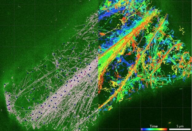
Tracking Lignocellulosic Breakdown by Anaerobic Fungi and Fungal Cellulosomes..........................................................................................15
Understanding Plant Signaling via Innovations in Probe Delivery and Imaging..................................................................................................17
Spatiotemporal Dynamics of Photosynthetic Metabolism in Single Cells at Subcellular Resolution............................................................18
Quantum Dot Toolkit for Multimodal Hyperspectral Bioimaging...............................................................................................................................19
Live-Cell, Quantum Dot-Based Tracking of Plant and Microbial Extracellular Vesicles........................................................................................20
orrelative Imaging of Enzyme and Metabolome Dynamics for Yield and Titer Co-Optimization
C
in Biofuel-Producing Microorganisms...................................................................................................................................................................................22
evelopment and Implementation of an In Situ High-Resolution Isotopic Microscope
D
for Measuring Metabolic Interactions in Soil Mesocosms.............................................................................................................................................24
E xpanding the Utility and Range of Quantum and Polymer Dots for Multiplexed
Super-Resolution Fluorescence Imaging in Plants...........................................................................................................................................................25
Hyperspectral Light-Sheet Raman Imaging of Leaf Metabolism................................................................................................................................26
Metaoptics-Enabled Multifunctional Imaging...................................................................................................................................................................27
Multiparametric Optical Label-Free Imaging to Analyze Plant Cell Wall Assembly and Metabolism............................................................28
Detecting Chemical Signals in the Soil with 4DMAPS, an Integrated Aptasensor Assembly...........................................................................30
A Quantum-Enhanced X-ray Microscope.............................................................................................................................................................................31
evelopment of a Full-Field X-ray Fluorescence Imaging System for Near Real-Time
D
Trace Element Microanalysis of Complex Biological Systems......................................................................................................................................33
T he 3DQ Microscope: A Novel System Using Entangled Photons to Generate Volumetric
Fluorescence and Scattering Images for Bioenergy Applications..............................................................................................................................34
I lluminating the Rhizosphere: Developing an Adaptive Optics, Multiphoton Microscope
for 3D Label-Free Live Imaging of Microbes and Organic Matter in Soil and Roots.............................................................................................35
Quantum Ghost Imaging of Water Content and Plant Health with Entangled Photon Pairs............................................................................36
I ntrinsically Coregistered Chemical Imaging of Living Plant and Microbial Systems
via 3D Nonlinear Optical Mapping and In Situ-Liquid Extraction-Mass Spectrometry........................................................................................38
Probing Photoreception with New Quantum-Enabled Imaging.................................................................................................................................39
Multimodal Chemical Imaging Across Scales to Visualize Metabolic Pathways in Live Plants and Microbial Systems...........................40
References...............................................................................................................................................................................................................................41
ii
2021 PI Meeting Proceedings

Preface
As part of the 2021 Biological Systems Science Division (BSSD) Principal Investigator (PI) Meeting, the Bioimaging
Science program (BSP), within BSSD's Biomolecular Characterization and Imaging Science portfolio, held its
annual PI meeting virtually February 22–23.

BSP's mission is to understand the translation of genomic information into the mechanisms that power living
cells, communities of cells, and whole organisms. The goal of BSP is to develop new imaging and measurement
technologies to visualize the spatial and temporal relationships of key metabolic processes governing pheno-
typic expression in plants and microbes.

BSP convenes annual PI meetings to bring together its contributing investigators to review progress and current
state-of-the-art bioimaging research. Holding the BSP meeting as part of the broader BSSD PI meeting allowed
researchers to interact with the extended Genomic Science program community. This convergence provided a
platform for networking and exchange of ideas, helping to forge new multidisciplinary collaborations among
investigators from the two sister programs.

An important highlight of the BSP meeting was the keynote presentation “Enhancing Fluorescence Microscopy
with Computation” by Dr. Hari Shroff of the NIH National Institute of Biomedical Imaging and Bioengineering. All
the BSP PIs made presentations describing their research focus and progress, and these were followed by round-
table discussions of each project. The meeting’s proceedings provide an outline of the program’s current state
and potential future directions and opportunities.

Prem C. Srivastava, Ph.D.
Program Manager
Biological Systems Science Division
Office of Biological and Environmental Research
Office of Science
U.S. Department of Energy
301.903.4071; prem.srivastava@science.doe.gov

                                                                                                                     iii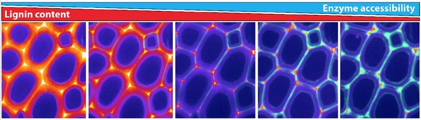
2021 PI Meeting Proceedings
Bioimaging Science Program Projects
Development and Implementation of an In Situ High-
Universities Resolution Isotopic Microscope for Measuring Metabolic
Multimodal Single-Cell/Particle Imaging and Engineering Interactions in Soil Mesocosms
for Energy Conversion in Bacteria Elizabeth A. Shank, University of Massachusetts Medical School
Peng Chen, Cornell University
Expanding the Utility and Range of Quantum and Polymer
Plasmonics-Enhanced Optical Imaging Systems for Dots for Multiplexed Super-Resolution Fluorescence Imaging
Bioenergy Research in Plants
Tuan Vo-Dinh, Duke University Gary Stacey, University of Missouri–Columbia
Real-Time Imaging and Quantification of Plant Cell Wall Hyperspectral Light-Sheet Raman Imaging of
Constituents Using Cavity-Dumped Stimulated Raman Leaf Metabolism
Scattering (cdSRS) Microscopy Keith Lidke, David Hanson, Jerilyn Ann Timlin, and Jamey Young
Shi-You Ding, Michigan State University University of New Mexico
Single-Molecule Imaging of Lignocellulose Deconstruction by Metaoptics-Enabled Multifunctional Imaging
SCATTIRSTORM Microscopy Paul Bohn, Anthony Hoffman, and Joshua Shrout
William Hancock, The Pennsylvania State University University of Notre Dame
Time-Resolved 3D Multi-Resolution Microscopy for Real-Time Multiparametric Optical Label-Free Imaging to Analyze Plant
Cellulase Actions In Situ Cell Wall Assembly and Metabolism
Haw Yang, Princeton University Marisa S. Otegui and Kevin W. Eliceiri, University of
Wisconsin–Madison
In Planta Multimodal Single-Molecule Imaging to Study
Real-Time Turnover Dynamics of Polysaccharides and
Associated Carbohydrate Metabolites National Laboratories
Sang-Hyuk Lee, Rutgers University Detecting Chemical Signals in the Soil with 4DMAPS,
an Integrated Aptasensor Assembly
Development of Broadband Infrared Nanospectroscopy of Marit Nilsen-Hamilton, Ames Laboratory
Biological Materials in Fluid
Tina Jeoh, University of California–Davis A Quantum-Enhanced X-ray Microscope
Sean McSweeney, Brookhaven National Laboratory
Inorganic Voltage Nanosensors as Tools for Bioelectricity
Studies in DOE-Relevant Bacteria and Their Communities Development of a Full-Field X-ray Fluorescence Imaging
Shimon Weiss, University of California–Los Angeles System for Near Real-Time Trace Element Microanalysis of
Complex Biological Systems
Tracking Lignocellulosic Breakdown by Anaerobic Fungi and Ryan Tappero, Brookhaven National Laboratory
Fungal Cellulosomes
Michelle O’Malley, University of California–Santa Barbara 3DQ Microscope: A Novel System Using Entangled Photons to
Generate Volumetric Fluorescence and Scattering Images for
Understanding Plant Signaling via Innovations in Probe Bioenergy Applications
Delivery and Imaging Ted A. Laurence, Lawrence Livermore National Laboratory
Jean T. Greenberg, The University of Chicago
Illuminating the Rhizosphere: Developing an Adaptive Optics,
Spatiotemporal Dynamics of Photosynthetic Metabolism in Multiphoton Microscope for 3D Label-Free Live Imaging of
Single Cells at Subcellular Resolution Microbes and Organic Matter in Soil and Roots
Jeffrey Cameron, University of Colorado–Boulder Peter K. Weber, Lawrence Livermore National Laboratory
Quantum Dot Toolkit for Multimodal Quantum Ghost Imaging of Water Content and Plant Health
Hyperspectral Bioimaging with Entangled Photo Pairs
Prashant Nagpal, University of Colorado–Boulder James Werner, Los Alamos National Laboratory
Live-Cell, Quantum Dot–Based Tracking of Plant and Intrinsically Coregistered Chemical Imaging of Living Plant
Microbial Extracellular Vesicles and Microbial Systems via 3D Nonlinear Optical Mapping and
Jeffrey L. Caplan, University of Delaware In Situ–Liquid Extraction–Mass Spectrometry
John F. Cahill, Oak Ridge National Laboratory
Correlative Imaging of Enzyme and Metabolome Dynamics
for Yield and Titer Co-Optimization in Biofuel-Producing Probing Photoreception with New Quantum-Enabled Imaging
Microorganisms James E. Evans, Pacific Northwest National Laboratory
Andreas E. Vasdekis, University of Idaho
Multimodal Chemical Imaging Across Scales to Visualize
Metabolic Pathways in Live Plants and Microbial Systems
Scott Lea, Pacific Northwest National Laboratory
iv
2021 PI Meeting Proceedings

Project Map

      University of
      California—Davis
                                                                    The University         University of
                                         University of                 of Chicago          Notre Dame
             Pacific Northwest           Colorado–Boulder (2)
             National Laboratory (2)                                                                    Rutgers                 University of
                                                             University of                            University                Massachusetts
                                                       Wisconsin–Madison                                                        Medical School
                      University                                                              Michigan State
                      of Idaho                             Ames                               University
                                                       Laboratory
                                                                                                  Cornell
                                                                                                University

                                                                                                                                 Brookhaven National
                                                                                                                                 Laboratory (2)
                                                                                                                           Princeton
                                                                                                                           University
                                                                                                                          University
                                                                                                                          of Delaware
                                                                                                                          The Pennsylvania
                                                                                                                          State University

                                                                                                                   Duke
                                                                                                                   University

          University of
          California—Los Angeles

       University of California—
       Santa Barbara                                                                               Oak Ridge
                                             Los Alamos                                            National Laboratory
                                             National Laboratory               University of
                                                                               Missouri—Columbia
 Lawrence Livermore
 National Laboratory (2)               University of
                                       New Mexico

                                                                    University Project               National Laboratory Project

                                                                                                                                             v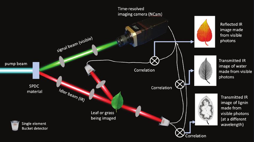
2021 PI Meeting Proceedings
Executive Summary
T he U.S. Department of Energy’s Bioimaging
Science program (BSP) supports fundamental
research to develop and apply new and enhanced
and nonlinear optical techniques. These techniques
include surface-enhanced Raman scattering (SERS),
stimulated Raman scattering (SRS), hyperspectral
bioimaging and measurement capabilities that stimulated SRS (hsSRS), and tip-enhanced Raman
enable scientists to study the biological functions of scattering (TERS), as well as nano-Fourier transform
plant and microbial systems relevant to bioenergy infrared (FTIR) and X-ray microscopies.
research. The program—within the Biomolecular
Characterization and Imaging Science portfolio BSP researchers are developing spectroscopic
of DOE’s Office of Biological and Environmental techniques to image dynamic events and molecular
Research (BER)—currently sponsors multidisciplinary processes in situ, enhancing various combinations
research at 9 national laboratories and 19 universi- of nondestructive and destructive approaches to
ties (see List of Funded Projects and map, pp. vi–vii) image laboratory-prepared or fixed samples, and
with the goal of understanding the mechanisms that creating inorganic voltage nanosensors to study
power living cells, communities of cells, and whole bacterial communities. Optical modalities are non-
organisms. BSP researchers are developing instru- invasive and include infrared/ultraviolet absorption
ments and imaging systems from the ground up and and adaptive optics multiphoton microscopy, fluo-
are enhancing existing capabilities with new or trans- rescence, and Raman techniques (e.g., conventional,
formational improvements. These novel capabilities, nonlinear, and plasmonics-enhanced). Recently,
design-based technologies, and improved or inno- BSP added quantum-enabled bioimaging science
vative uses of established methodologies will enable research projects at national laboratories. These
new fundamental discoveries and provide solutions projects encompass state-of-the-art quantum-based
to challenges in plant and microbial systems biology. techniques such as quantum-enhanced X-ray micros-
These challenges cross a range of scales—from single copy, quantum ghost imaging, three-dimensional
molecules to small unicellular organisms to complex (3D) quantum microscopy, and quantum-enabled
microbial and fungal community interactions with imaging using entangled photons. Individual research
plants. Together, BSP-supported researchers are programs focused on multidisciplinary projects are
creating an extensive and versatile toolbox enabling complemented by research and development at
real-time dynamic imaging of metabolic pathways, DOE-sponsored user facilities, which are building and
material transport within and between cellular organ- applying various technologies, such as ion micros-
elles, plant-root and organism interactions, enzyme copy and full-field X-ray fluorescence imaging.
functions, and cellular structures.
BSP researchers are further enhancing co-application
of mass spectrometry and spectrochemical imaging
Overview of Current BSP Research capabilities to yield highly selective, sensitive, and
Expansion of New and Existing Technologies quantitative chemical maps that identify intra- and
extracellular molecular gradients and the distribu-
BSP has significantly expanded since its inception in tions, abundances, and fates of stable isotopes, nat-
2015. The program recently added an extensive range ural elements, and metabolites. Using conventional
of novel bioimaging technologies and cutting-edge microscopies for correlated structural and chemical
sensing approaches, including super-resolution imaging, this work supports simultaneous observa-
microscopy, hyperspectral light-sheet imaging, adap- tion and interpretation of the biological function of
tive optics, code-aperture methods, quantum entan- living plant and microbial systems.
glement and quantitative phase imaging, correlative
imaging, and holographic force spectroscopy. These Researchers also are significantly expanding the
new technologies are complementary to and syner- performance and impact of label-based and label-free
gistic with ongoing developments in instrumentation sensing and imaging technologies by developing
involving molecular, optical, fluorescence, Raman, unique probes, such as quantum and polymer dots,
vi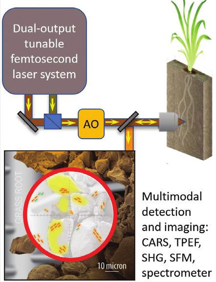
2021 PI Meeting Proceedings
as well as plasmonic nanoprobes equipped with images. While these imaging approaches focus on
various bioreceptors (e.g., antibodies, aptamers, and events inside cells, an alternative imaging approach
gene probes). These probes can specifically detect uses aptamers as sensors to image specific molecular
important biomarkers, including metabolites, pro- species present around cells. These imaging modali-
teins, and genomic markers, related to particular proc ties will be complemented by advanced technologies
esses and metabolic pathways in microbial and plant such as high-speed atomic force microscopy (AFM),
systems relevant to bioenergy research. Development interferometric scattering microscopy, infrared, and
of these unique probes and sensors is expanding the vibrational sum frequency generation. Researchers
applicability of the new instrumentation by enabling also are applying plasmonic infrared nanofocusing
researchers to dynamically track targeted cells, organ- gratings combined with microfluidics to map cellu-
elles, enzymes, biomarkers, and small molecules and lose surface fibrils with cellulose at the nanoscale.
to test and validate cellular processes and genome-
based models of cellular metabolism. Raman and Mass Spectrometry–Based Approaches
With the new instrumentation and optical probes Other important portfolio components are various
developed under BSP sponsorship, these investiga- Raman spectroscopy–based approaches, including
tions are expected to result in a better understanding spontaneous, far-field sub-diffraction, TERS, coherent
of the spatial and temporal distributions of metab- anti-Stokes (CARS), SRS, SERS, spatially offset Raman
olites associated with growing microbial and plant spectroscopy (SORS), shifted-excitation Raman differ-
systems. Also anticipated are new insights into the ence spectroscopy (SERDS), and cavity-dumped SERS.
fundamental biology of many macro events, such as BSP researchers also have developed a multimodal
nutrient utilization and community and ecosystem microscope integrating CARS, SRS, and two-photon
interactions that include soil water retention caused excitation systems and adaptive optics. The combi-
by the presence or absence of particular organisms or nation of SERDS with hyperspectral Raman imaging
biomass. This comprehensive portfolio will improve (HSRI) demonstrated the possibility of directly
understanding of the molecular underpinnings imaging microRNA biotargets in intact living plants
of a diverse array of biological and environmental under ambient light conditions.
processes.
Added to these imaging modalities will be a capa-
Multimodal Microscopy Techniques bility that enables researchers to capture samples
for profiling metabolites using several forms of mass
New BSP instruments span a wide range of modalities.
spectrometry, including laser ablation electrospray
Microscopy approaches include optical methods,
ionization mass spectrometry (LAESI-MS) and LAESI-
such as luminescence, confocal, adaptive optics
Fourier-transform ion cyclotron resonance mass
multiphoton, fluorescence scattering, reflected/
spectrometry (FTICR-MS) using a 21 Tesla magnet. To
transmitted light extinction spectroscopy, entangled
provide 3D spatiotemporal chemical information in
photon, and total internal reflection fluorescence
bulk and at the interfaces of biological systems, BSP
(TIRF). Also included are full-field X-ray fluorescence,
researchers developed a nonlinear optical mapping
imaging, polarimetry, entangled X-ray imaging, and
and in situ liquid extraction–mass spectrometry
novel single-molecule sensing methods, such as
stochastic optical reconstruction microscopy (STORM) (LE-MS) capability utilizing a porous membrane
and photo-activated localization microscopy (PALM). microfluidic surface in combination with a continuous
In addition, BSP researchers are increasing imaging LE sampling probe. Also developed was a wide-field
throughput rates and resolution of single cells CARS microscope for rapid and simultaneous acquisi-
through quantitative phase imaging (QPI) combined tion of CARS images across an entire field of view.
with light-sheet fluorescence microscopy-based
optical quasi-lattice technology. Dark-field fluores-
Imaging Using Nucleic Acids
cence-based hyperspectral imaging is enabling the In a different approach, BSP researchers are devel-
collection of high signal-to-noise images and will oping electrochemical impedance spectroscopy with
allow multiplex collections of multi-fluorophore nucleic acid aptamer sensors. This technology will
vii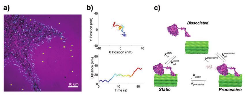
2021 PI Meeting Proceedings
enable scientists to monitor nutrient transformations cultivate and analyze biosystems from single cells to
and microbial metabolic activities in the rhizosphere complex communities.
that contribute to plant growth and health and to
investigate plant-microbe interactions that involve Characterizing Diverse Molecules Across Scales
chemical communications that travel through the rhi- BSP research teams are developing capabilities to
zosphere. Other applications of nucleic acids to image study molecular signatures and processes that are
plant and microbial activities include the detection highly diverse and cover a broad range of length
of microRNAs using silver-coated gold nanorods and scales. These include atomic isotopes, metabolites,
SERS sensing, as well as the detection of riboswitches plant hormones, silica, trace elements, redox metabo-
that act as metabolite reporters using quantitative lism, microbial electron transfer, membrane potential,
phase imaging that leverages a light-sheet fluores- intercellular trafficking, cellulose and lignin synthesis
cence technique and Raman imaging. and degradation, microRNAs that regulate lignifica-
tion, enzymes and other proteins secreted by plants,
Tracking Molecules In Situ and in Real Time and quorum-sensing molecules.
BSP researchers are focusing on understanding a
variety of biological systems for better controlling The functional dimensional scales in biological sys-
plant health and growth to improve bioenergy tems are vast, spanning molecules to multiorganismal
resources. The subject organisms include plants, systems. Because these systems are hierarchical in
bacteria, fungi, and their combinations. Teams are nature, activities on longer length and time scales are
developing sophisticated instruments to image built on activities and structures on shorter length
metabolism in a single organism, gene expression, and time scales. Therefore, processes must be fully
and regulatory molecules (e.g., microRNAs, quorum- explained at the molecular level to be fully under-
sensing molecules, and protein kinases) that operate stood at the organismal or multiorganismal level.
in intact organisms or are involved in communica- Recognizing this need, BSP supports some innovative
tion between organisms. Several approaches are cross-scale imaging approaches that include plas-
underway to capitalize on the advantageous spec- monic nanoprobes to track single molecules. Also
troscopic properties offered by semiconductors, supported is 3D tracking with high-speed AFM and
polymers, and quantum dots. Nuclear-based imaging optical tweezers to control molecules or microbes,
technologies such as Positron Emission Tomography enable force measurements, or track molecules such
enable scientists to visualize and quantify the move- as cellulose synthase as it moves along the mem-
ment of radiolabeled nutrients, plant hormones, and brane or cellulase as it moves along cell walls. These
other signal molecules within intact live plants. How- studies will answer important questions regarding the
ever, the widespread use of these technologies has mechanisms of cellulose synthesis and degradation.
been hampered by access to the limited number of Understanding such mechanisms will, in turn, enable
facilities that have the unique capabilities to produce the development of biomass feedstocks that more
these specialized agents. BSP-supported researchers readily can be converted to biofuels and bioproducts.
are developing instruments that will address the
critical need of measuring these features directly, Quantum-Enabled Techniques
enabling a future in which molecular signatures can BSP has recently added state-of-the-art quantum-
be tracked in real time and over time periods consis- enabled bioimaging projects at national laboratories.
tent with the biological processes under study. These Quantum-enhanced X-ray microscopy uses entangled
developments will include the capability of visual- X-rays beams. With the ghost imaging technique,
izing biosystems as they respond to external stressors samples are illuminated using less-intense beams with
and perturbations such as nutrient starvation and energy more suitable for maintaining biological integ-
chemical exchanges. BSP researchers also are using rity. Furthermore, the quantum nature of the imaging
synthetic rhizosphere microhabitats, transparent soil process enables visualization of details impossible to
microcosms, and versatile nanofluidic and microflu- detect with classical methods. BSP researchers also are
idic imaging and sampling devices simultaneously to developing a new microscope using entangled photon
viii2021 PI Meeting Proceedings
pairs to visualize water, lignocellulose, and lipids in struction, or boosting feedstock sustainability and
plants. To probe samples, this system uses a wave- plant drought tolerance. Organisms under study
length that can be in the near- or mid-infrared range include:
where vibrational fingerprinting to identify key molec-
ular species is possible. Detection and imaging are • Living plants — Arabidopsis thaliana, Medicago
then performed with visible light using high-efficiency truncatula, Brachypodium distachyon, Populus sp.,
and low-noise imaging detectors. Pinus taeda, and Zea mays.
Research teams are developing a high-quality 3D • Microbial chemotrophs — Bacillus subtilis, Yarrowia
imaging modality that uses quantum-entangled lipolytica, and Pantoea sp.
photon pairs to obtain more information on fluores- • Microbial phototrophs — Cyanothece sp.,
cence and scattering events than is available with Rhodopseudomonas palustris, Ostreococcus tauri,
standard fluorescence or scattering measurements. Chlamydomonas reinhardtii, and Synechococcus sp.
The system uses two separate 2D detectors to obtain
three- and four-dimensional information about the • Systems for studying plant-microbe interactions
same photon, providing 3D optical imaging at high — Arbuscular mycorrhizal symbioses, Glycine
frame rates to monitor dynamic host-bacterial interac- max with Bradyrhizobium japonicum, and Suillus
tions in bioenergy algal pond and plant systems. brevipes with P. taeda.
Also under development is a hybrid quantum-enabled
imaging platform that combines advances in adap-
Research Challenges and
tive optics, quantum entanglement, coincidence Future Opportunities
detection, ghost imaging, quantum phase-contrast Multidisciplinary Research Teams
microscopy, and multidimensional nonlinear coherent
(nonentangled) photons and four-wave mixing. This Biological imaging is inherently transdisciplinary,
system will enable researchers to visualize photore- and successful teams need to continue to reflect this
ception in phytotropin and phytochrome proteins and approach to advance BSP programmatic goals. Mul-
other quantum coherent processes that occur natu- tidisciplinary teams are needed to integrate imaging
rally within biosystems, improving the ability to track results with the corresponding genomic, proteomic,
ultrafast protein dynamics and the flow of metabolites lipidomic, and metabolomic changes within cells to
between biological compartments in real time. further understand biological complexity and hetero-
geneity. Achieving this understanding requires com-
Moving Toward More Complex Systems bining the expertise from researchers in conventional
and quantum-enabled imaging technology, nano-
Although many of the initial samples BSP researchers
science, computer science, structural biology, bio-
use to test new instruments and methods may be
chemistry, plant physiology, microbiology, genomic
from canonical model systems, the program should
science, ecology, soil science, and biogeochemistry.
continue to evaluate and adapt to real-world biosys-
This cross-disciplinary approach will be a critical step
tems as well. For example, label-free identification of
toward connecting phenotypes with genotypes and
microbes obtained from the environment remains
translating laboratory-developed technologies into
a grand challenge in biology, so extending BSP- the natural environment.
developed label-free approaches to such microbes in
the long term is a next frontier. Further Integration of Technologies into
In addition, while focusing on high-resolution Multimodal Hybrid Instruments
imaging, some BSP-supported projects are applicable BSP-supported development of individual imaging
to more complex biological systems and challenges techniques is making significant strides. These
relevant to bioenergy and the environment, such technologies range from complementary targeted
as understanding quorum sensing, improving lipid and untargeted methods to destructive and nonde-
feedstock yields, enhancing lignocellulosic decon- structive imaging modalities (e.g., optical, scanning
ix2021 PI Meeting Proceedings
probe, mass spectrometric, X-ray, and ion-based important advances are integrated data processing
approaches) that cover a wide range of spatial and algorithms, visualizations, and modeling, which are
temporal scales. The recent addition of cutting-edge, key components for properly interpreting the diverse
sophisticated and laboratory-based imaging methods sets of imaging data, omics-based organismal models,
(e.g., quantum entanglement and super-resolution and other information emanating from BER genomics
techniques such as STORM and PALM) strongly com- research.
plement the sensing and imaging approaches more
suitable for general laboratory and field use. Advances in Data Management and Analytics
To enable effective extraction of critical biological
In addition to pursuing advances within each of these
and environmental information from experimental
techniques, a major programmatic focus moving
data, major advances are needed in data storage,
forward should be on making the developments
processing, and visualization. BSP’s long-term goal
robust, easy to use, and accessible to the BER research
is to develop enabling capabilities that can generate
community. One approach toward meeting this goal
could be to further integrate these different and spatially and time-resolved snapshots of relevant
complementary approaches into hybrid all-in-one cellular metabolism, including both primary and
instruments. Multimodal spectral imaging in a single secondary metabolites as well as genomic biomarkers
and user-friendly setup across nano-, micro-, meso-, and internal and secreted compounds. Achieving
and macroscopic spatial domains will be a useful and real-time data collection and interpretation of these
versatile tool for future users. There is also a need to integrated data will lead to major advances in bio-
develop highly specialized, sophisticated instrumen- imaging technology that will improve monitoring
tation for fundamental research in the laboratory as and phenotyping of plant and microbial systems and
well as portable and easy-to-use instrumentation expand the understanding of molecular and genomic
for large-scale monitoring applications in the field. pathways, in both the laboratory and in complex nat-
Previously unachievable studies of microbes, plants, ural environments. These advancements will require
and other species in their environments will be pos- new methods and algorithms to handle increasingly
sible due to the new capabilities provided by these challenging volumes of data, along with automated
instruments. Results of these studies are expected to and machine learning approaches to rapidly analyze
reveal new insights on how to optimize development this data and identify biologically and environmen-
of sustainable bioenergy resources. tally meaningful signals.
Of interest is a central clearinghouse for archiving
Cross-Platform Data Fusion and Integration
experimental and simulation data that incorporates
With BSP’s expansion and the rapid increase in mon- a standardized output and imaging framework for
itoring modalities, data integration across multiple different and potentially widely adoptable analytical
technologies and approaches remains a high priority. modalities. Such a data repository could be indepen-
Data fusion (i.e., linking complementary data from dif- dent or integrated with the DOE Systems Biology
ferent techniques) will produce a more holistic picture Knowledgebase (KBase) and take advantage of
and better understanding of the biological systems advances in artificial intelligence to extract patterns
being imaged. Facilitating cross-platform bioimaging from raw data for improved organization, interpreta-
systems will require indexing and registering images tion, and representation.
(e.g., multifunctional tracers, probes, and sensors to
serve as cross-platform fiducial markers) and mean- Another opportunity for improving data interpret-
ingfully co-referencing and co-registering disparate ability is to leverage computer science (CS) graduate
datasets for the same sample but of different for- programs to help accelerate image processing or
mats, magnifications, or resolutions. Also needed data analysis pipelines for the large datasets collected
are models capable of integrating multimodal data within BSP. Many CS programs require students to
spanning a wide range of spatial and temporal scales gain access to and experience with real-world data by
to effectively extract causality from observations and building new software or other algorithms for more
understand complex biological phenomena. Other effective analytics. Using the plethora of BSP data,
x2021 PI Meeting Proceedings
principal investigators (PIs) could sponsor CS grad- Field-Deployable Capabilities for Whole Organisms
uate students to develop the next frontier of bioim- and Complex Communities
aging analytics tools.
Another important challenge for the near term is
New Probes and Quantum-Enabled Techniques the extension of laboratory-based approaches into
to Expand Investigations applications for whole organisms and plants in their
natural environments and under field settings. This
In parallel with BSP’s instrumentation development expansion will require incorporating the dynamics
efforts, there is also a critical need for probe devel- of microbially driven biogeochemistry (e.g., within
opment that enables identification, sensing, and the rhizosphere, biofilms, and other key biological
functional imaging of various targets within complex interfaces) into the imaging process. Although there
biological systems, ranging from key metabolites has been progress in imaging genomic biotargets
to molecular and genomic biotargets (e.g., mRNA, in living plants, advances are needed in imaging
microRNA, proteins, and regulatory small mole- complex native microbial communities to decipher
cules). Relevant key advances would include the their organization and the multiple metabolic pro-
simultaneous marking, spatially resolved tracking, cesses occurring simultaneously in space and time.
and sensing of multiple players (e.g., elements, Concerted efforts will also be needed to develop the
isotopes, enzymes, metabolites, and other molec- ability to probe inherent signals within nontractable
ular biomarkers) in a given biological system. BSP’s microbes in the environment and to create pathways
wide range of biosensing and imaging capabilities that enable in situ microbial synthesis of probes for
are expected to provide the essential flexibility to assaying function and activity. Furthermore, in addi-
broaden the scope of investigations, opening new tion to sophisticated lab-based analytical methods,
possibilities to discover yet-unknown key biomarkers portable instrumentation and practical techniques
or intermediates. will allow the detection of weak optical signals from
Probing a sample inherently perturbs it, yet methods whole-organismal data containing strongly inter-
based on selective probe-induced perturbations fering background signals such as fluorescence,
of key biotargets or metabolic pathways of specific ambient light, vibrations, and fixed-pattern noise
organisms could provide opportunities to investigate encountered under field conditions.
and understand biological processes that otherwise
would be difficult to unveil. BSP researchers are also
Correlative Frozen or Fixed-Sample Imaging
pursuing an approach to minimize perturbation: Finally, it is important to realize the benefits of com-
the incorporation of quantum-enabled science and bining additional approaches that may be destructive
technologies. The potential of using ghost imaging or applicable only to frozen or fixed samples, which
for bioimaging applications is intriguing because this are typically outside the scope of the BSP portfolio.
approach can image a sample by detecting a photon Many current BSP capabilities are based on optical
that never interacted with the sample. Furthermore, approaches that empower real-time or in situ obser-
the ability of quantum-entangled two-photon vations of living systems, but they do not provide
imaging to provide higher detection efficiency and a complete picture of the sample or a whole-cell
decrease the total photon flux needed to observe a context. Some science questions require more holistic
high-contrast image, and thereby permit very low imaging and analysis to decipher complex associ-
dose imaging that could minimize photodamage ations within or between living cells. Combining
effects, would facilitate longer-term, time-resolved current BSP approaches with sequential downstream
imaging of biosystems. Deeper penetration by X-rays frozen or fixed-sample correlative imaging (such as
combined with X-ray-entangled imaging will enable cryo-electron microscopy or nano-secondary ion
imaging in thicker biological samples. The develop- mass spectrometry) can provide additional spatial,
ment and integration of these and other quantum- ultrastructural, or chemical context needed for critical
enabled imaging technologies or sensors into the scientific breakthroughs related to cellular sensing
BSP portfolio could significantly expand the range of and metabolite response, flow, and fate. Such multi
scientific questions the program addresses. modal and correlative imaging approaches should
xi2021 PI Meeting Proceedings
be encouraged within BSP to accelerate the under- an approach would also facilitate continued tech-
standing of biosystem complexity and organization nological developments through the important
and their impact on dynamics. user-developer feedback loop and the synergistic
interactions between imaging scientists and facil-
Summary of Opportunities and Needed ities. These interactions would expand the scope
Developments of research being conducted using BSP-developed
capabilities.
In summary, several advances are needed in key areas:
• Integrating bioimaging techniques with advanced Bioimaging Science Program Annual Meeting
probes and delivery mechanisms that expand the BSP’s annual PI meeting provides an important
monitoring capability for important biotargets avenue for the program to increase the cross-platform,
ranging from key metabolites to molecular and cross-disciplinary, and multiscale synergies needed to
genomic biomarkers. achieve its goals. Scheduling this meeting proximal
• Developing new or improved conventional or to the DOE Genomic Science program (GSP) annual PI
quantum-enabled imaging technologies capable meeting creates invaluable opportunities for syner-
of monitoring biological systems in their natural gistic interactions with that community. Furthermore,
states or as they respond to environmental pertur- inviting imaging experts external to BSP as keynote
bations and stressors. speakers injects novel perspectives and approaches
into discussions during the program’s annual meeting.
• Developing new or improved biosensing and Additional interactions across BSP’s research teams
bioimaging approaches that enable real-time data (e.g., through teleconferencing or web conferencing)
collection across the full range of relevant spatial could help maintain this interactive momentum and
scales in the laboratory and under field conditions. catalyze new directions of investigation.
• Correlating multimodal dynamic and static “snap-
shot” imaging methods, both destructive and
Additional Cross-Program Interactions and
nondestructive, to provide a holistic understanding Community Engagement
of chemical-structural-functional linkages. A new mechanism that allows supplemental funding
could foster even more direct cross-fertilization and
• Establishing cross-platform protocols for sample
interaction between the GSP and BSP research com-
preparation, calibration, indexing and spatial
munities. The envisioned new class of funding could
registration, data verification, and correlation to
supplement the travel and supply costs of embed-
increase the suite of complementary analyses
ding a graduate student or postdoctoral researcher
that can be conducted on a given sample or suite
from a GSP-funded research group into a BSP-funded
of samples.
research group for 1 to 6 months. This arrangement
• Developing methods to increase throughput for would stimulate more direct collaboration and cross-
more mature imaging technologies that can be talk between the two programs, yielding benefits for
used for new applications. both. For GSP researchers, this collaboration would
give them access to cutting-edge technology that
Expanding BSP’s Impact and Interactions otherwise may have been beyond reach, leading
to new scientific discoveries. For BSP researchers, it
Community Access to BSP-Developed Technologies would provide access to new science and samples
Through User Facilities they could use for adapting, benchmarking, and
User accessibility to new BSP technologies and evaluating the performance of their newly developed
approaches will be a key factor for the program’s instrumentation and methods. This funding mecha-
success and longevity. Deploying some BSP imaging nism would be very similar to DOE’s Office of Science
capabilities to DOE scientific user facilities would Graduate Student Research opportunity. However,
expand the research community’s access to these instead of enabling researchers to pursue part of their
technologies, thereby increasing their impact. Such graduate thesis research at a DOE national labora-
xii2021 PI Meeting Proceedings
tory or user facility, it would support GSP researchers nity of scientists who could use the program’s
who want to visit and use the new technologies bioimaging approaches. As part of outreach to BER
developed by BSP-funded groups at universities and researchers, the portal would detail BSP’s diverse
national laboratories. technological approaches, highlight the applications
for which they are best suited, and provide a forum
Finally, the creation of a bioimaging capability portal for information dissemination, tutorials, and training
could enhance BSP’s impact on a wider commu- opportunities.
xiii2021 PI Meeting Proceedings xiv
2021 PI Meeting Proceedings
Abstracts
Multimodal Single-Cell/Particle Imaging and Engineering for Energy Conversion in Bacteria
Principal Investigators: Peng Chen (PI), Tobias Hanrath,
and Buz Barstow
Institution: Cornell University
Email: pc252@cornell.edu
Research Plans and Progress: This project’s research
aims to combine quantum materials synthesis, bacterial
synthetic biology, and multimodal single-entity imaging
to quantitatively study how hybrid quantum dot (QD)–
bacteria systems convert light to value chemicals at the
single- to subcell level, with the overall goal of gaining
insights to guide the engineering of QDs and bacterial In this schematic of bacterial cells sitting on a layer of
genetics for more efficient bioenergy conversion. quantum dots (QDs) on top of a transparent electrode,
focused laser beams excite local regions on the QD
On quantum materials, the project focused on developing layer. The excited electrons can be donated to the
semiconductor cadmium sulfide (CdS) thin films on indium bacteria for subsequent reduction of CO2 to biomass,
tin oxide (ITO) as photosensitizers; for that, CdS’s energy in which a photoelectrochemical current is generated.
The QDs and the bacterial and photoelectrochemical
gap and redox potential can be tuned by its size and processes can all be imaged. Courtesy Tobias Hanrath,
surface chemistry. The project examined partial surface Cornell University.
oxidation and ligand-exchange processes and character-
ized them using spectroscopy and photoelectrochemical
measurements. The project also studied using PEDOT:PSS
between the ITO electrode and the QD thin film to ensure
and/or PhaP1 that decorates the surface of biomass PHB
that photoexcited electrons flow toward the microbe, thus
granules, with a (photoactivatable) fluorescent protein.
focusing on photoreduction (rather than photooxidation)
Under H2/CO2/air lithoautotrophic growth, researchers
processes in the QD/microbe hybrids.
determined: (1) the intracellular concentrations of MBH
On bacterial biology, the team has completed a systematic and SH; (2) MBH and SH concentrations both have strong
survey of thermodynamic constraints on electromicrobial positive correlations with PHB accumulation, with SH
conversion of CO2 and electricity to bioproducts, encom- having slightly stronger correlation; and (3) biomass
passing microbes that uptake electricity by H2-oxidation accumulation remains unchanged upon deleting MBH
(Ralstonia eutropha) and by extracellular electron uptake but decreases by ~95% upon deleting SH, suggesting
(Shewanella oneidensis). Team members demonstrated SH’s role in supplying reducing equivalents toward bio-
that both methods of electron uptake have comparable mass synthesis.
high maximum conversion efficiencies. This theoretical
Team members further examined the photoelectrochem-
analysis allowed for building a 10-point roadmap for the
ical current across single semiconductor-cell interfaces
development of electromicrobial production technology.
for individual R. eutropha cells in contact with a semi-
The project has also identified genes encoding an electron
conductor film. With CdS (n-type), researchers measured
uptake pathway in S. oneidensis. Using a high-throughput
single-interface photoelectrochemical currents at anodic
screening, researchers discovered 150 genes that affect
conditions to quantify the cells’ ability to accept pho-
electron uptake; four of them are indispensable. This set
togenerated holes (i.e., donate electrons). Many cells
of genes provides a portable electron uptake module,
show enhanced or suppressed photocurrent relative
transferrable to highly engineerable microbes to enable
to CdS films alone, suggesting pronounced cell-to-cell
electron uptake and power CO2 fixation.
heterogeneities and highlighting the need of single-
On multimodal single-entity imaging in the R. eutropha entity experiments. Researchers examined one-on-one
chromosome, researchers have tagged the membrane- correlations between cell-induced photocurrent changes
bound hydrogenase (MBH), the soluble hydrogenase (SH) and the characteristics of the associated single cells (e.g.,
12021 PI Meeting Proceedings
cell size/shape, the amounts of hydrogenase/PHB). Team aper: Salimijazi, F., et al. 2020. “Constraints on
3. P
members observed a clear correlation between cell the Efficiency of Engineered Electromicrobial
induced photocurrent changes and cell size. The team Production,” Joule 4(10), 2101–30. DOI: 10.1016/j.
also employed Cu2WS4 (p-type) and measured single- joule.2020.08.010.
interface photocurrents at cathodic conditions. Many cells
4. P
reprint: Rowe, A. R., et al. 2021. “Identification of a
are associated with cathodic photocurrent enhancement,
Pathway for Electron Uptake in Shewanella oneiden-
indicating that under this condition most cells exhibit
sis,” bioRxiv. DOI: 10.1101/2021.01.12.426419.
strong electron-accepting capabilities.
Potential Benefits and Applications: This research will
Current and/or Anticipated Accomplishments and
provide quantitative knowledge to understand the basic
Deliverables:
materials and biological factors as well as guiding princi-
1. Image analysis software to find electron uptake ples to engineer and improve such systems. If successful,
genes in S. oneidensis: github.com/barstowlab/ this research will transform the study of hybrid inorganic
macroscope-imageanalyzer. bacterial systems for energy and chemical conversions.
2. C
ode for calculating electromicrobial production The proposed experiments should break new scientific
efficiency: github.com/barstowlab/rewiredcarbon. grounds and open unforeseen opportunities.
22021 PI Meeting Proceedings
Plasmonics-Enhanced Optical Imaging Systems for Bioenergy Research
Principal Investigators: Tuan Vo-Dinh1 (PI), Tai-Ping (iMSs) that can be monitored using surface-enhanced
Sun,1 and Kenneth Kemner2 Raman scattering (SERS). The team is currently develop-
Institutions: 1Duke University and 2Argonne National ing innovative imaging technologies for visualization and
Laboratory quantitative characterization of biomarkers related to
Email: tuan.vodinh@duke.edu molecular processes and cellular function within living
plants, namely Multimodal Optical Sensing and Imaging
The goal of this project is aimed at addressing the DOE Combinatory (MOSAIC) System. The advanced MOSAIC
Funding Opportunity Announcement need to develop system will provide much-needed biofuel research tools
innovative and improved imaging instrumentation that such as elucidating the regulation of the pathway to
can enable visualization and quantitative characterization synthesize photosynthetic terpenes more efficiently for
of biomarkers and their dynamic role in cellular functions biofuel production and tracking pathways of carbon
in living plants relevant to DOE bioenergy programs. fixation in plants.
Research Plans and Progress, Including Objectives Current and/or Anticipated Accomplishments/
and Goals for the Project Period: Monitoring gene Deliverables for the Project Period: The project
expression in whole plants is a key requirement in has developed a strategy for efficient delivery of iMS
many important fields, ranging from fundamental plant nanoprobes into plant cells using silver-coated gold
biology to biofuel development. However, current nanostars (AuNR@Ag) for SERS sensing. Figure panels
methods to monitor gene expression in plants cannot be A and B show the transmission electron microscopy
performed directly in vivo. To overcome these limita- (TEM) image of AuNR@Ag (A) and the SERS detec-
tions, the project has developed in vivo imaging and tion of microRNAs (miRNAs) (B) using AuNR@Ag-iMS
biosensing of nucleic acid biotargets using plasmonic nanoprobes. Also shown is the confocal imaging coregis-
nanoprobes referred to as inverse molecular sentinels tration of iMS nanoprobes inside tobacco cells (C).
(D)
(A) TEM image of silver-coated gold nanostars (AuNR@Ag). (B) SERS spectra of AuNR@Ag-iMS in the presence
(bottom spectrum) or absence (top spectrum) of target miRNAs. (C) Representative confocal microscopy images
of Cy3-labeled AuNR@Ag-iMS infiltrated into tobacco plants expressing GFP fluorophore in the cytoplasm.
(D) Overlay of gold (Au; green), zinc (Zn; blue), and manganese (Mn; red) distributions in XRF confocal image of
200 μ × 200 μ area of leaf treated with AuNR@Ag. White lines drawn as an aid to identify four leaf cells within the
field of view. Dashed white lines drawn to delineate where the confocal plane of the image transitions from inside
to outside of the cell. Courtesy Tuan Vo Dinh, Duke University; and Ken Kemner, Argonne National Laboratory.
3You can also read